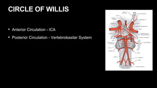
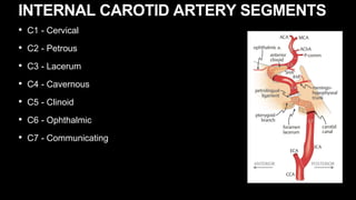
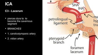
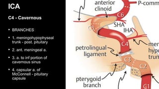
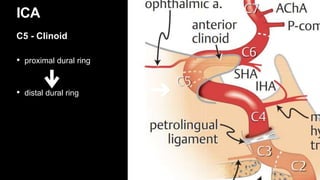
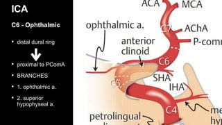
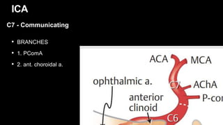
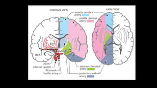
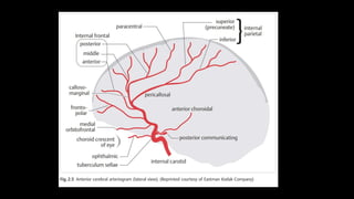
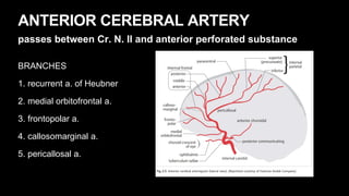
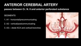
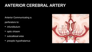
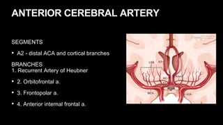
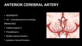
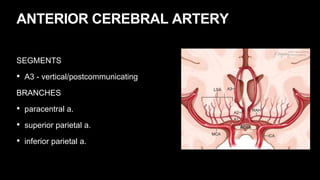
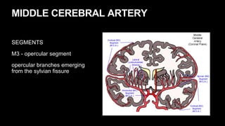
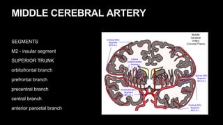
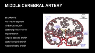
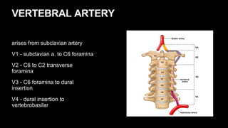
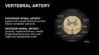
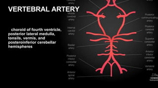
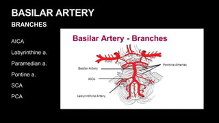
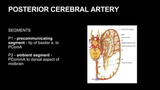
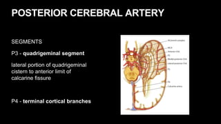
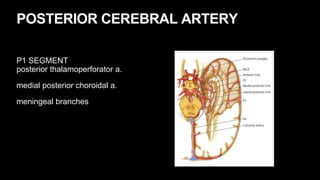
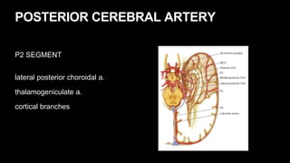
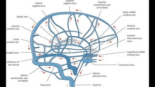
Mark Milo Asug, MD
VASCULAR ANATOMY
BLOOD SUPPLY TO THE BRAIN
• 2% of body weight
• receives 20% of CO
CEREBRAL BLOOD FLOW
CBF ml/100g/min
Normal 50
Ischemic Penumbra 8-23
Irreversible neuronal death <8
CIRCLE OF WILLIS
• Anterior Circulation - ICA
• Posterior Circulation - Vertebrobasilar System
INTERNAL CAROTID ARTERY
• C1 - Cervical
• C2 - Petrous
• C3 - Lacerum
• C4 - Cavernous
• C5 - Clinoid
• C6 - Ophthalmic
• C7 - Communicating
INTERNAL CAROTID ARTERY SEGMENTS
• carotid bifurcation to entrance to
petrous bone
• encircled with postganglionic
sympathetic nerves
ICA
C1 - Cervical
• ends at the posterior edge
of the foramen lacerum
• encircled with
postganglionic
sympathetic nerves
ICA
C2 - Petrous
• pierces dura to to
become the cavernous
segment
• BRANCHES
• 1. caroticotympanic artery
• 2. vidian artery
ICA
C3 - Lacerum
• S-shaped course
• enters subarachnoid
space adjacent to the
anterior clinoid
• pierces distal dural ring
ICA
C4 - Cavernous
• BRANCHES
• 1. meningohypophyseal
trunk - post. pituitary
• 2. ant. meningeal a.
• 3. a. to inf portion of
cavernous sinus
• 4. capsular a. of
McConnell - pituitary
capsule
ICA
C4 - Cavernous
• proximal dural ring
• distal dural ring
ICA
C5 - Clinoid
• distal dural ring
• proximal to PComA
• BRANCHES
• 1. ophthalmic a.
• 2. superior
hypophyseal a.
ICA
C6 - Ophthalmic
• proximal to PComA
origin
• travels b/w CN II
and III
• bifurcates into ACA
and MCA
ICA
C7 - Communicating
• BRANCHES
• 1. PComA
• 2. ant. choroidal a.
ICA
C7 - Communicating
ANTERIOR CEREBRAL ARTERY
passes between Cr. N. II and anterior perforated substance
BRANCHES
1. recurrent a. of Heubner
2. medial orbitofrontal a.
3. frontopolar a.
4. callosomarginal a.
5. pericallosal a.
ANTERIOR CEREBRAL ARTERY
passes between Cr. N. II and anterior perforated substance
SEGMENTS
1. A1 - horizontal/precommunicating
2. A2 - vertical/postcommunicating
3. A3— distal ACA and cortical branches
ANTERIOR CEREBRAL ARTERY
SEGMENTS
A1 - horizontal/precommunicating
• medial lenticulostriate a. - perforators to:
• optic nerve
• optic chiasm
• septum pellucidum
• anterior commissure
• pillars of the fornix
ANTERIOR CEREBRAL ARTERY
passes between CN II and anterior perforated substance
Anterior Communicating a.
perforators to:
• infundibulum
• optic chiasm
• subcallosal area
• preoptic hypothalamus
ANTERIOR CEREBRAL ARTERY
Anterior Communicating a.
perforators to:
• infundibulum
• optic chiasm
• subcallosal area
• preoptic hypothalamus
ANTERIOR CEREBRAL ARTERY
SEGMENTS
• A2 - distal ACA and cortical branches
BRANCHES
1. Recurrent Artery of Heubner
• 2. Orbitofrontal a.
• 3. Frontopolar a.
• 4. Anterior internal frontal a.
ANTERIOR CEREBRAL ARTERY
• SEGMENTS
• A3 - vertical/postcommunicating
BRANCHES
• Callsomarginal a.
• Pericallosal a.
• Middle internal frontal a.
• posterior internal frontal a.
ANTERIOR CEREBRAL ARTERY
SEGMENTS
• A3 - vertical/postcommunicating
BRANCHES
• paracentral a.
• superior parietal a.
• inferior parietal a.
MIDDLE CEREBRAL ARTERY
SEGMENTS
M1 - horizontal segment
extends horizontally and laterally
beneath the anterior perforated
substance, proximal to the
bifurcation (50%) or trifurcation
(25%).extends horizontally and
laterally beneath the anterior
perforated substance, proximal to
the bifurcation (50%) or
trifurcation (25%).
MIDDLE CEREBRAL ARTERY
SEGMENTS
M2 - insular segment
bifurcation to the genu
MIDDLE CEREBRAL ARTERY
SEGMENTS
M3 - opercular segment
opercular branches emerging
from the sylvian fissure
MIDDLE CEREBRAL ARTERY
SEGMENTS
M4 - cortical segments
MIDDLE CEREBRAL ARTERY
SEGMENTS
M1 - horizontal segment
uncal a.
temporopolar a.
ant. temporal a.
lateral lentriculostriate a.
MIDDLE CEREBRAL ARTERY
SEGMENTS
M2 - insular segment
SUPERIOR TRUNK
orbitofrontal branch
prefrontal branch
precentral branch
central branch
anterior paroetal branch
MIDDLE CEREBRAL ARTERY
SEGMENTS
M2 - insular segment
INFERIOR TRUNK
posterior parietal branch
angular branch
temporo-occipital branch
posterotemporal branch
middle temporal branch
POSTERIOR CIRCULATION
VERTEBRAL ARTERY
arises from subclavian artery
V1 - subclavian a. to C6 foramina
V2 - C6 to C2 transverse
foramina
V3 - C6 foramina to dural
insertion
V4 - dural insertion to
vertebrobasilar
VERTEBRAL ARTERY
BRANCHES
1. Extracranial
2. Posterior spinal a.
3. Anterior spinal a.
4. PICA
VERTEBRAL ARTERY
Three branches:
1. Segmental spinal branches
2. Muscular branch anastomoses with muscular branch of ECA (ascending
pharyngeal, thyrocervical, and costocervical)
VERTEBRAL ARTERY
3. Meningeal branch
Anterior meningeal branch → dura around the foramen magnum
Posterior meningeal branch → falx and posterior fossa dura
Blood supply to the falx cerebelli = posterior meningeal branches, occipital,
ascending pharyngeal, and PICA arteries
VERTEBRAL ARTERY
POSTERIOR SPINAL ARTERY -
gracile and cuneate fasciculi and the
inferior cerebellar peduncle
ANTERIOR SPINAL ARTERY -
pyramid, medial lemniscus, medial
longitudinal fasciculus, olive and
vagal and hypoglossal nuclei
VERTEBRAL ARTERY
choroid of fourth ventricle,
posterior lateral medulla,
tonsils, vermis, and
posteroinferior cerebellar
hemispheres
BASILAR ARTERY
BRANCHES
AICA
Labyrinthine a.
Paramedian a.
Pontine a.
SCA
PCA
POSTERIOR CEREBRAL ARTERY
SEGMENTS
P1 - precommunicating
segment - tip of basilar a. to
PComA
P2 - ambient segment -
PCommA to dorsal aspect of
midbrain
POSTERIOR CEREBRAL ARTERY
SEGMENTS
P3 - quadrigeminal segment
lateral portion of quadrigeminal
cistern to anterior limit of
calcarine fissure
P4 - terminal cortical branches
POSTERIOR CEREBRAL ARTERY
P1 SEGMENT
posterior thalamoperforator a.
medial posterior choroidal a.
meningeal branches
POSTERIOR CEREBRAL ARTERY
P2 SEGMENT
lateral posterior choroidal a.
thalamogeniculate a.
cortical branches
POSTERIOR CEREBRAL ARTERY
P3 SEGMENT
posterior temporal a.
internal occipital a.
parieto-occipital a.
calcarine a.
posterior pericallosal a.
VENOUS DRAINAGE
BRANCHES
√
VENOUS DRAINAGE
BRANCHES
√

Neurovascular Anatomy.pptx