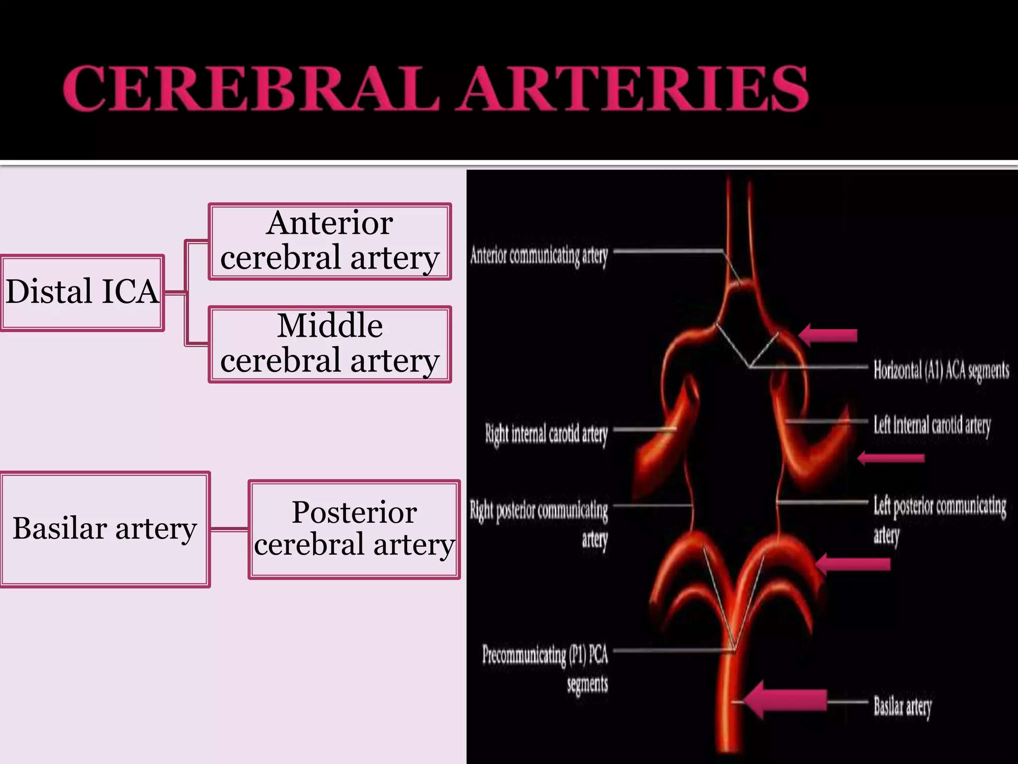
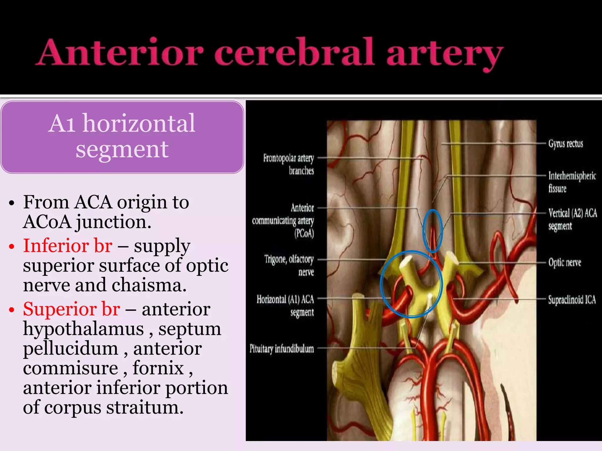
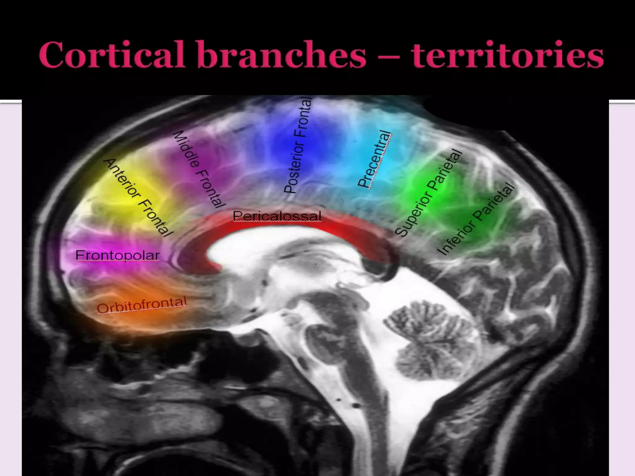
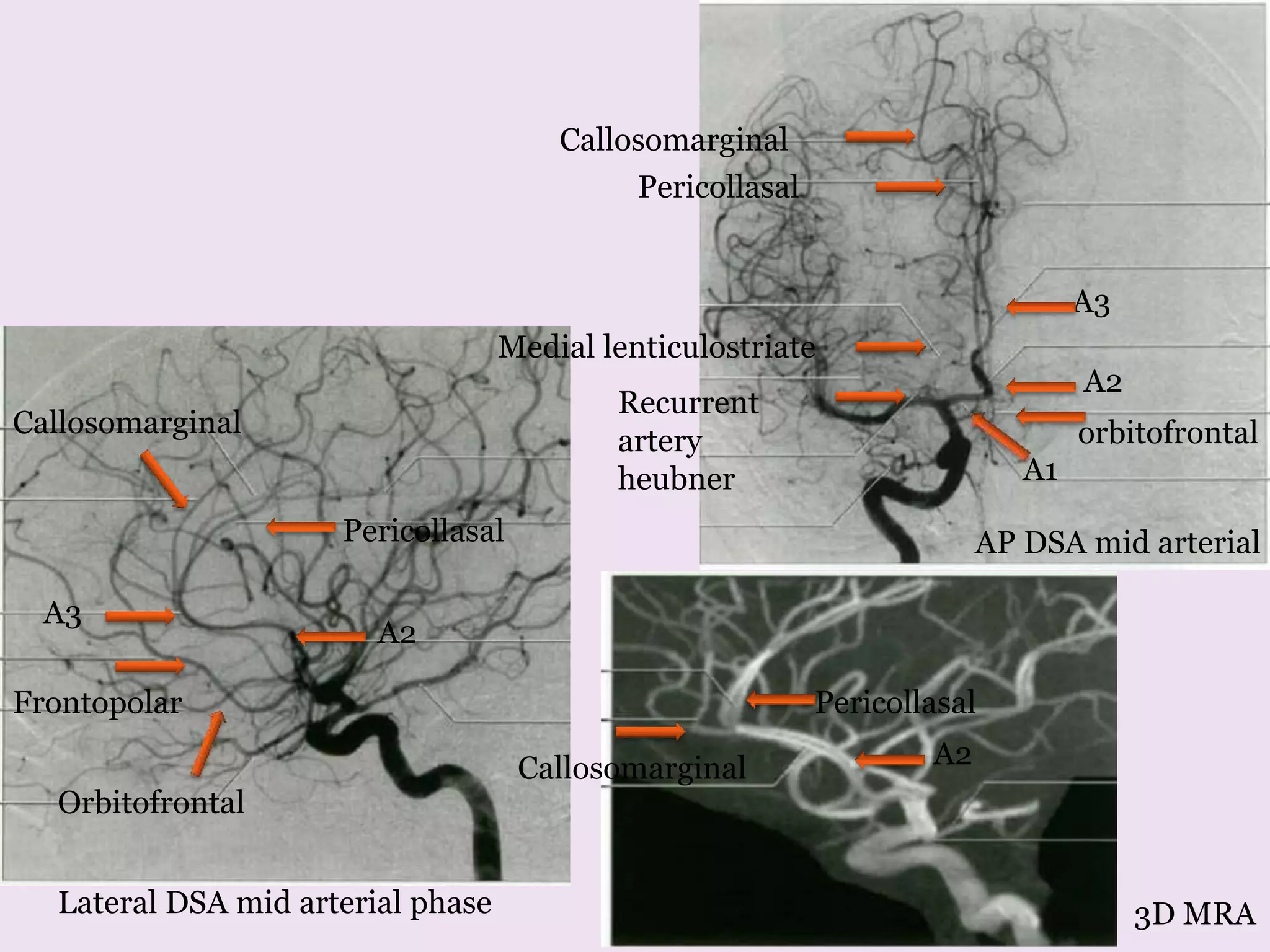
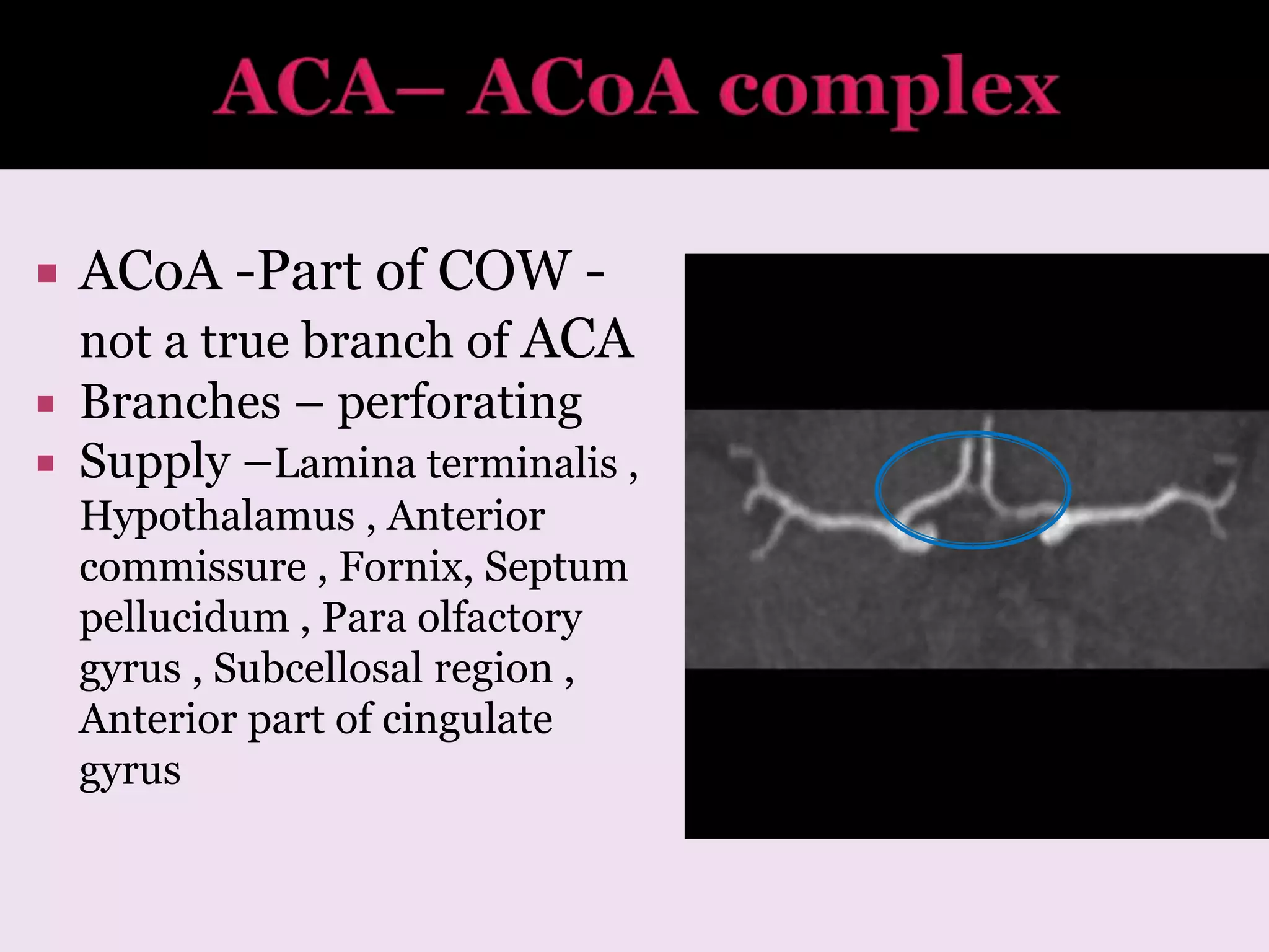
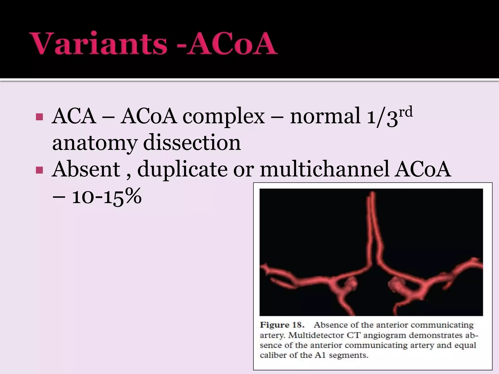
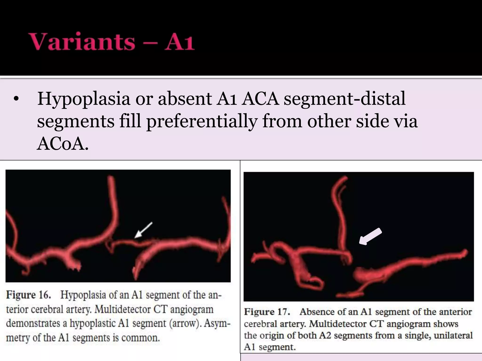
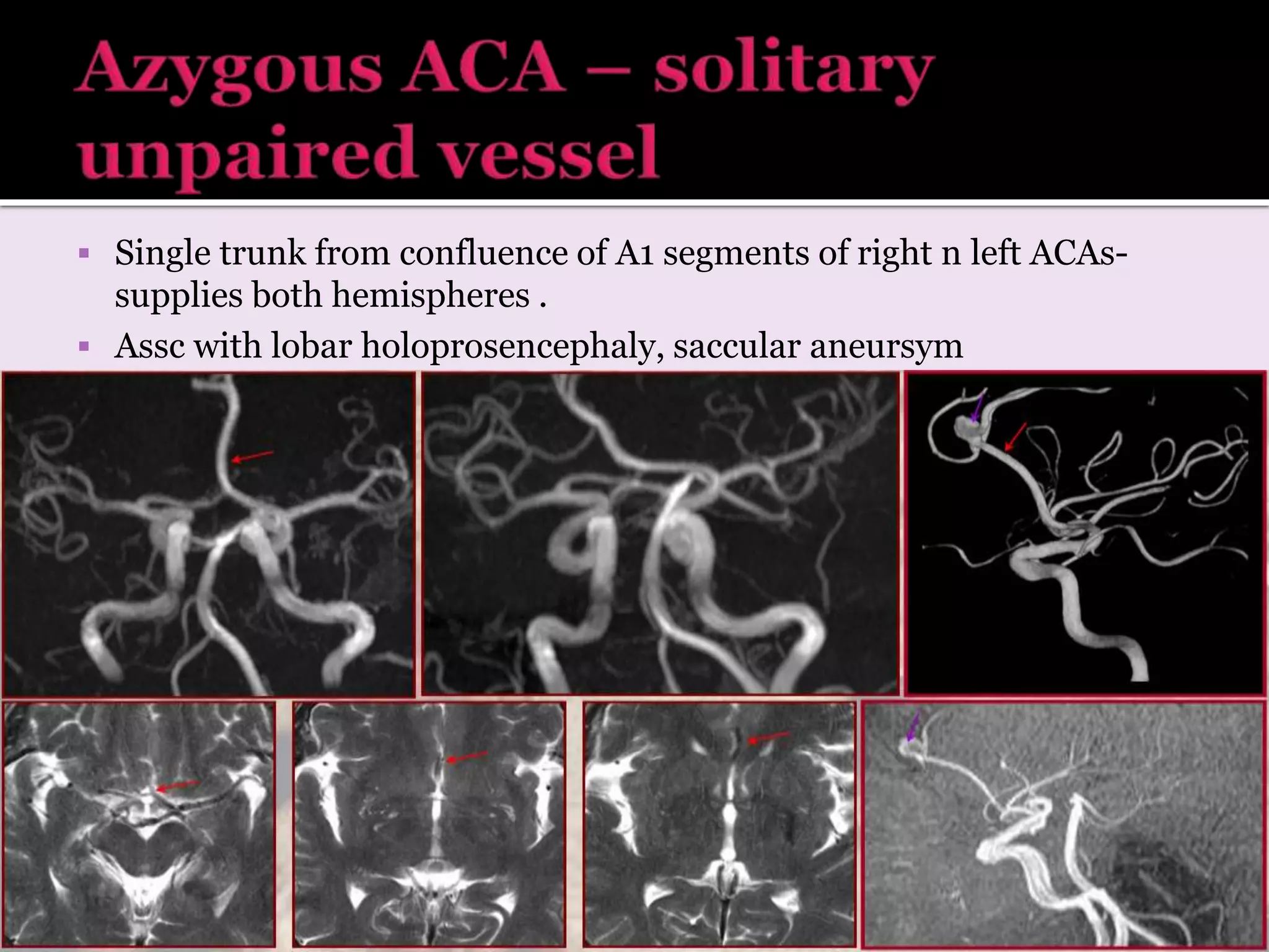
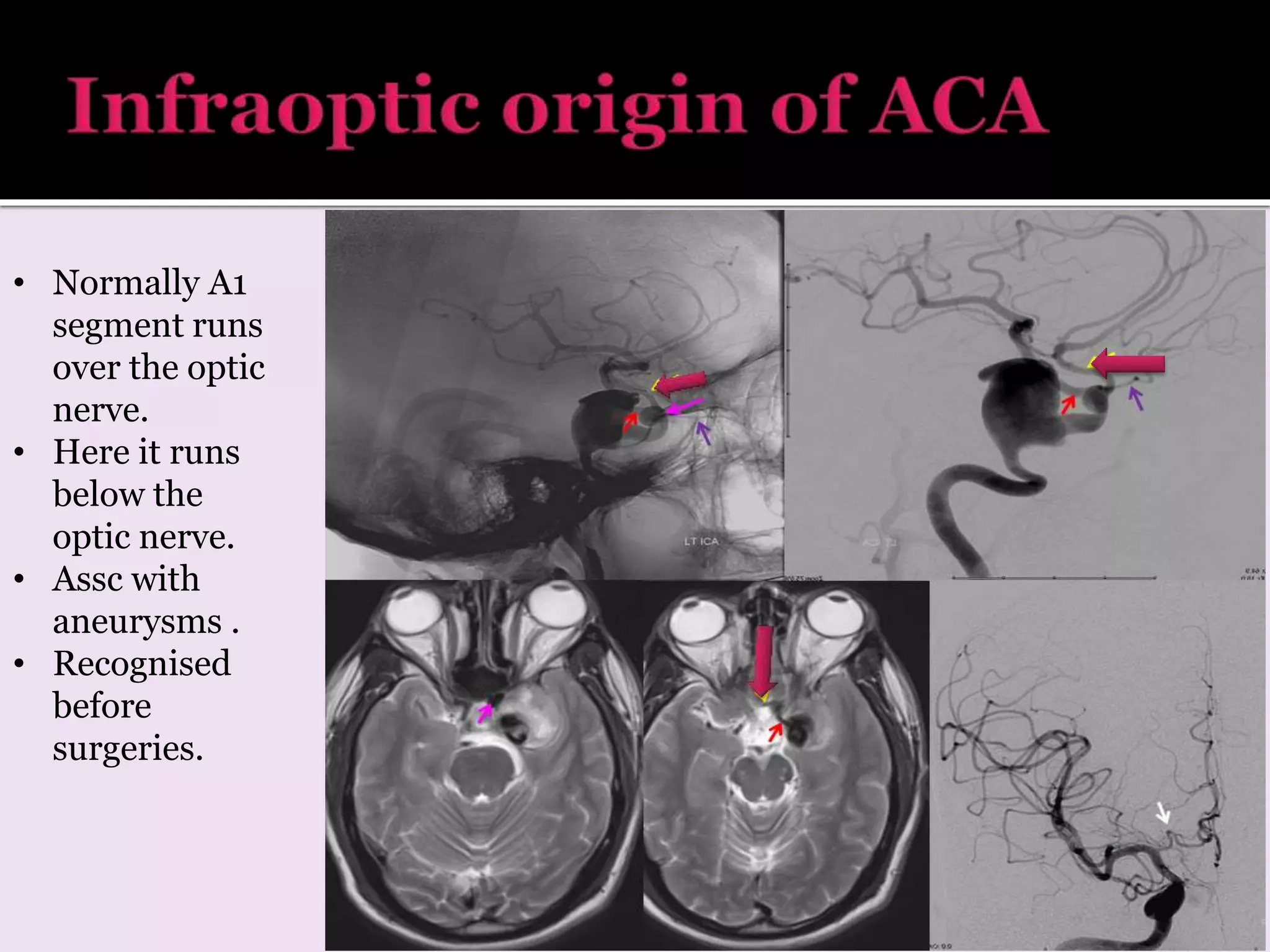
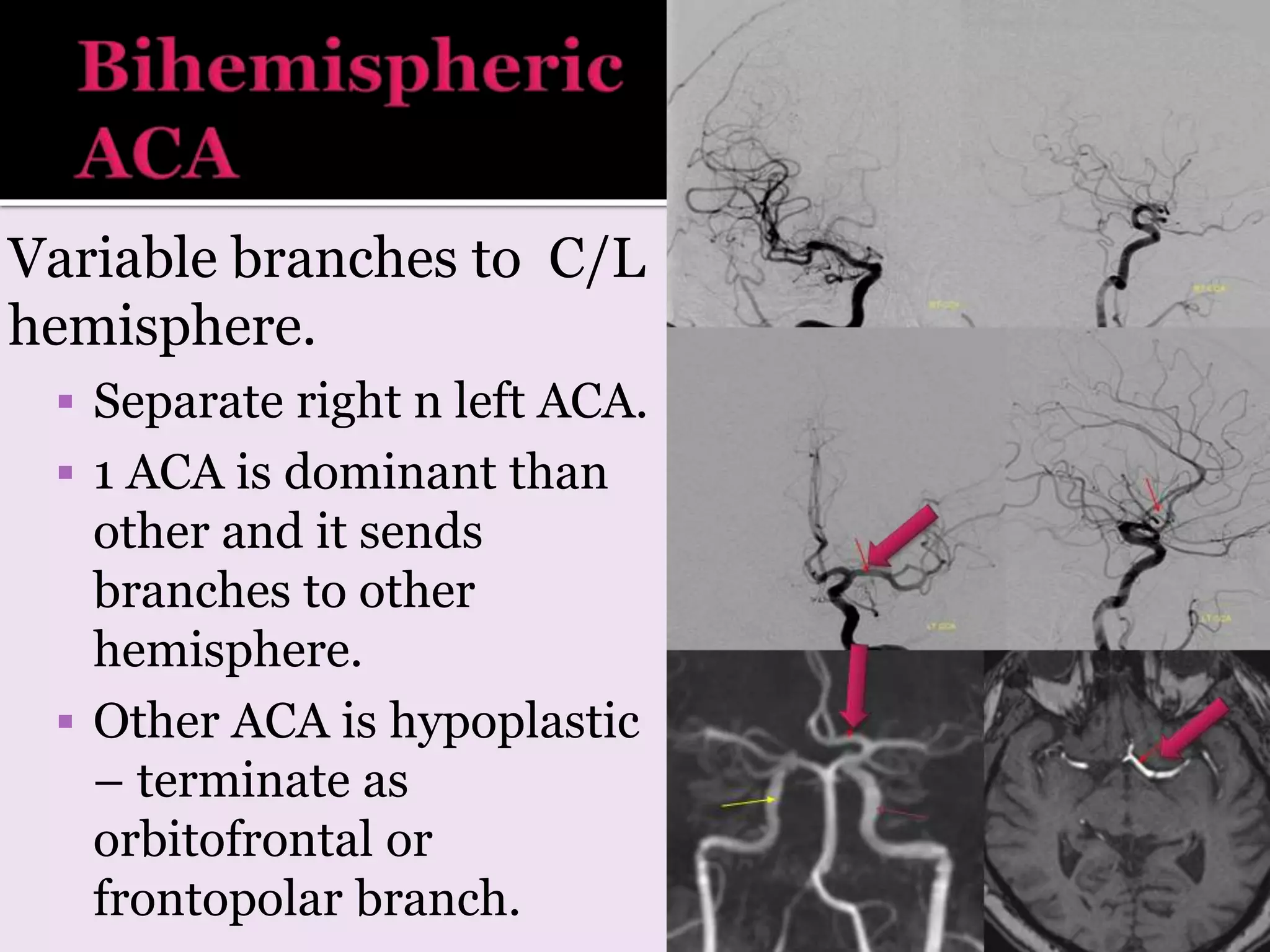
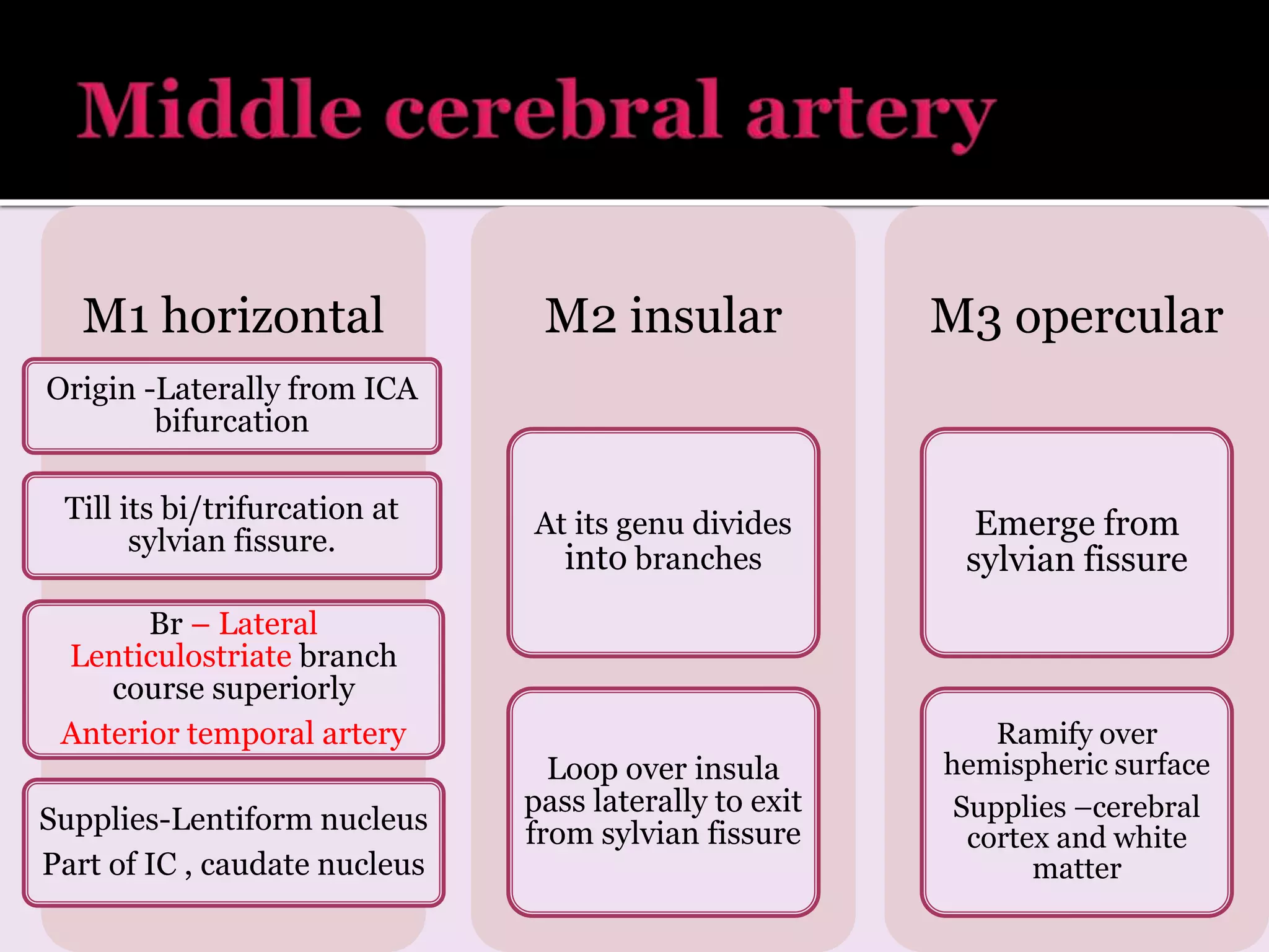
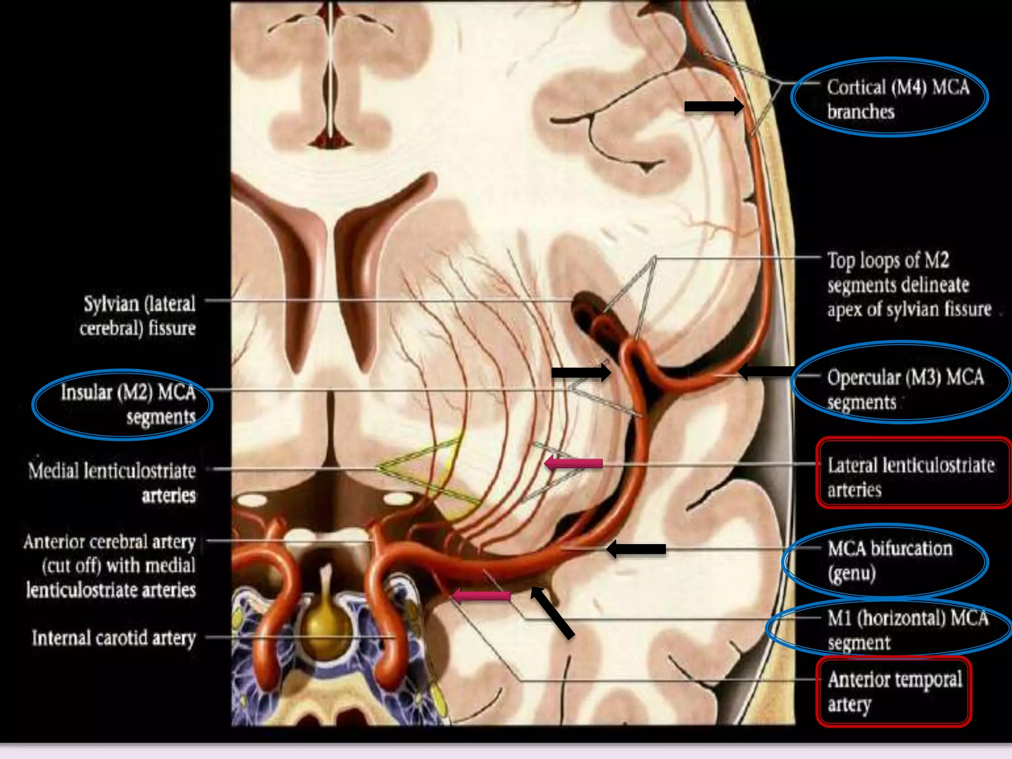
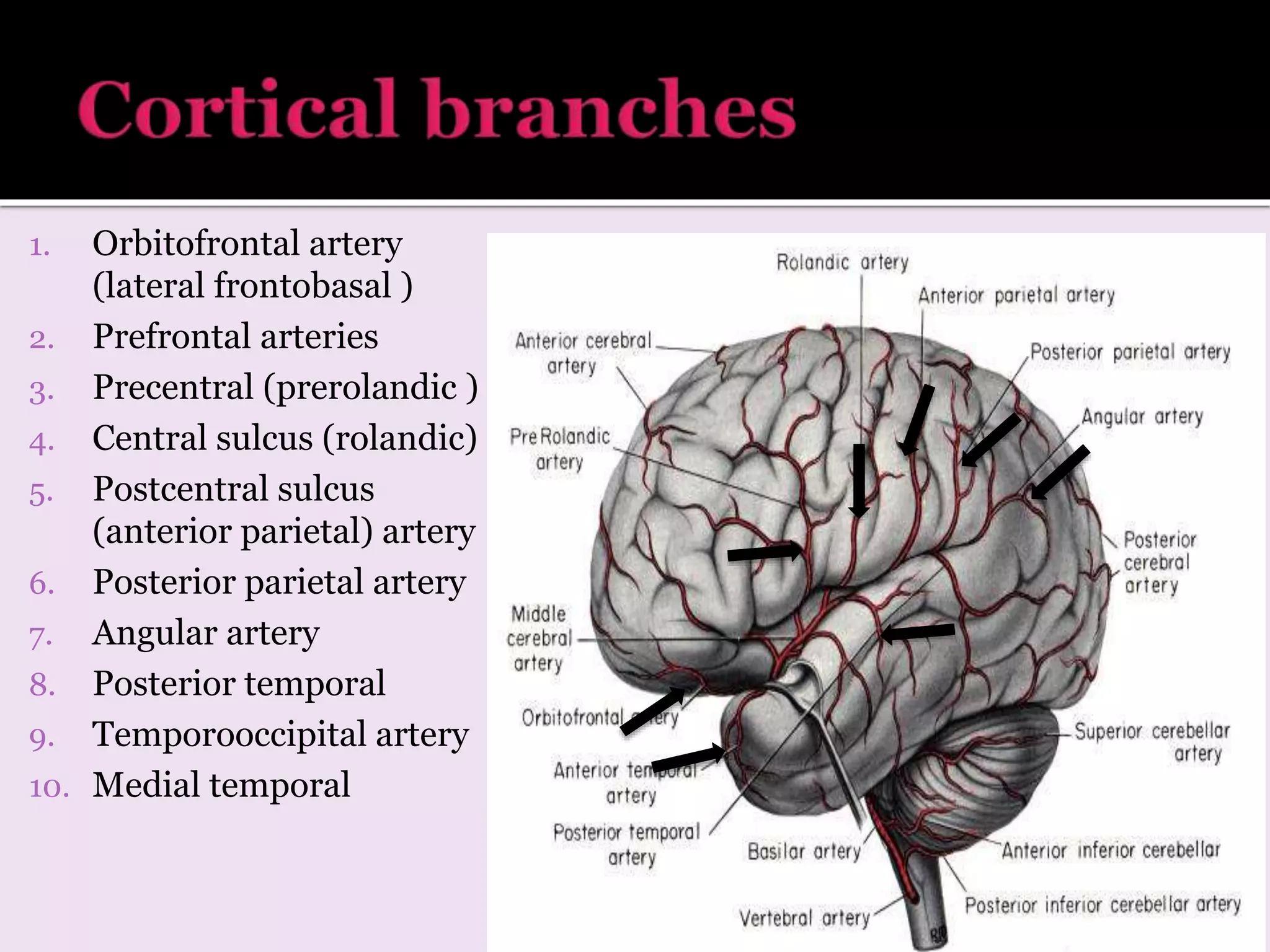
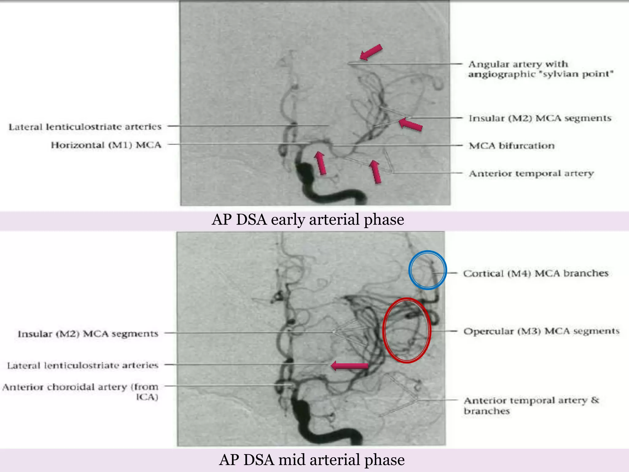
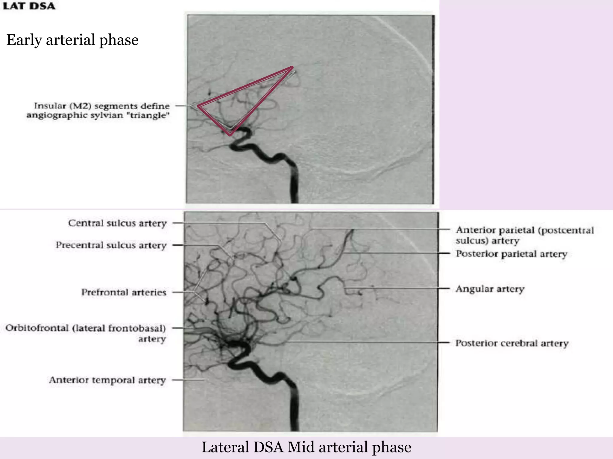
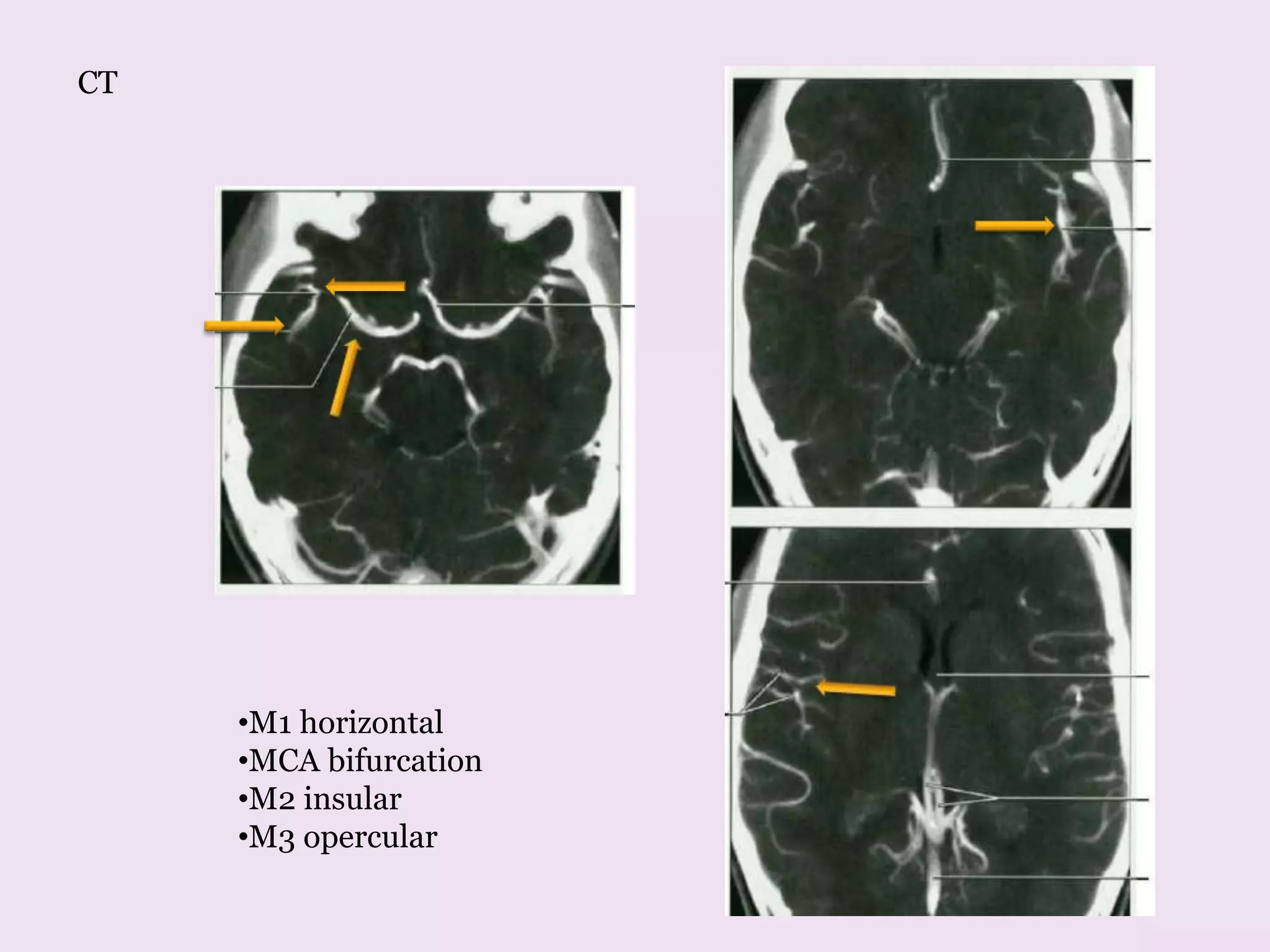
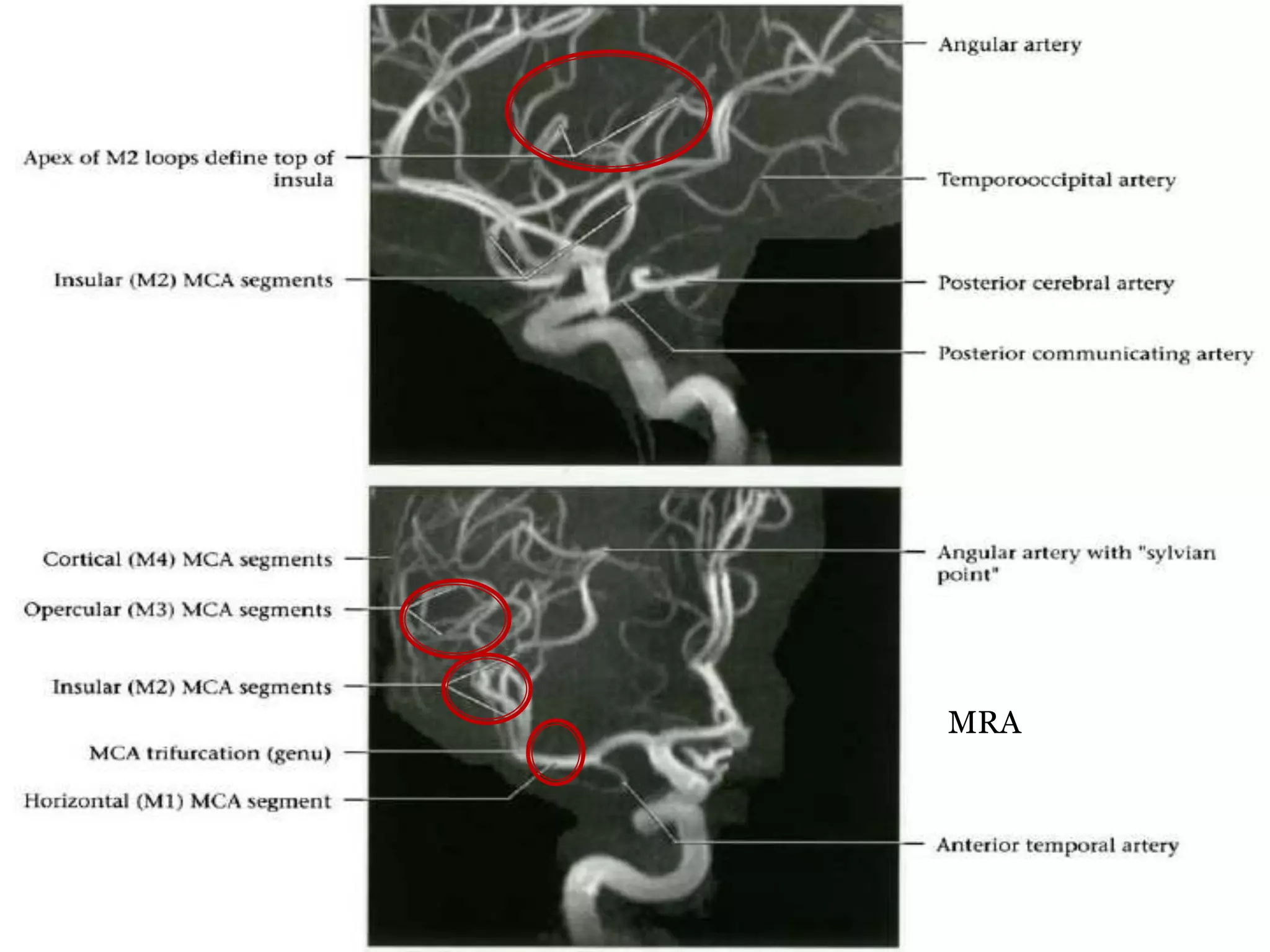
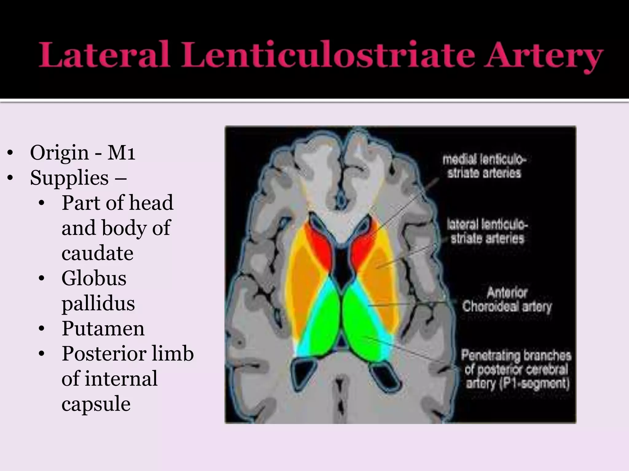
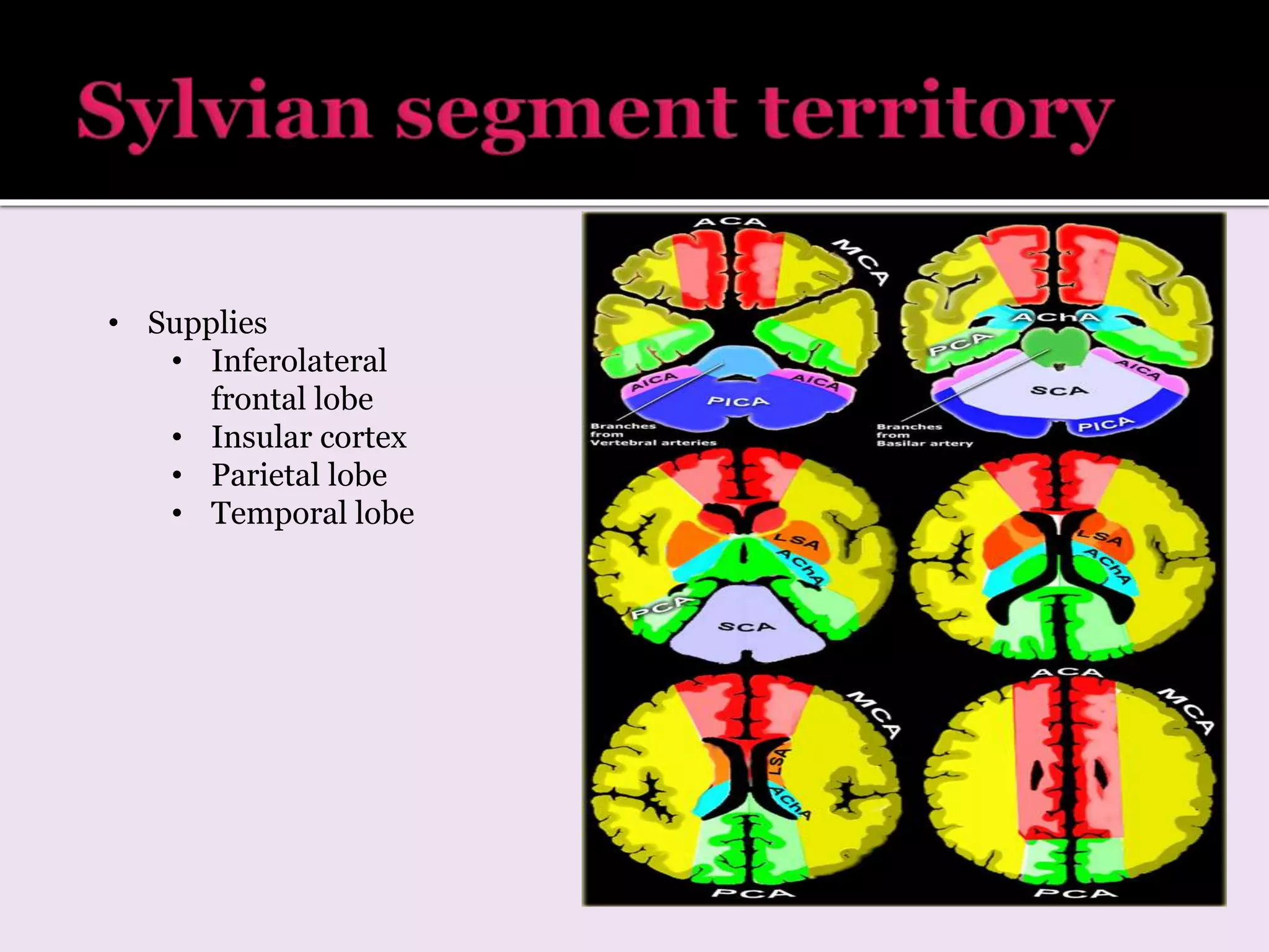
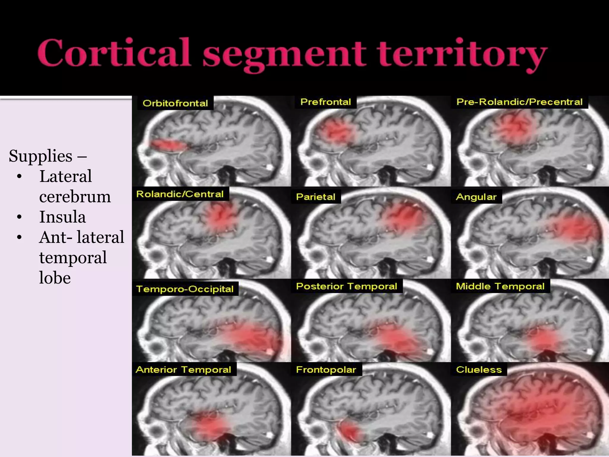
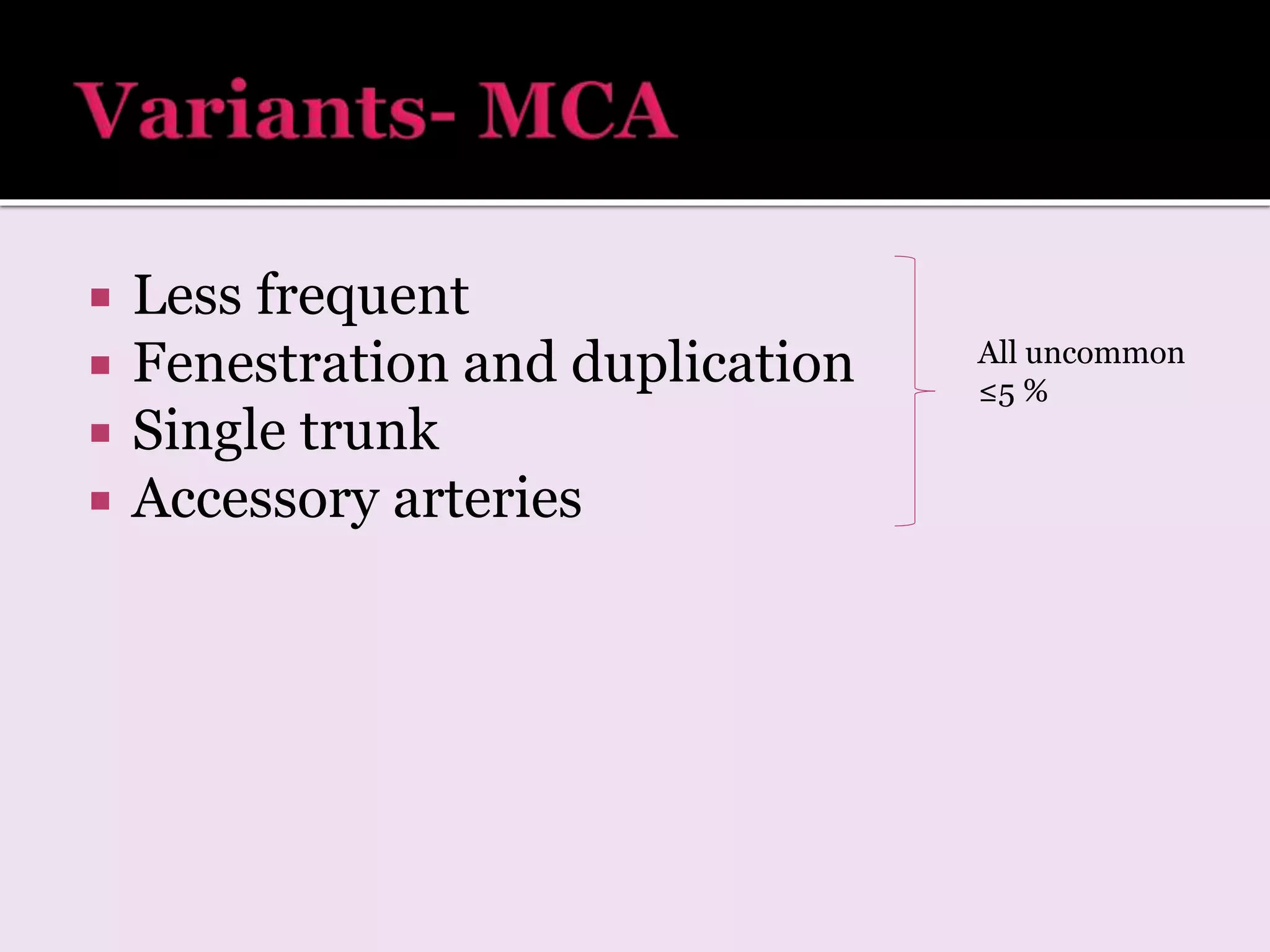
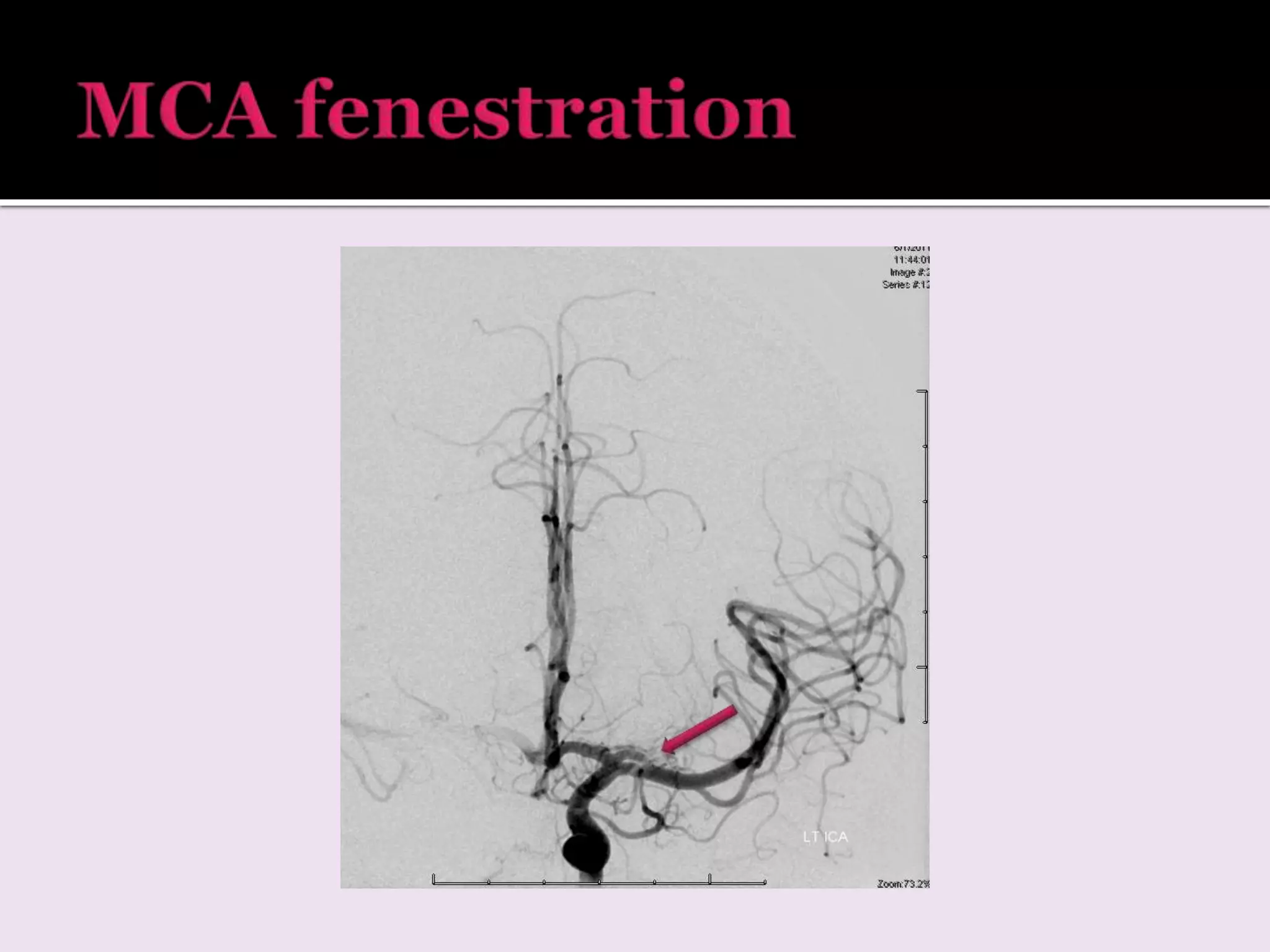
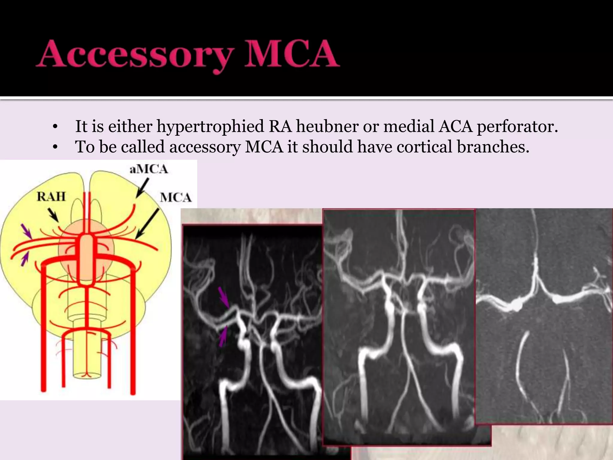
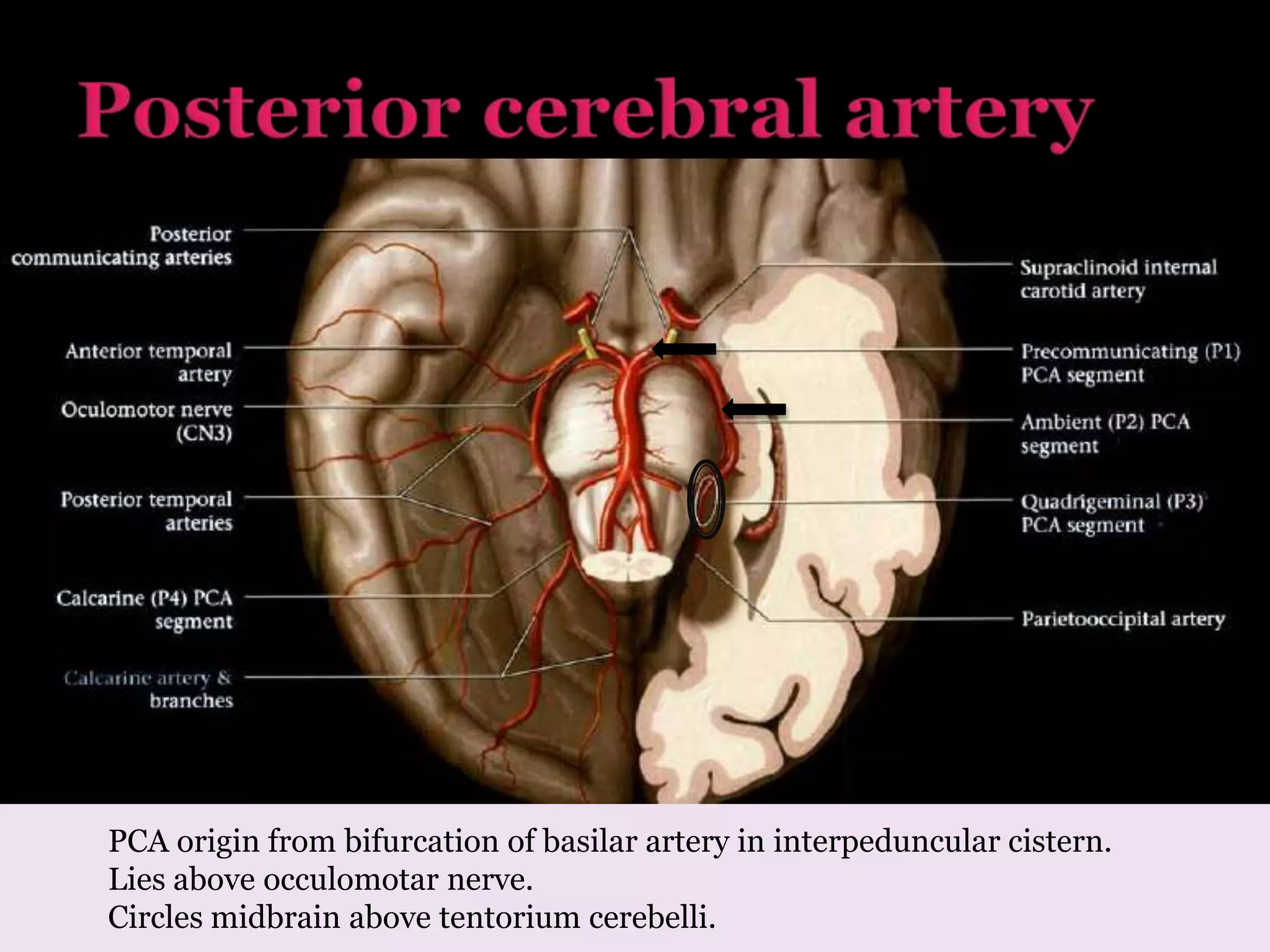
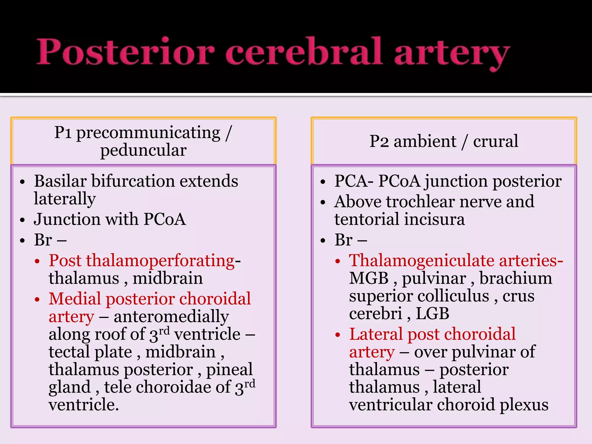
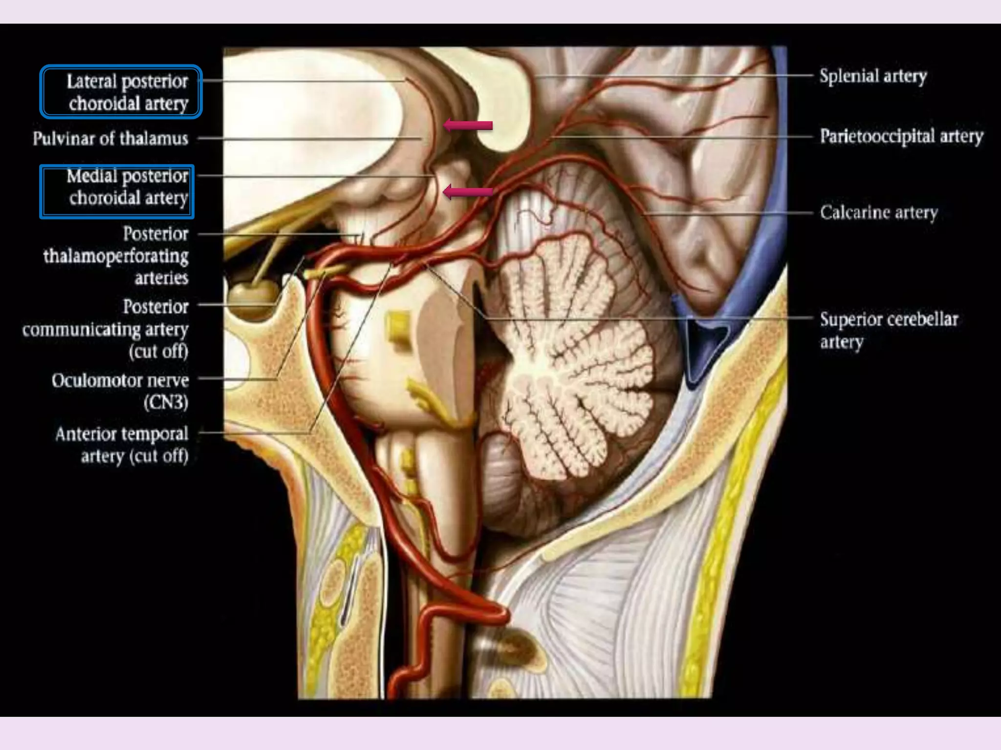
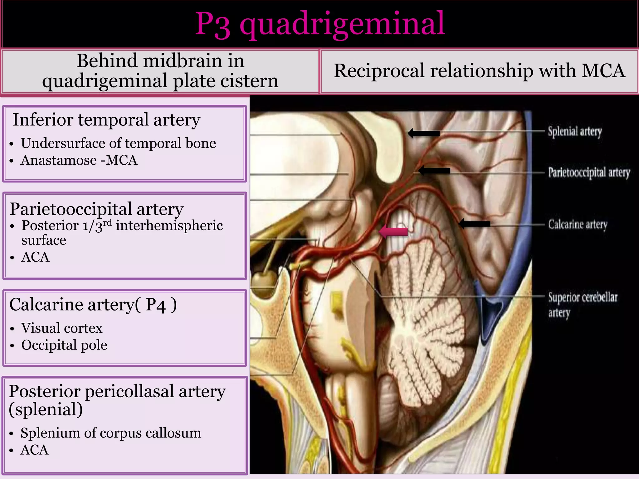
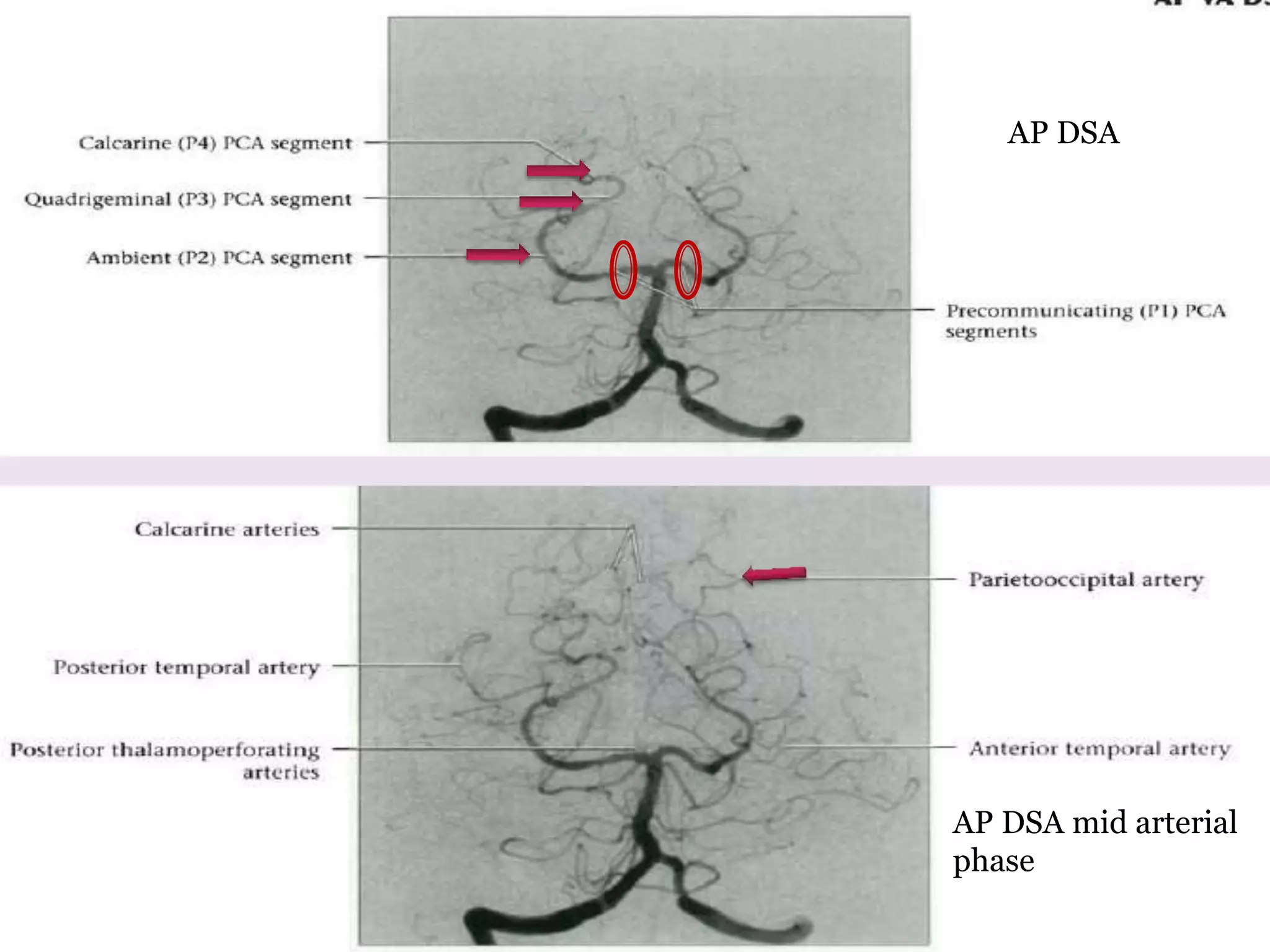
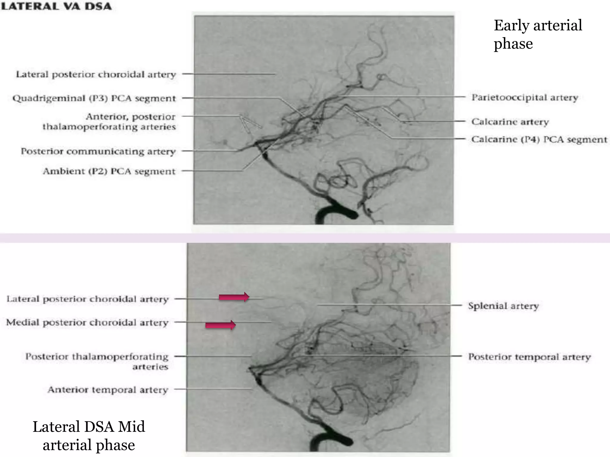
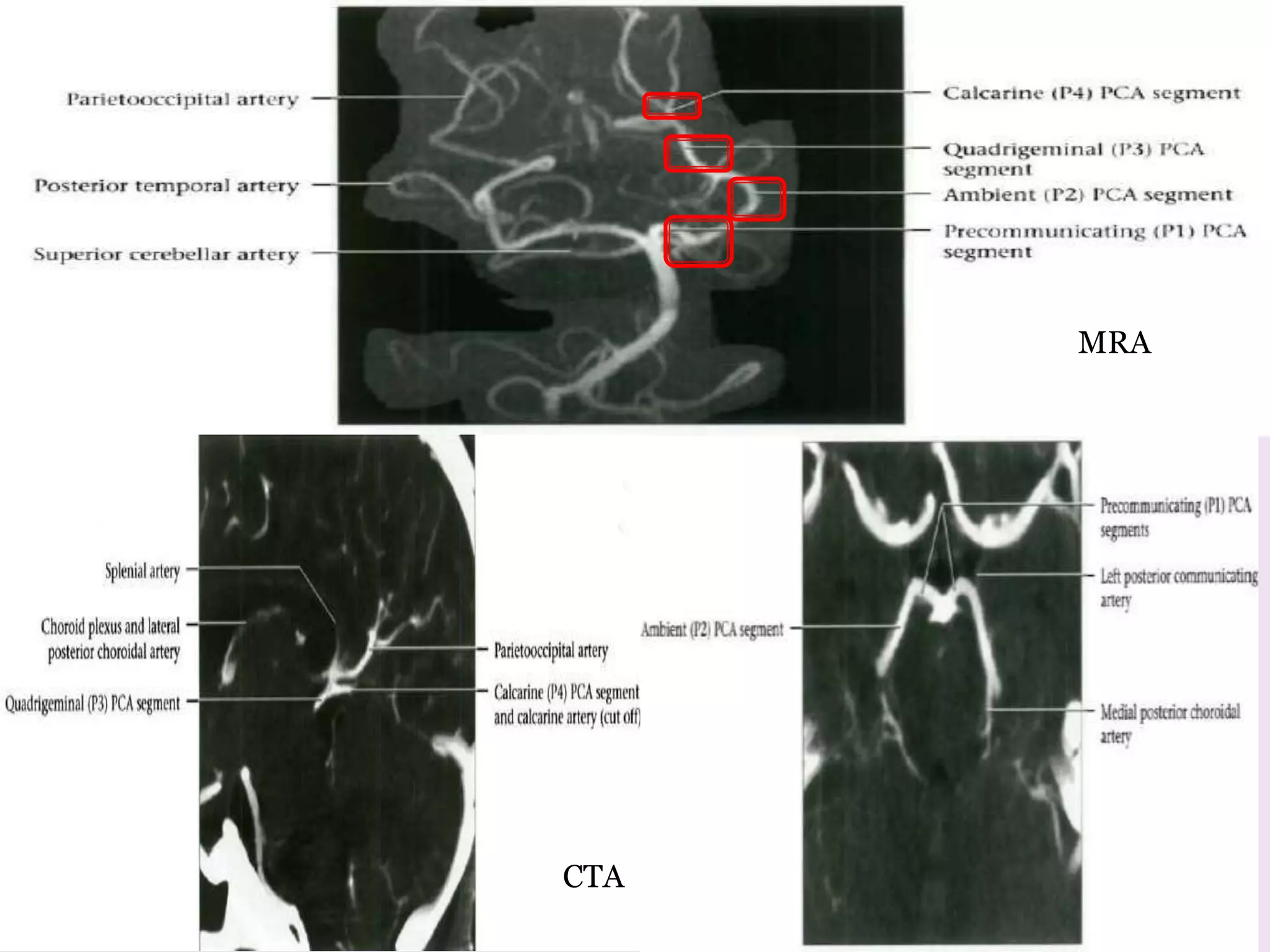
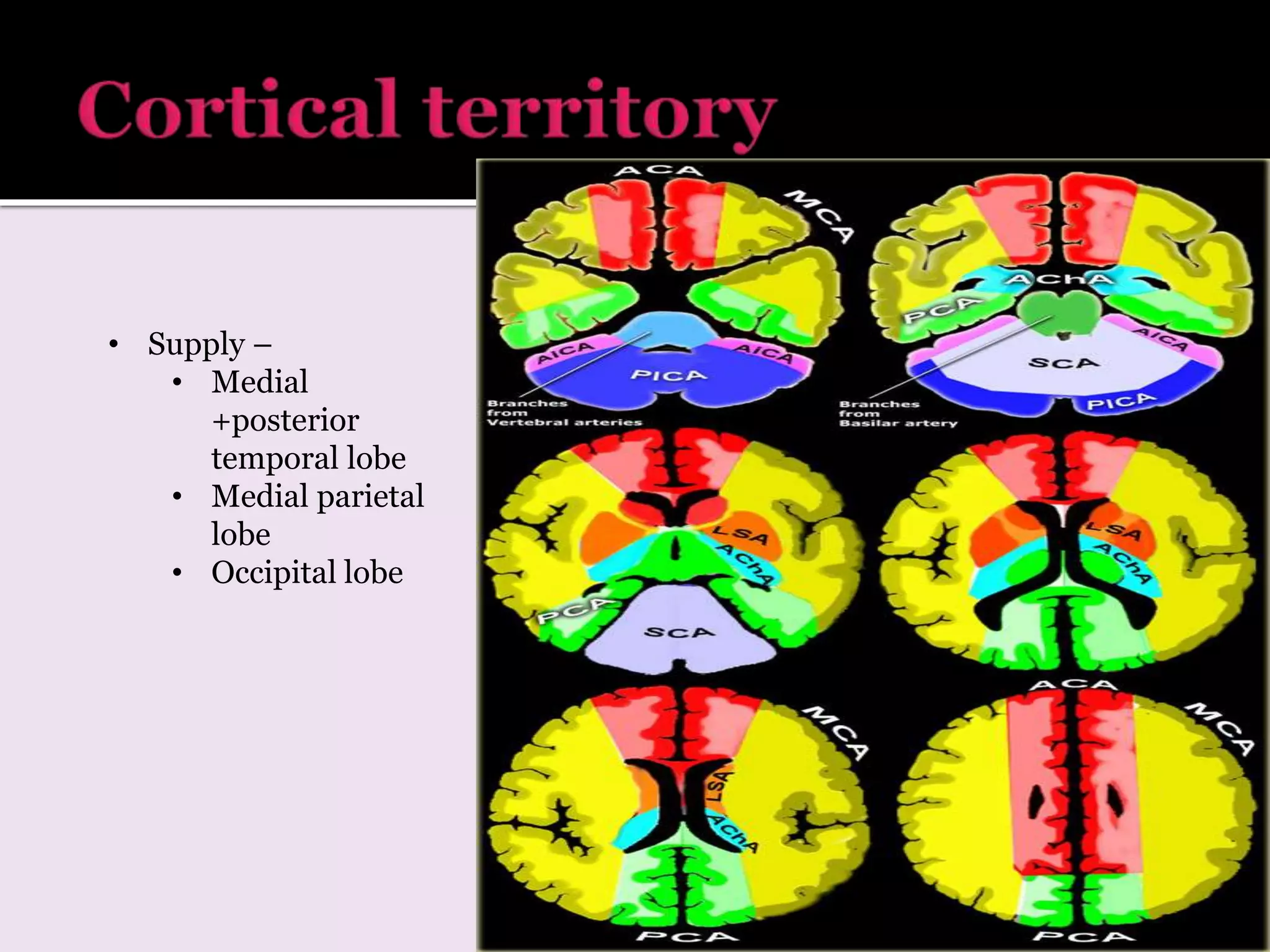
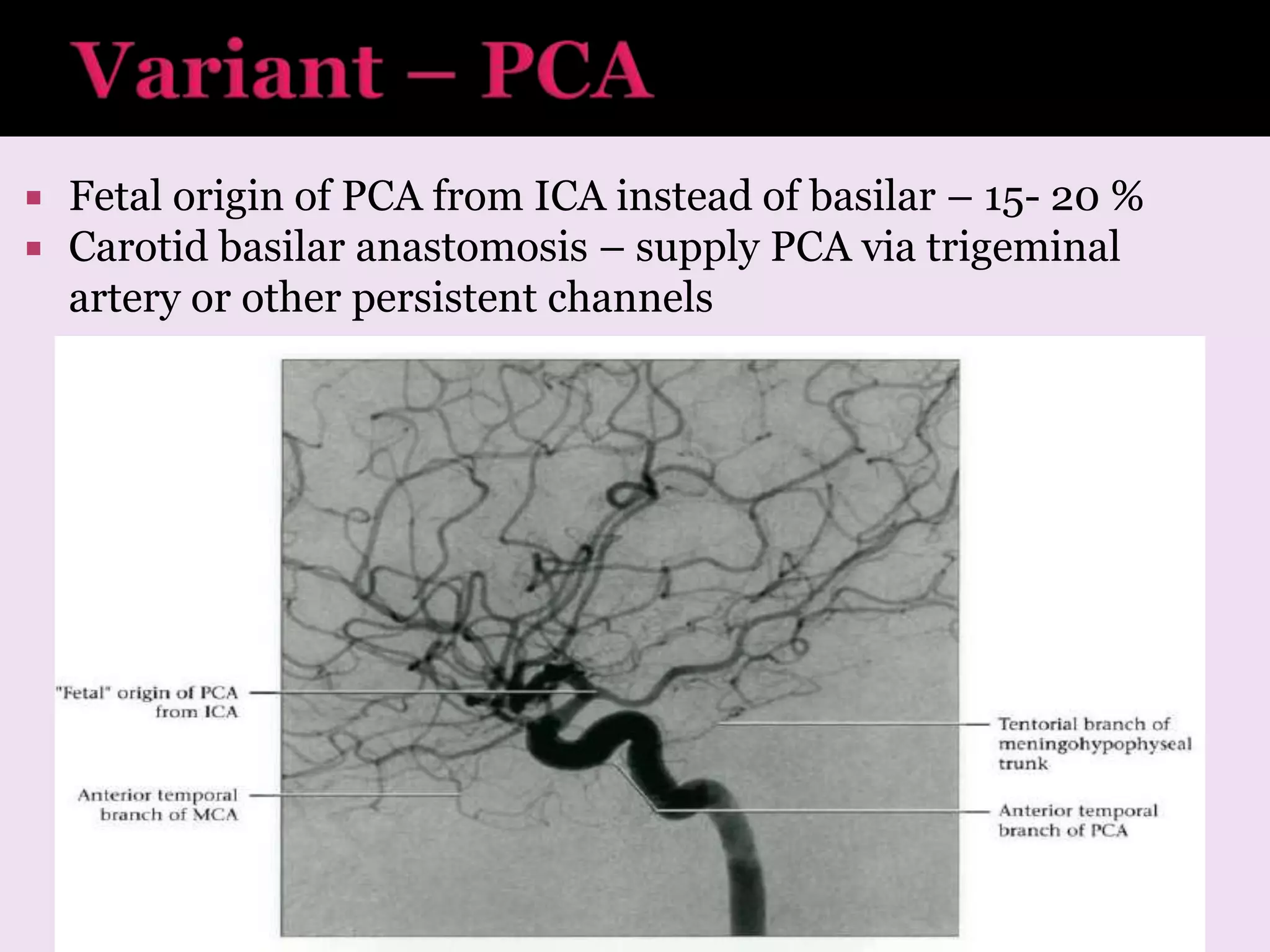
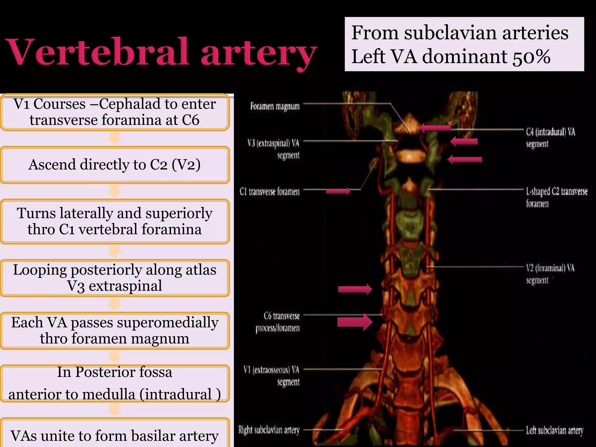
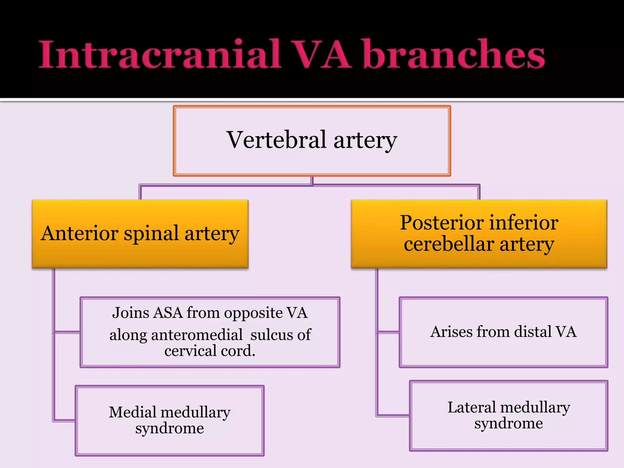
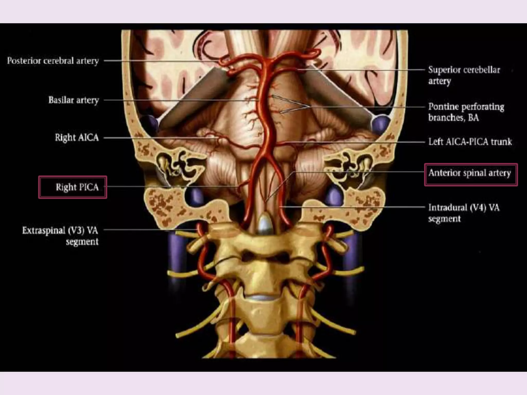
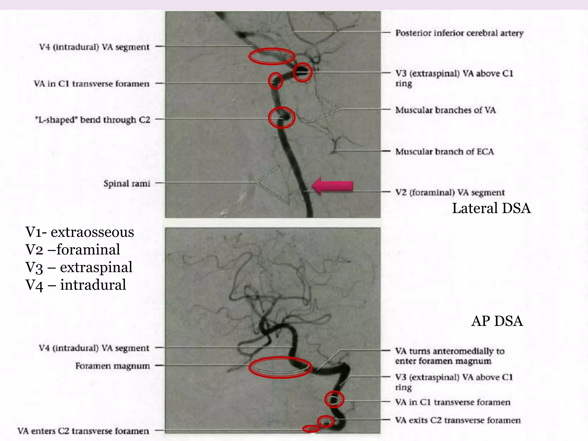
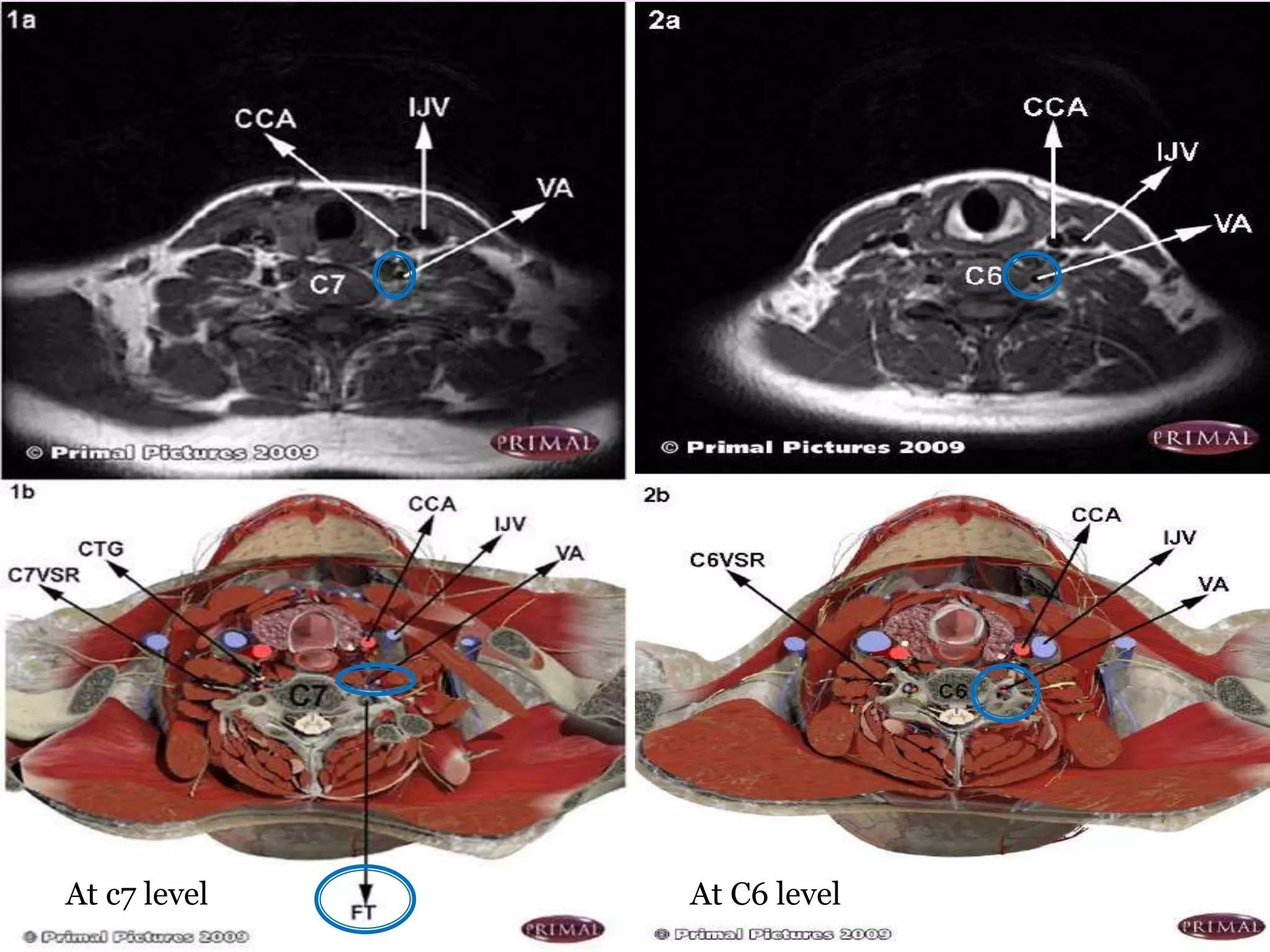
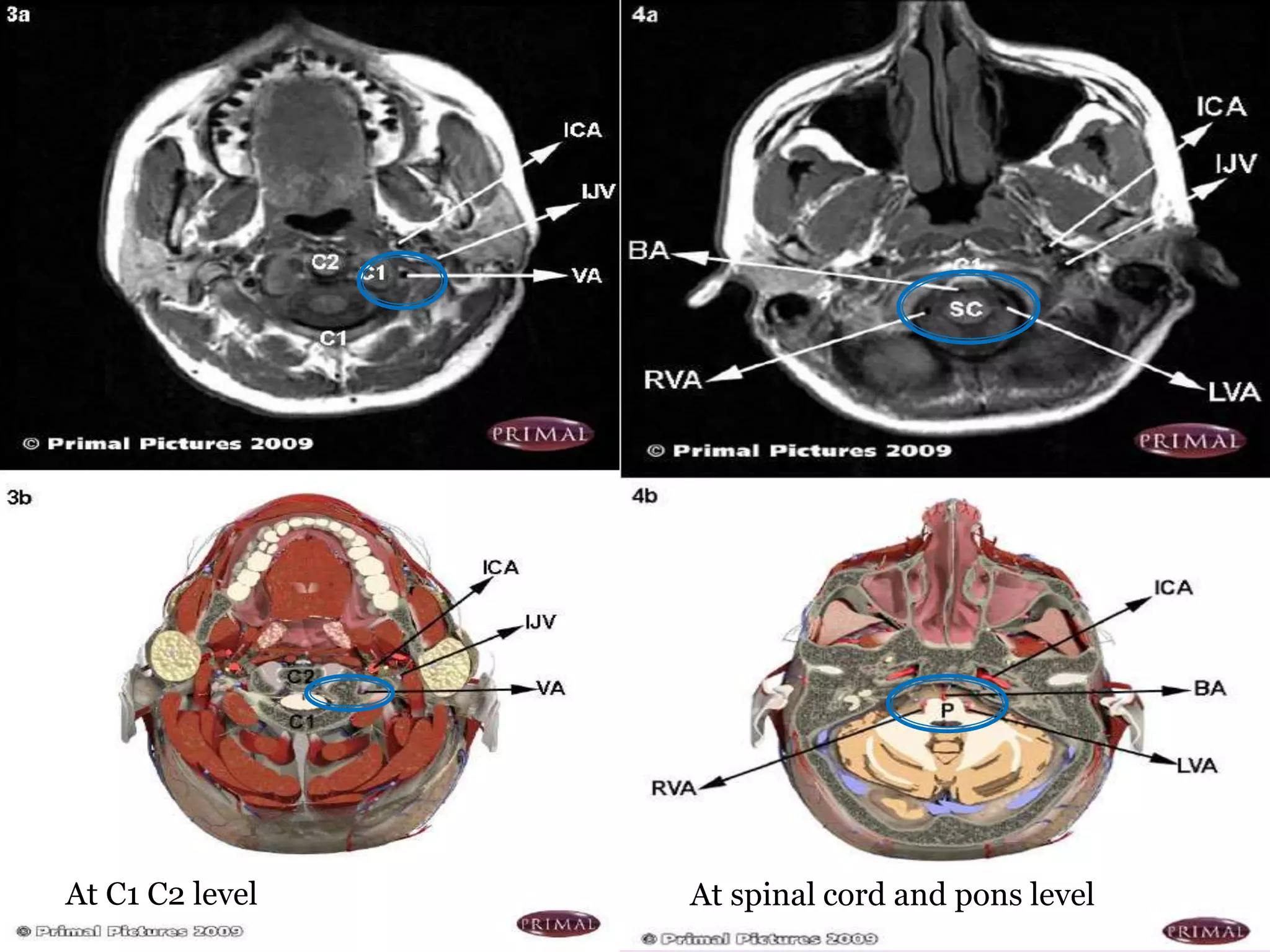
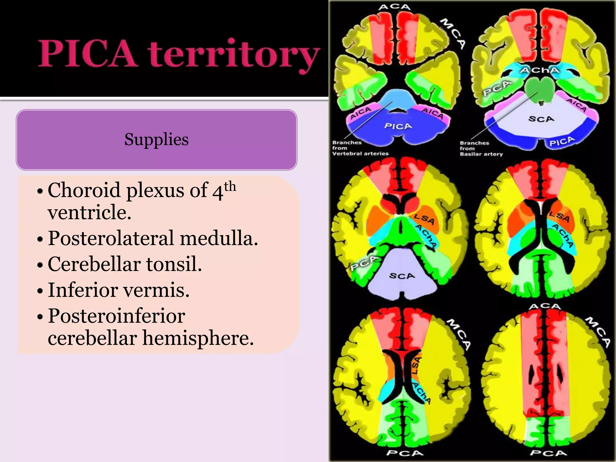
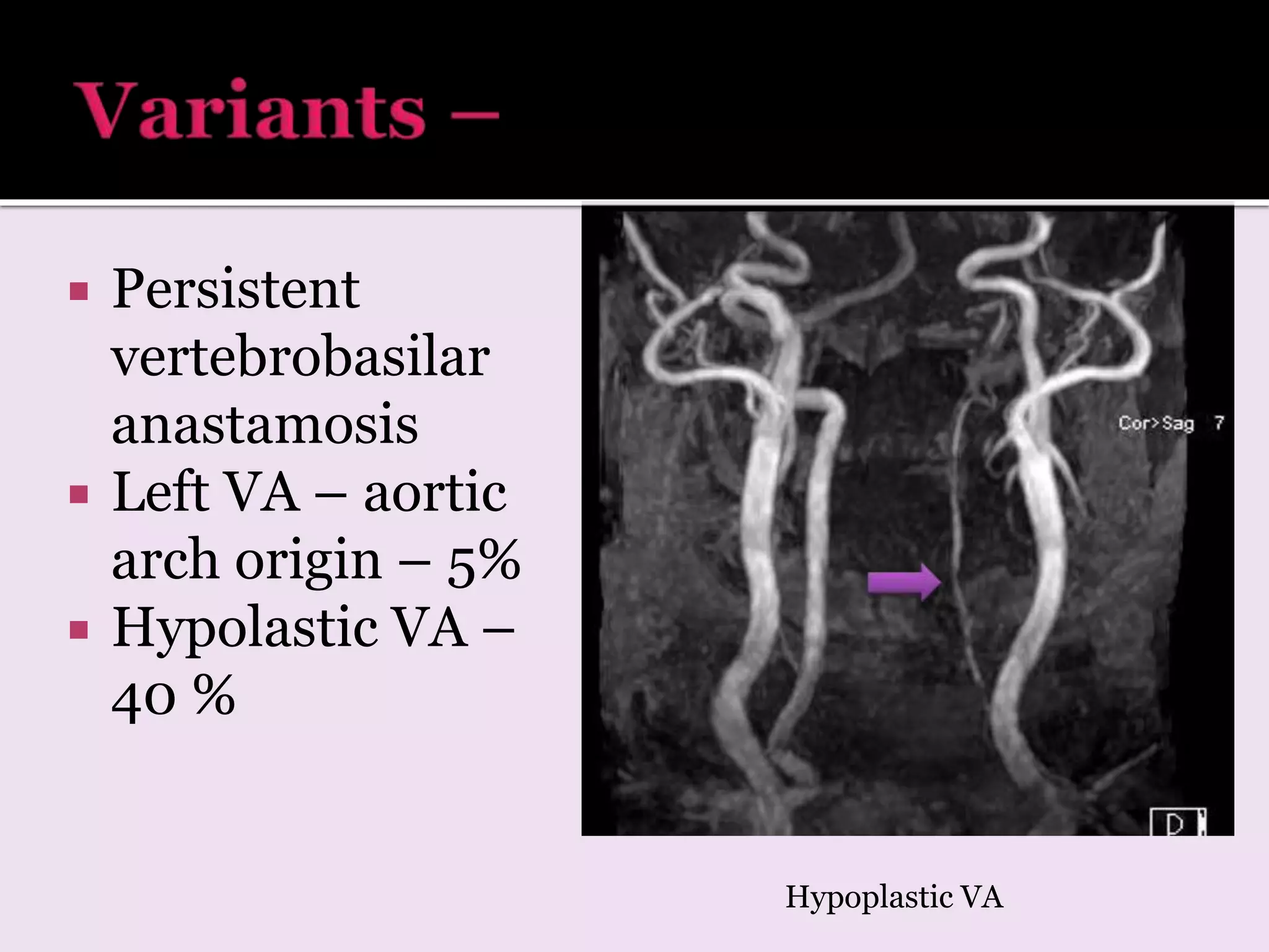
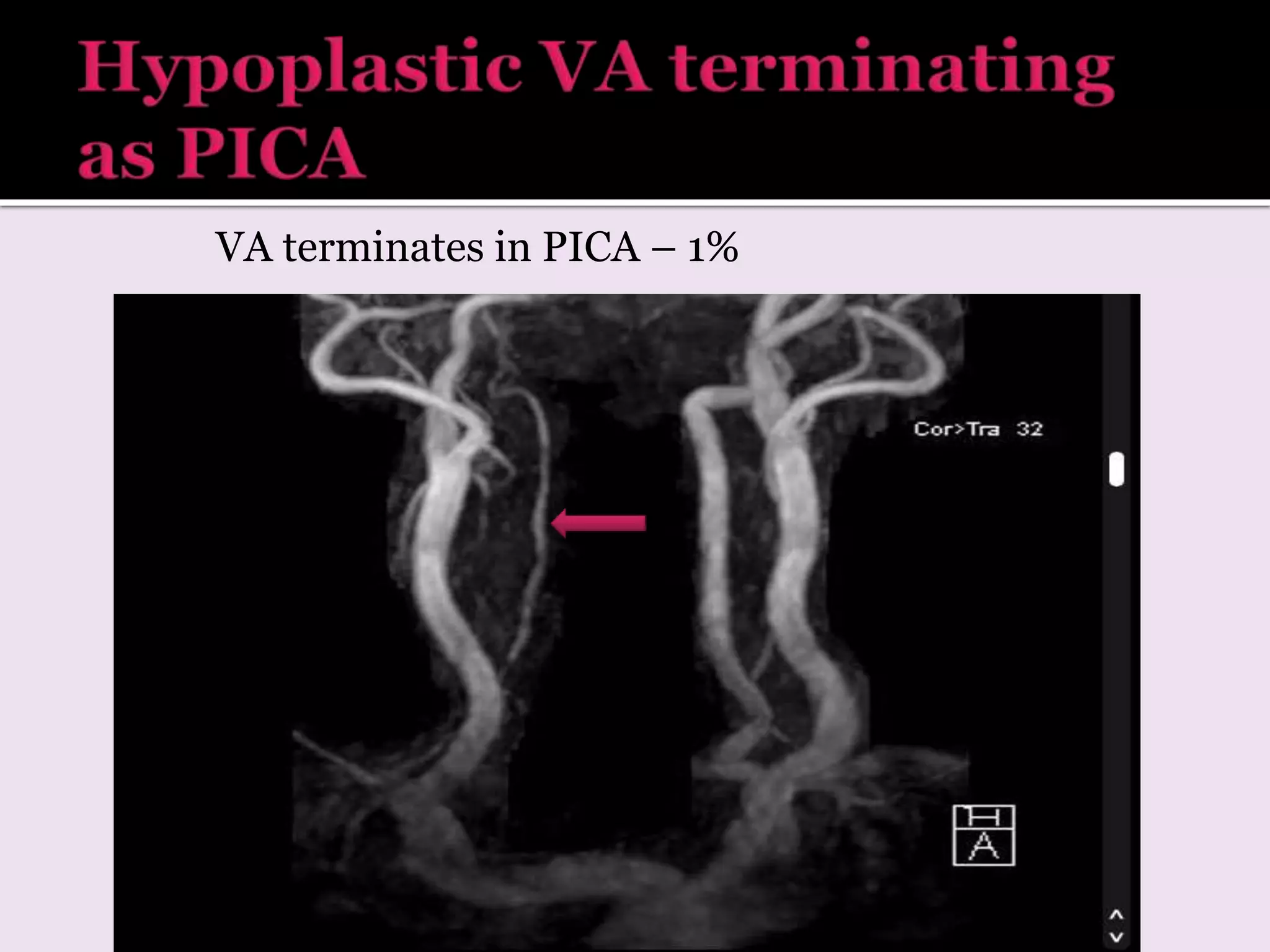
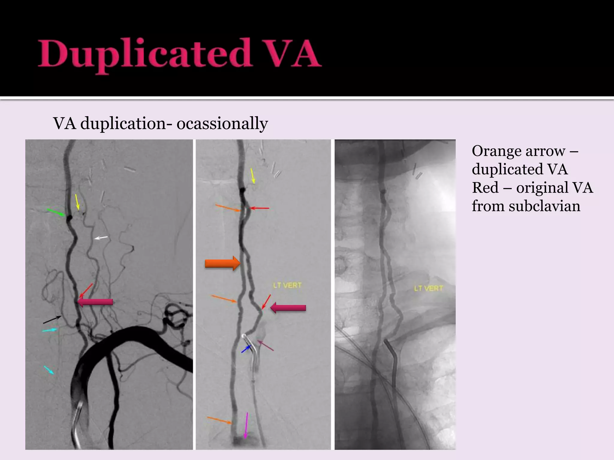
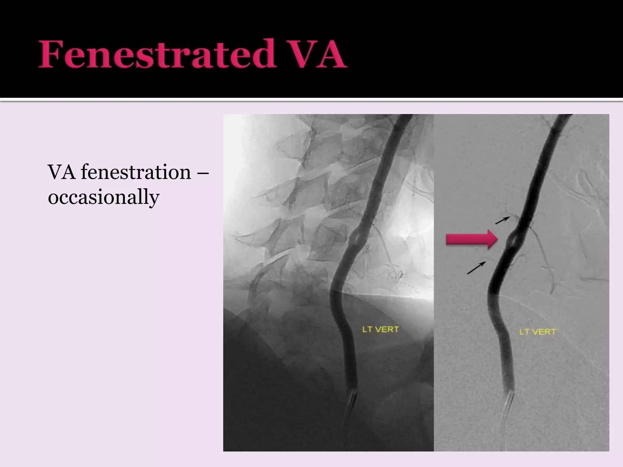
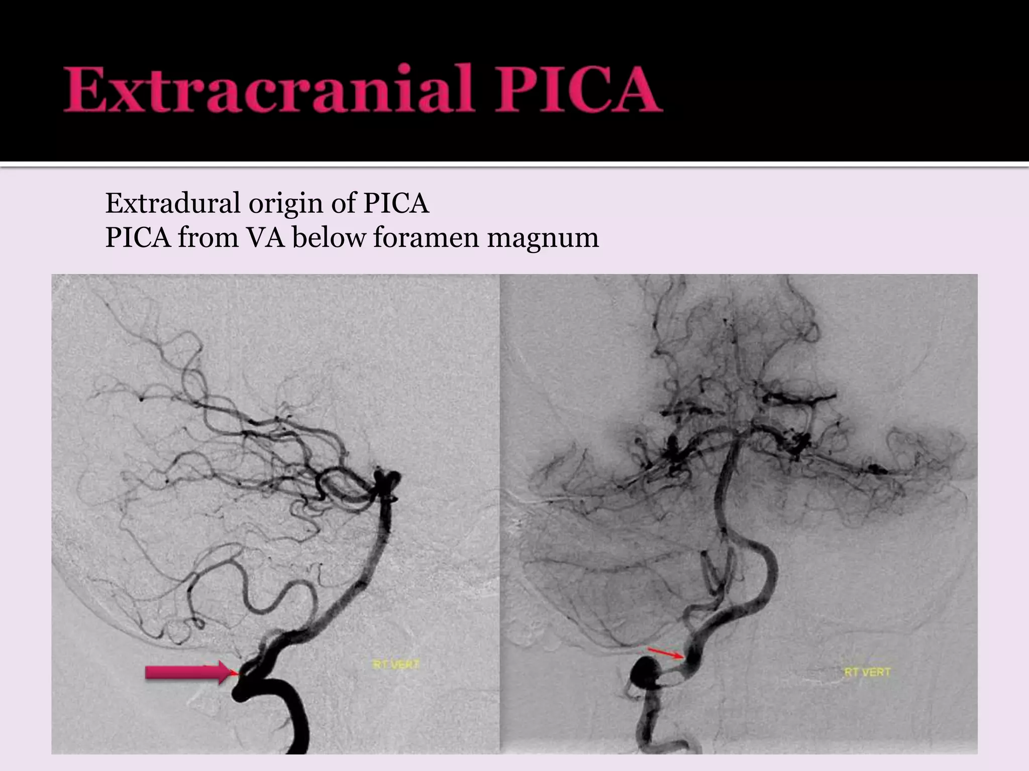
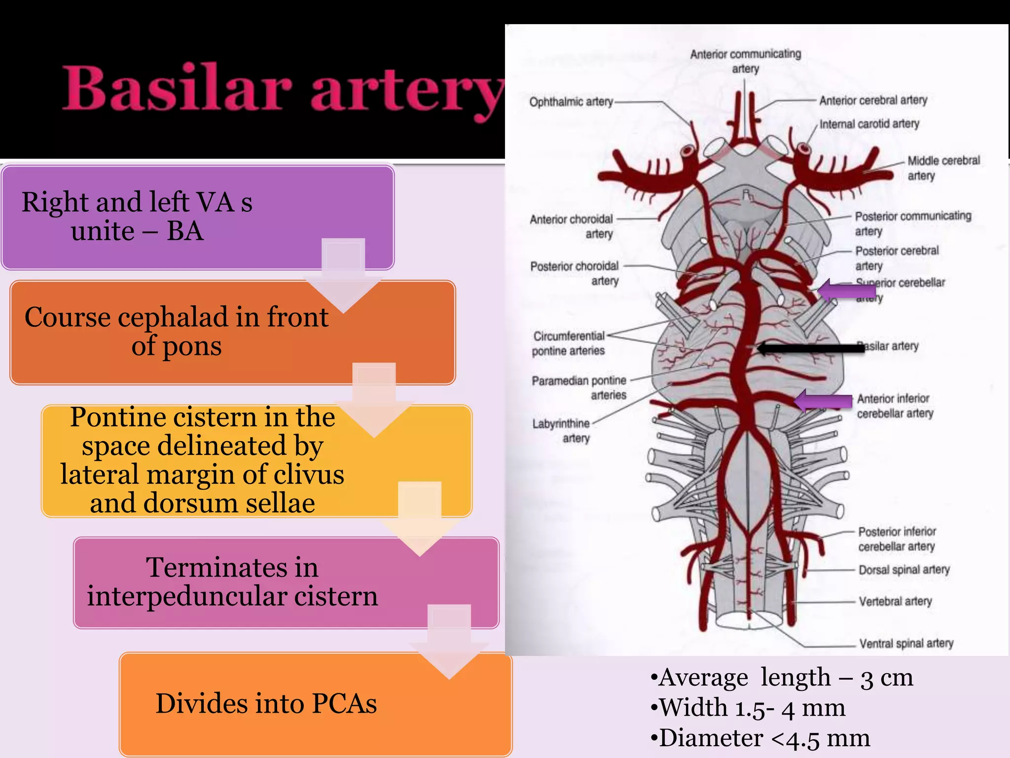
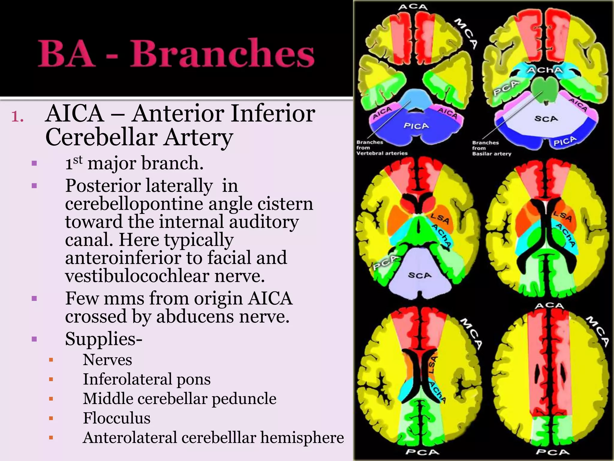
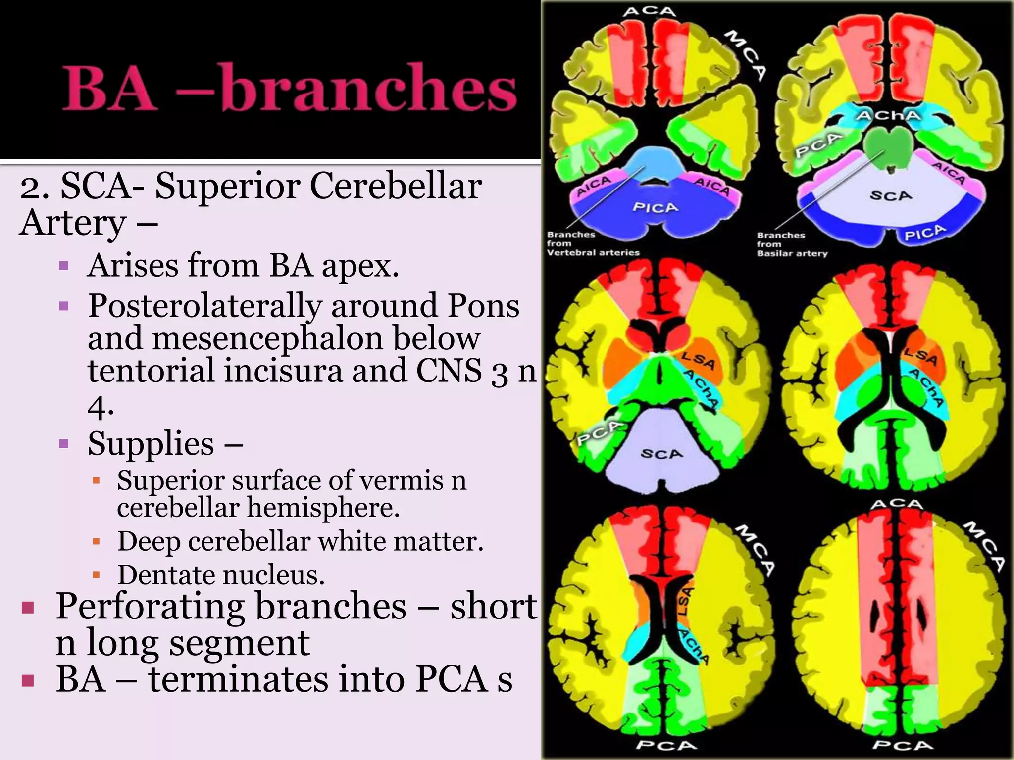
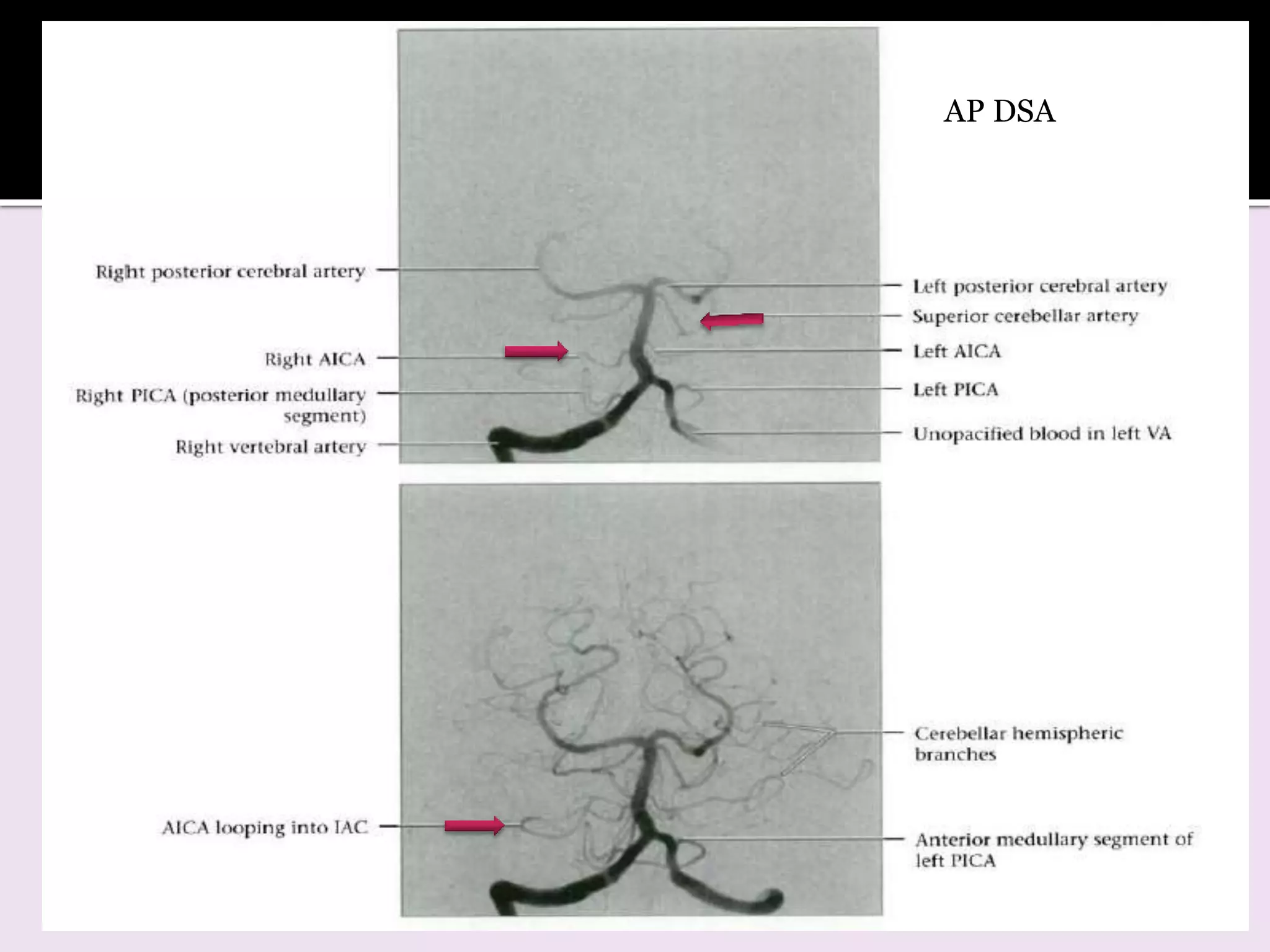
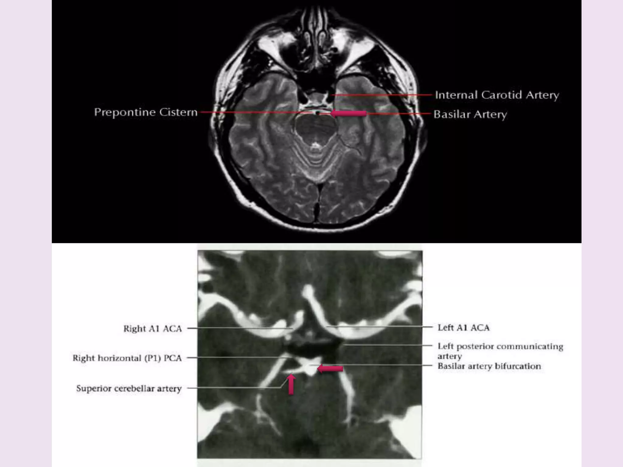
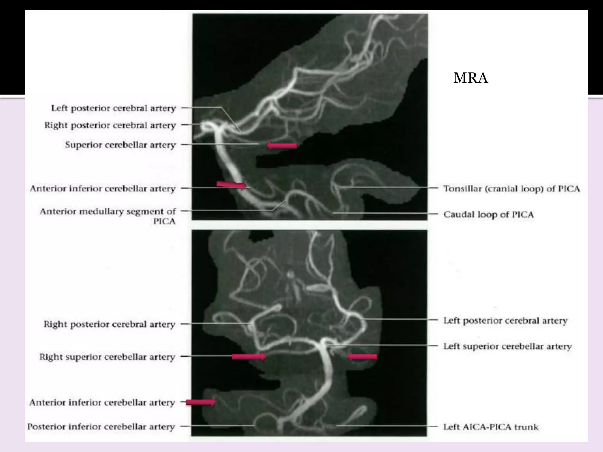
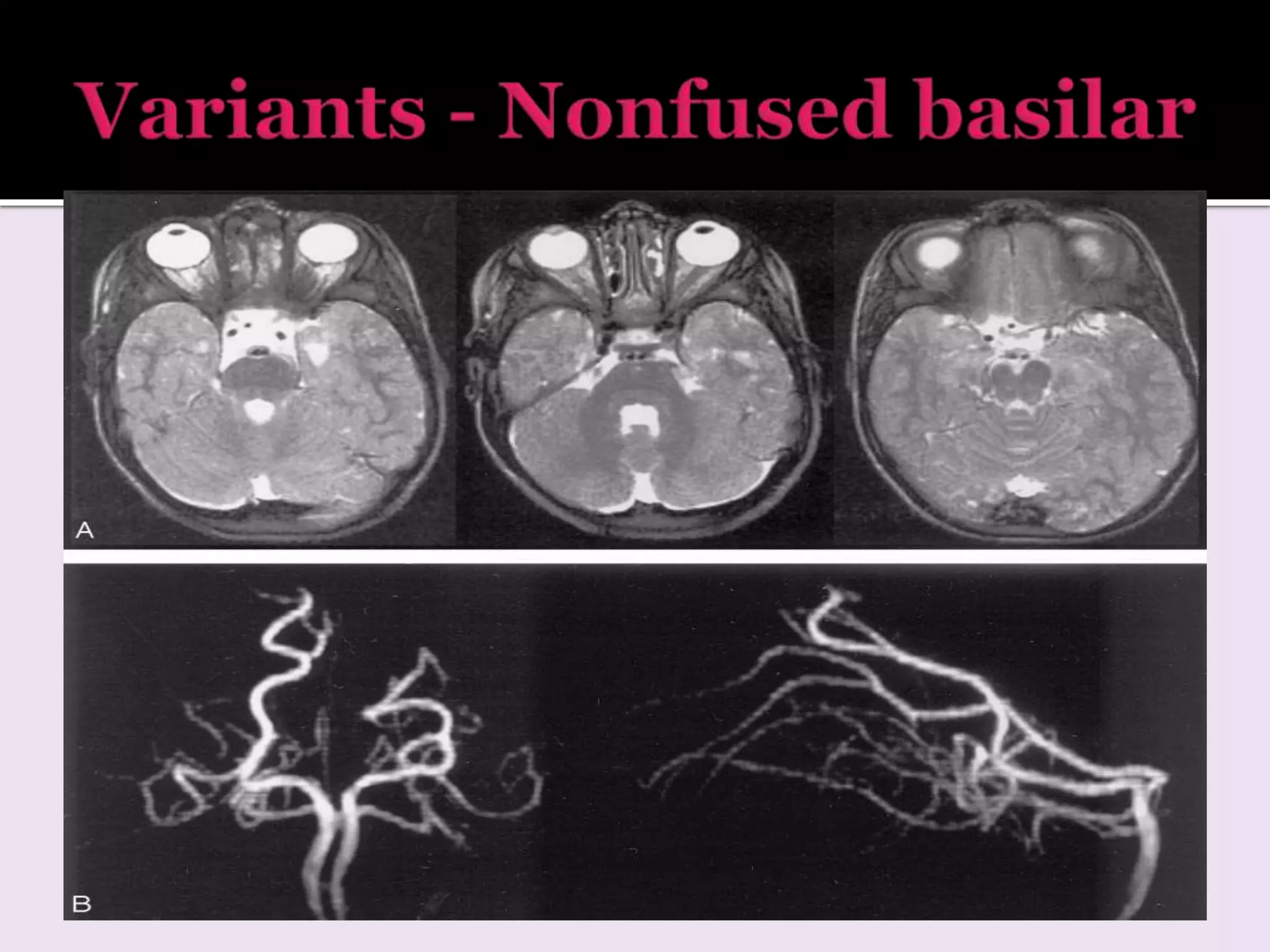
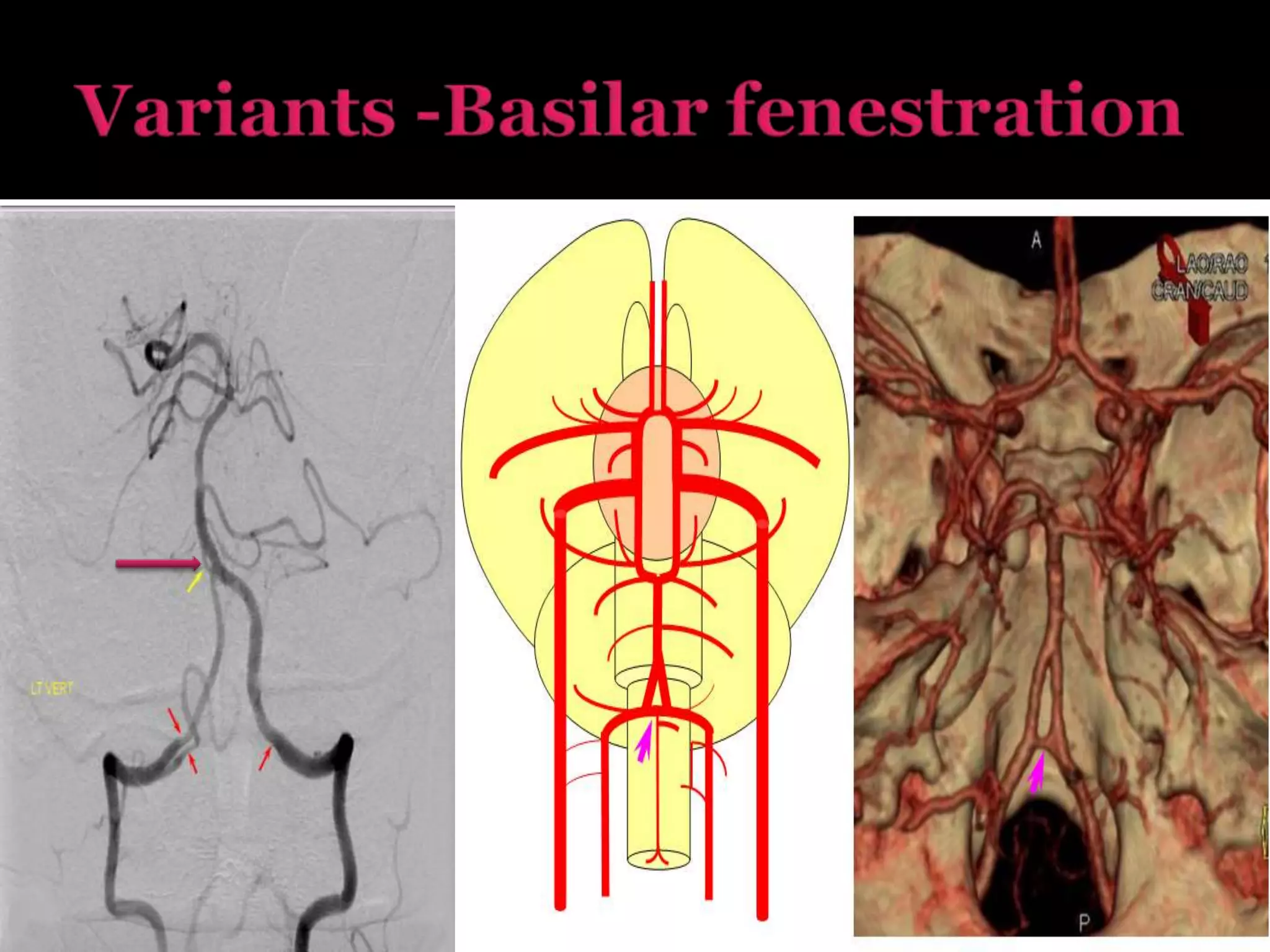
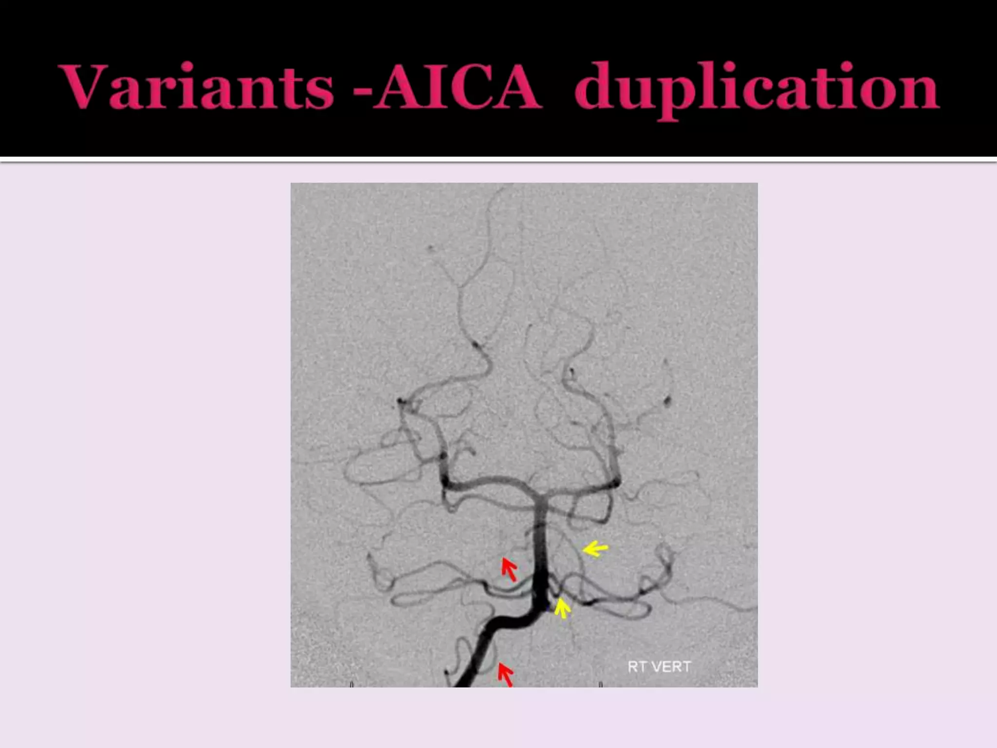
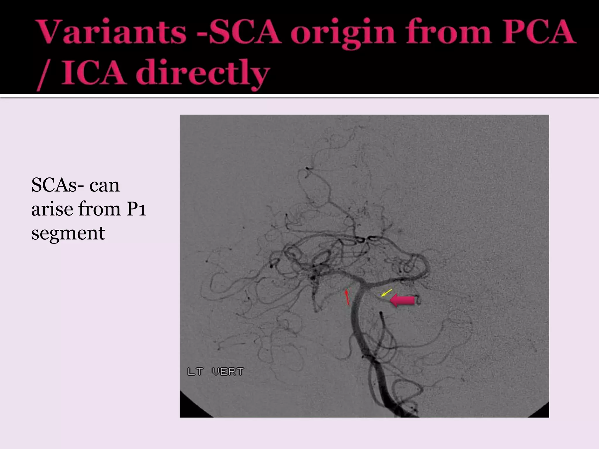
1. The document describes the anatomy and branches of the major cerebral arteries including the anterior, middle, and posterior cerebral arteries.
2. It discusses the typical branching patterns and territories supplied by each artery and their segments.
3. Variations in arterial anatomy are also summarized such as fenestrations, duplications, hypoplasia.
This presentation provides a comprehensive review of major sulci of brain which help in defining the different lobes of brain.Very useful for first year residents.
Its important to recognise the myelination pattern in neonates and infants. This presentation talks about the myelination pattern and imaging of white matter diseases in children.
Anatomy of Brain by MRI
In this presentation we will discuss the cross sectional anatomy of brain. Then we will discuss the Most common diseases to be evaluated by brain imaging.
In my opinion this presentation is a road map for beginars.
Frequently Asked Questions about the Locked-In SyndromeFemke Nijboer
When I tell people I investigate the Locked-In Syndrome (LIS), I often get the same questions. I thought I could start gathering these Frequently Askes Questions and asnwering them to provide a better understanding of LIS and what it is like to live with LIS. I hope to take away some prejudicies about this condition.
Disclaimer: I am not a medical expert. I am a neuropsychologist. I have seen and interviewed many people with LIS, but they might not be representative of all people with LIS. These answers are not neccesarily the truth, but an educated guess.
Magnetic resonance venography & venous ultrasosnography for diagnosisng deep ...Prof. Shad Salim Akhtar
Method of detecting thrombosis in deep leg veins. Use of magnetic resonance venography in comparison to venous ultrasonography. A comparative blinded trial.
anterior choroidal artery course, clinical implications, angiography and surgical importance
clinical features of aneurysm, AVM involving the anterior choridal artery
A review of the growth of the Israel Genealogy Research Association Database Collection for the last 12 months. Our collection is now passed the 3 million mark and still growing. See which archives have contributed the most. See the different types of records we have, and which years have had records added. You can also see what we have for the future.
Introduction to AI for Nonprofits with Tapp NetworkTechSoup
Dive into the world of AI! Experts Jon Hill and Tareq Monaur will guide you through AI's role in enhancing nonprofit websites and basic marketing strategies, making it easy to understand and apply.
Biological screening of herbal drugs: Introduction and Need for
Phyto-Pharmacological Screening, New Strategies for evaluating
Natural Products, In vitro evaluation techniques for Antioxidants, Antimicrobial and Anticancer drugs. In vivo evaluation techniques
for Anti-inflammatory, Antiulcer, Anticancer, Wound healing, Antidiabetic, Hepatoprotective, Cardio protective, Diuretics and
Antifertility, Toxicity studies as per OECD guidelines
Read| The latest issue of The Challenger is here! We are thrilled to announce that our school paper has qualified for the NATIONAL SCHOOLS PRESS CONFERENCE (NSPC) 2024. Thank you for your unwavering support and trust. Dive into the stories that made us stand out!
Acetabularia Information For Class 9 .docxvaibhavrinwa19
Acetabularia acetabulum is a single-celled green alga that in its vegetative state is morphologically differentiated into a basal rhizoid and an axially elongated stalk, which bears whorls of branching hairs. The single diploid nucleus resides in the rhizoid.
4. A1 horizontal
segment
• From ACA origin to
ACoA junction.
• Inferior br – supply
superior surface of optic
nerve and chaisma.
• Superior br – anterior
hypothalamus , septum
pellucidum , anterior
commisure , fornix ,
anterior inferior portion
of corpus straitum.
5. Arise from A1
segment- perforating
branches.
• Pass cephalad
thro anterior
perforated
substance.
• Supply head of
caudate nucleus
and anterior
limb of IC,
putamen .
6. • Largest of the perforating
branches.
• May arise from A1 or A2
segment.
• A1 – 44%
• Proximal A2 – 50%
• ACoA – less common
• Derives its name from the
fact that it doubles back
on its parent artery at an
acute angle to join
lenticulostriate vessel.
• Lies parallel to A1 .
7. From ACoA junction
Ascend in front of 3rd ventricle in
cistern of lamina terminalis
br –Orbitofrontal, frontopolar
Curves around corpus callosum
genu gives terminal branches
A2 terminal
branches-
Pericollasal
Collasomarginal
8. • Supply the anterior
2/3rds of medial
hemispheric surface +
small superior area over
the convexities.
• Callosomarginal a.– lies
in cingulate gyrus
supplies medial frontal
lobe
• Pericallosal a.– course
along the posterior aspect
of corpus callosum and
supplies it and medial
parietal lobe
15. Single trunk from confluence of A1 segments of right n left ACAs-
supplies both hemispheres .
Assc with lobar holoprosencephaly, saccular aneursym
16. • Normally A1
segment runs
over the optic
nerve.
• Here it runs
below the
optic nerve.
• Assc with
aneurysms .
• Recognised
before
surgeries.
17. Variable branches to C/L
hemisphere.
Separate right n left ACA.
1 ACA is dominant than
other and it sends
branches to other
hemisphere.
Other ACA is hypoplastic
– terminate as
orbitofrontal or
frontopolar branch.
18. M1 horizontal
Origin -Laterally from ICA
bifurcation
Till its bi/trifurcation at
sylvian fissure.
Br – Lateral
Lenticulostriate branch
course superiorly
Anterior temporal artery
Supplies-Lentiform nucleus
Part of IC , caudate nucleus
M2 insular
At its genu divides
into branches
Loop over insula
pass laterally to exit
from sylvian fissure
M3 opercular
Emerge from
sylvian fissure
Ramify over
hemispheric surface
Supplies –cerebral
cortex and white
matter
40. Fetal origin of PCA from ICA instead of basilar – 15- 20 %
Carotid basilar anastomosis – supply PCA via trigeminal
artery or other persistent channels
41. V1 Courses –Cephalad to enter
transverse foramina at C6
Ascend directly to C2 (V2)
Turns laterally and superiorly
thro C1 vertebral foramina
Looping posteriorly along atlas
V3 extraspinal
Each VA passes superomedially
thro foramen magnum
In Posterior fossa
anterior to medulla (intradural )
VAs unite to form basilar artery
From subclavian arteries
Left VA dominant 50%
48. • Front of medulla
Anterior medullary
segment
• Along side of medulla caudally to level of CN 9-11Lateral medullary segment
• Around inferior half of cerebellar tonsilTonsilomedullary segment
• Cleft btw tela choridae and inferior medullary velum
rostrally and superior pole of tonsil caudallyTelovelotonsillar segment
Cortical / hemispheric
segment
59. Right and left VA s
unite – BA
Course cephalad in front
of pons
Pontine cistern in the
space delineated by
lateral margin of clivus
and dorsum sellae
Terminates in
interpeduncular cistern
Divides into PCAs
•Average length – 3 cm
•Width 1.5- 4 mm
•Diameter <4.5 mm
60. 1. AICA – Anterior Inferior
Cerebellar Artery
1st major branch.
Posterior laterally in
cerebellopontine angle cistern
toward the internal auditory
canal. Here typically
anteroinferior to facial and
vestibulocochlear nerve.
Few mms from origin AICA
crossed by abducens nerve.
Supplies-
▪ Nerves
▪ Inferolateral pons
▪ Middle cerebellar peduncle
▪ Flocculus
▪ Anterolateral cerebelllar hemisphere
61. 2. SCA- Superior Cerebellar
Artery –
Arises from BA apex.
Posterolaterally around Pons
and mesencephalon below
tentorial incisura and CNS 3 n
4.
Supplies –
▪ Superior surface of vermis n
cerebellar hemisphere.
▪ Deep cerebellar white matter.
▪ Dentate nucleus.
Perforating branches – short
n long segment
BA – terminates into PCA s