2. NERVOUS SYSTEM
CENTRAL
NERVOUS SYSTEM
PERIPHERAL
NERVOUS SYSTEM
AFFERENT DIVISION
(SENSORY)
EFFERENT DIVISION
(MOTOR)
SOMATIC
SYSTEM(VOLUNTARY) AUTONOMIC SYSTEM
(INVOLUNTARY)
SYMPATHETIC SYSTEM
(THORACO LUMBER) PARASYMPATHETIC
SYSTEM
(CRANIO SACRAL)
ENTERIC SYSTEM
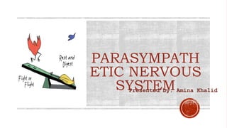
3. What is Parasympathetic Nervous
System?
PSNS is one of two major
divisions of the larger
autonomic system in your
body that keeps the basic
functions of your body
working as they should.
It relaxes your body after
periods of stress or
danger.
“rest and digest” or “feed
and breed.”
4. Parasympathetic
nervous system
function:
PSNS starts in the brain and extends out via
long fibers that connect with special neurons
near the organ they intend to act on.
The areas the PSNS acts on include:
•eyes
•lacrimal glands that produce tears
•parotid glands that also produce saliva
•salivary glands that produce saliva
•nerves in the stomach and trunk
•nerves that go to the bladder
•nerves and blood vessels responsible for the
male erection
5. Neurons of the
Parasympathetic Nervous
System
The axons of preganglionic PNS neurons are much longer
than those of the SNS and synapse with the postganglionic
neurons in the ganglia at or near the effector organs. The
very short postganglionic axons then relay signals to the
cells of the effector organs.
6. CRANIAL NERVES
CN III oculomotor nerve
CN VII facial nerve
CN IX glossopharyngeal nerve
CN X vagus nerve
SPINAL NERVE
Sacral 2,3,4 pelvic nerve
Nerves responsible in PSNS:
(CRANIO SACRAL)
The nerves are composed of pre- and postganglionic
neurons that act on effector organs.
Preganglionic neurons of the PNS come from
brainstem nuclei and the sacral spinal cord
7. • The autonomic nerves arise from the
lateral horn of the spinal cord.
• The nerves of PNS arise from the lateral
horn of the spinal cord in the sacral
region.
• PNS nerve fibers are fewer as compared
to SNS so the lateral horn in the
parasympathetic is not prominent as in
the sympathetic nervous system.
8.
9. THE OCULOMOTOR NERVE
(CN III)
“Oculo” means “pertaining to the
eye” and “motor” means
“producing movement”.
The oculomotor nerve innervates
muscles that move the eye itself or
components of the eye.
It supplies 5 of 7 extrinsic muscles
that move the eye and two
intrinsic muscles that control
pupil constriction and lens
accommodation.
10. THE FACIAL NERVE ( CN
VII):
The facial nerve is responsible for providing motor
innervation to these facial muscles, enabling you to
smile or frown.
• It innervates to muscles of facial expression.
• Innervates the anterior two-thirds of the tongue
and palate and conveys taste sensation.
• Innervates lacrimal, nasal, and palatine glands as
well as the submandibular and sublingual glands.
• Sensation to parts of the skin around the external
acoustic meatus and the retro auricular region.
11. THE GLOSSOPHARYNGEAL
NERVE (CN IX)
• The glossopharyngeal nerve innervates the salivary
gland left i.e., the parotid gland.
• It carries special sensory information from the
posterior one-third of the tongue that was left by the
facial nerve.
• It also innervated palatine tonsils, oropharynx, mucosa
of the middle ear, pharyngotympanic tube and the
mastoid air cells, carotid body
12. Gastro-Intestinal System
The sends branches to the
esophagus, stomach, and most
of the intestinal tract – up to
the splenic flexure of the large
colon.
It stimulates smooth muscle
contraction and glandular
secretions in these organs.
THE VAGUS NERVE (
CNX):
The Heart
The right vagus nerve supplies the
SA node and slows its pacemaker.
The left vagus innervates the AV
node and slows its conduction of
the cardiac impulse to the bundle
of His.
13. The pelvic splanchnic nerves are sacral nerves 2,3,
&4 provide parasympathetic innervation for most of
the pelvic organs, including the urinary bladder,
hindgut (descending colon, sigmoid
colon, rectum), ureter, prostate, urethra,
and penis/clitoris.
More specifically, activation of parasympathetic
fibers in the pelvic splanchnic nerves leads
to vasodilation of the erectile tissues in the penis and
clitoris, secretion in the hindgut, and motor activity
in the hindgut and urinary bladder.
THE PELVIC SPLANCNIC NERVE (
S2,S3,S4):