X-rays from a Central “Exhaust Vent” of the Galactic Center Chimney
JBEI Research Highlights - August 2017
1. Droplet Microfluidics for Synthetic Biology
Outcomes
• This review highlighted recent advances in droplet microfluidics
in the realm of synthetic biology.
• Microfluidic systems overcome many of the drawbacks of both
manual and robotic systems, as they are capable of high
throughput, low reagent consumption, and automation.
• Droplet-based microfluidics, in which sub-microliters to
picoliters of aqueous phase are encapsulated into
monodisperse droplets, are especially useful for applications
requiring parallel experiments at minimal reagent costs.
Gach, P. et al. (2017) “Droplet Microfluidics for Synthetic Biology,” Lab on a Chip,
DOI: 10.1039/C7LC00576H.
Background
• Most synthetic biology experiments are performed manually
and are very labor-intensive, consume large amounts of
expensive reagents such as enzymes and synthetic DNA, are
limited in throughput, and have poor reproducibility.
• Robotic liquid-handling stations can overcome the throughput
and reproducibility limitations, however they are very
expensive, hard to maintain, and consume the same amount
of reagents as manual experiments
Significance
• This review highlights the most compelling advances and remaining
challenges that must be addressed in order for droplet microfluidic
devices to reach optimal performance and impact.
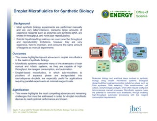
Molecular biology and analytical steps involved in synthetic
biology using droplet microfluidic systems. Biological
design/build/test engineering cycles include key steps such as
DNA synthesis, DNA assembly, DNA transformation, cell
culture, and phenotypic analysis, which often require costly and
labor-intensive manual processes. Microfluidic systems have
the potential to overcome such drawbacks, through enabling
high-throughput automated processing with low reagent
consumption requirements
2. The Molecular Basis for Binding of an Electron
Transfer Protein to a Metal Oxide Surface
Outcomes
• We showed that α-Fe2O3 nanoparticles bind to MtrF specifically but
not tightly and the binding does not induce significant
conformational changes in the protein.
• We identified specific amino acid residues on MtrF likely to be
involved in electron transfer. These residues are separated in
primary sequence, but cluster into a small putative binding pocket.
• Together, our results show that binding of MtrF to α-Fe2O3 follows a
strategy that resembles the binding between donor-acceptor
electron transfer proteins.
Fukushima et al. (2017) “The Molecular Basis for Binding of an Electron Transfer
Protein to a Metal Oxide Surface,” J. Am. Chem. Soc., DOI:10.1021/jacs.7b06560
Background
• Understanding the molecular mechanism of electron transfer
between a living organism and material is a long standing challenge
in the field of bioelectronics.
• X-ray Footprinting and Mass Spectrometry (XFMS) methods can
directly quantify solvent accessibility information of amino acids in a
protein under native conditions. Consequently, this method in
combination with other biophysical techniques can probe protein
binding interactions at the molecular level.
Significance
• This research will help us better understand interactions between
protein and nanomaterials that will lead to new innovations such as
bio-based sensors that can diagnose diseases and detect
contaminants.
Approach
• We combined XFMS with protease footprinting, fluorescence
binding assay and mutational analysis to determine the binding
site of α-Fe2O3 nanoparticles to the extracellular electron transfer
protein MtrF from Shewanella oneidensis MR-1
XF-MS shows that amino acids located near heme 6-7 region and heme 10
of MtrF are protected by α-Fe2O3 nanoparticles
(A) Amino acid residues moderately (yellow) or strongly (red) protected by binding of the
nanoparticles at pH 4 (left) and 7 (right) as viewed from the front and back perspective. The
solvent accessible regions of the protein and heme groups of MtrF are shown as gray and light
red surfaces, respectively. (B, C) Ratio of the modification rate for different amino acids in MtrF
at pH 4 (B) and pH 7 (C). Grey bars indicate a modification rate ratio (MtrF alone /α-Fe2O3:MtrF)
less than 1.5, whereas yellow residues indicate moderately protected residues (R=1.5 to 1.7)
and red residues are strongly protected residues (R>1.7). The hemes are numbered in red
according to their position in primary sequence.
3. The Experiment Data Depot: a Web-based
Software Tool for Biological Experimental Data
Storage, Sharing, and Visualization
Outcomes
• In this paper, we describe EDD and showcase its utility for three different use
cases: storage of characterized synthetic biology parts, leveraging proteomics
data to improve biofuel yield, and the use of extracellular metabolite
concentrations to predict intracellular metabolic fluxes.
1) The Experiment Data Depot (EDD)
collects data from different instruments, stores
and visualizes them in an interactive way, and
enables downloading them in a standardized
format for use with a variety of modeling and
analysis techniques.
Morell, W. et al. (2017) “The Experiment Data Depot: a web-based software tool for biological
experimental data storage, sharing, and visualization,” ACS Synth. Biol., doi: 10.1021/acssynbio.7b00204
Background
• Although recent advances in synthetic biology allow us to produce biological
designs more efficiently than ever, our ability to predict the end result of these
designs is still nascent.
• Predictive models require large amounts of high-quality data to be parametrized
and tested, which are not generally available.
• Here, we present the Experiment Data Depot (EDD), an online tool designed as
a repository of experimental data and metadata. EDD provides a convenient
way to upload a variety of data types, visualize these data, and export them in a
standardized fashion for use with predictive algorithms.
Significance
• In the current world, where there is an increasingly strong trend to disclose
algorithms as open source code, but training data is viewed as extremely
valuable, EDD will provide significant value as more experiments are available.
EDD will help enabling reproducibility and predictability in the fields of metabolic
engineering and synthetic biology relevant to applications in bioenergy.
2) Experiment description on EDD. Example of
how a common experiment is described in EDD.
Approach
4. A Comparative Study of Sample Preparation
for Immunomicroscopy of Plant Cell Walls
Outcomes
• In order to help the plant community in understanding and selecting
adequate methods of embedding and sectioning for cell wall
immunodetection, we review in this article the advantages and limitations of
these three methods.
• Moreover, we offer detailed protocols of embedding for studying plant
materials through microscopy.
Comparision of the three methods used with arabidopsis stem
sections. The thick microtome and vibratome sections have more
exposed epitopes and give a stronger signal with the anti-xylan antibody
when using the same exposure time for all sections. Increased resolution
can be obtained in the vibratome sections by staining with toluidine blue.
Background
• Staining and immunodetection by light microscopy are methods widely
used to investigate plant cell walls.
• The two techniques have been crucial to study the cell wall architecture in
planta, its deconstruction by chemicals or cell wall-degrading enzymes.
• They have been instrumental in detecting the presence of cell types, in
deciphering plant cell wall evolution and in characterizing plant mutants
and transformants.
Significance
• From our experience, the use of a microtome appears to be the best option
for studies that include co-localization of epitopes as well as chemical and
enzymatic pretreatments.
• However, whenever possible, we advise beginning with the use of a
vibratome as the technique is fast, easy to master, offers the possibility to
study large specimens, large number of biological replicates, and is the best
method to preserve the antigenicity of the plant material.
Enzyme treatments can affect epitope detection.
Treatment with pectate lyase removes pectin epitopes as
expected. The treatment also reveals mannan epitopes that
had been masked prior to treatment. This effect can even be
seen in resin-embedded ultramicrotome sections although it
is more pronounced in the other sections.
Approach
• The success of immunolabeling relies on how plant materials are
embedded and sectioned. Agarose coating, wax and resin
embedding are, respectively, associated with vibratome,
microtome and ultramicrotome sectioning.
• Here, we have systematically carried out a comparative analysis
of these three methods of sample preparation when they are
applied for cell wall staining and cell wall immunomicroscopy.
Verhertbruggen, Y. et al. (2017) "A Comparative Study of Sample Preparation for Staining and Immunodetection of
Plant Cell Walls by Light Microscopy,” Frontiers in Plant Science, 8(1505). doi, 10.3389/fpls.2017.01505
5. Rhorix: An Interface between Quantum
Chemical Topology and the 3D Graphics
Program Blender
Outcomes
• Developed a canonical mapping for visual representation of
molecular structure and atoms in molecules using the quantum
mechanically determined electron density.
Mills, M. et al. (2017) “Rhorix: An Interface between Quantum Chemical Topology and the 3D
Graphics Program Blender,” Journal of Computational Chemistry, DOI: 10.1002/jcc.25054
Background
• Chemical research is assisted by the creation of
visual representations that map concepts (such as
atoms and bonds) to 3D objects.
• The method of Quantum Chemical Topology (QCT)
provides a parameter-free means to understand
chemical phenomena directly from quantum
mechanical principles.
• Representation of the topological elements of QCT
has lagged behind the best tools available.
Significance
• Allows chemists to use modern drawing tools and artists to
access QCT in the visual representation of chemical
phenomena, including enzymes relevant to bioenergy.
Approach
• Develop a general abstraction and corresponding file
format that permits the definition of mappings
between topological objects and their 3D
representations
• Implement a new Python “Add-On” named Rhorix for
the state-of-the-art 3D modeling program Blender.
Representations of the molecular structure of the PSMα3 fibril (showing atom-
atom interactions mediated by crystallographic water molecules and a ring
structure) and the outer atomic boundaries of the S8 molecule (mimicking the
more common empirical CPK representation of a molecule).
Stereoscopic (cross-eyed) view of the active site of the O-
demethylase enzyme LigM. This is the first published
stereoscopic QCT image, allowing visual appreciation of depth
in molecular structure.
6. Hybrid Biochemical Routes for the Conversion
of Lignin into Value-added Chemicals
Outcomes
• Route 1 yielded 0.69 g ccMA/g vanillin and 7.3 mg pyrogallol/g
syringate
• Route 2 produced 0.31 g ccMA / g PCA (0.45 mg ccMA /g Tobacco
stem) and the catechol yield at 0.79 mg catechol/g plant tissue
Weihua Wu, W. et al. (2017) "Lignin Valorization: Two Hybrid Biochemical Routes for the Conversion of
Polymeric Lignin into Value-added Chemicals,” Scientific Reports, 7, 8420, doi:10.1038/s41598-017-07895-1
Background
• Lignin is one of the major components of plant cell wall besides
cellulose and hemicellulose, accounting for 10–40 wt% (w/w) of
plant cell wall on weight basis.
• Despite intensifying research efforts, there remain very few viable
conversion pathways capable of converting complex lignin
substrates into biofuels and/or bioproducts.
• Modifying lignin in planta combined with biochemical conversion is
an intriguing route that is relatively unexplored.
Significance
• We have demonstrated the concept and feasibility of bioproduction
of high-value ccMA and pyrogallol as value-added chemicals from
lignin in E. coli, thereby serving as a promising route for lignin
valorization.
Approach
• Compared two different routes to lignin valorization
• Route 1: Engineered microbial pathway in E. coli for producing cis,
cis-muconic acid (ccMA) from vanillin, and pyrogallol from syringate
• Route 2: Engineered plant pathway in tobacco for production of
ccMA and protocatechuate (PCA)
Bioconversion of syringate into pyrogallol. (A) Synthetic pathway for the
bioconversion of syringate into pyrogallol; (B) pyrogallol and gallic acid
concentration without the presence of tetrahydrofolate in the fermentation
broth; (C) pyrogallol and gallic acid concentration with the addition of 100 μM
tetrahydrofolate in the fermentation broth; (D) pyrogallol and gallic acid
concentration in the whole cell bioconversion mixture.
Two conversion routes studied for lignin valorization