EXTERNAL PHOTON BEAMS THERAPY (PART 2)
•Download as PPTX, PDF•
41 likes•9,543 views

Central Axis Depth Doses In Water

Recommended
More Related Content
What's hot
What's hot (20)
Similar to EXTERNAL PHOTON BEAMS THERAPY (PART 2)
Similar to EXTERNAL PHOTON BEAMS THERAPY (PART 2) (20)
More from Nik Noor Ashikin Nik Ab Razak
More from Nik Noor Ashikin Nik Ab Razak (13)
Recently uploaded
Recently uploaded (20)
EXTERNAL PHOTON BEAMS THERAPY (PART 2)
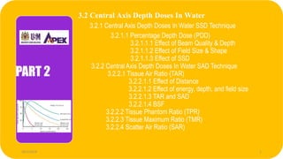
- 1. 3.2 Central Axis Depth Doses In Water 3.2.1 Central Axis Depth Doses In Water SSD Technique 3.2.1.1 Percentage Depth Dose (PDD) 3.2.1.1.1 Effect of Beam Quality & Depth 3.2.1.1.2 Effect of Field Size & Shape 3.2.1.1.3 Effect of SSD 3.2.2 Central Axis Depth Doses In Water SAD Technique 3.2.2.1 Tissue Air Ratio (TAR) 3.2.2.1.1 Effect of Distance 3.2.2.1.2 Effect of energy, depth, and field size 3.2.2.1.3 TAR and SAD 3.2.2.1.4 BSF 3.2.2.2 Tissue Phantom Ratio (TPR) 3.2.2.3 Tissue Maximum Ratio (TMR) 3.2.2.4 Scatter Air Ratio (SAR) PART 2 28/1/2018 1
- 2. 3.2.1 Central Axis Depth Doses In Water SSD Technique 228/1/2018 Dr. Nik Noor Ashikin Bt Nik Ab Razak
- 3. SOURCE TO SKIN DISTANCE Part VIII.3.5 Determination of Dose to a Patient-I Slide 3 SSD=80cm What is SSD?
- 4. 28/1/2018 Dr. Nik Noor Ashikin Bt Nik Ab Razak 4 What is SSD? Kilovoltage machines are typically fixed and therefore treatment is based using a constant source – surface distance (SSD). SSD calculations use percentage depth dose curves and are easier to measure in a phantom. With more modern treatments using multiple fields, the use of a constant SSD technique leads to frequent patient repositioning between treatments.
- 5. Part VIII.3.5 Determination of Dose to a Patient-I Slide 5 •SSD is the acronym of Source to Skin distance. It is the distance between the source and the patient skin •Machines have Standard SSD at which output and PDD are measured •50 cm for a Cs 137 Unit •80 – 100 cm for Co 60 unit •100 cm for Linear accelerator What is SSD?
- 6. 28/1/2018 Dr. Nik Noor Ashikin Bt Nik Ab Razak 6
- 7. Why is SSD important? • The dose calibration of the External beam unit is at the SSD (or Isocentre – explained later) • Any change in this will vary the dose by ‘inverse square’ factor Part VIII.3.5 Determination of Dose to a Patient-I Slide 7 What is SSD?
- 8. 28/1/2018 Dr. Nik Noor Ashikin Bt Nik Ab Razak 8 3.2.1.1 Percentage Depth Dose (PDD) 3.2.1.1.1 Effect on Beam Quality & Depth 3.2.1.1.2 Effect on Field Size & Shape 3.2.1.1.3 Effect on SSD
- 9. 1. What it is? • Attenuation factors • Simple to measure and used for dose calculation • Measured at SSD 3.2.1.1 Percentage Depth Dose (PDD)
- 10. • • 2. PDD Measurement Photon beam hits a rectangular phantom Photon beam get attenuated by the phantom material. The beam intensity falls as the beam is attenuated by the phantom • 3.2.1.1 Percentage Depth Dose (PDD)
- 11. • Absorbed dose at any depth: Dd • Absorbed dose at a fixed reference depth: Dd0 100 0 d d D D P collimator surface phantom D d0 D d d d0 3.1.3 Percentage Depth Dose (PDD) Quotient expressed as percentage, of the absorbed dose at any depth d to the absorbed dose at dmax along the central axis of the beam 3.2.1.1 Percentage Depth Dose (PDD)
- 12. Dose distribution 3.1.3 Percentage Depth Dose (PDD)3.2.1.1 Percentage Depth Dose (PDD)
- 13. For higher energies, the reference depth is at the peak absorbed dose ( d 0= d m) D max : maximum dose of the given dose For orthovoltage (up to 400 kVp) and lower energy X-rays, the reference depth is usually the surface (d 0= 0(position)) 3.1.3 Percentage Depth Dose (PDD)3.2.1.1 Percentage Depth Dose (PDD)
- 14. Part VIII.3.7 Operational Considerations – Planning of physical treatment Slide 14 3.1.3 Percentage Depth Dose (PDD)3.2.1.1 Percentage Depth Dose (PDD) 3. Dose deposition from a megavoltage photon beam in a patient
- 15. Beam enter the patient Deliver surface dose, Ds Dose rise rapidly after the surface dose , build up region Reach maximum value at depth zmax Decrease exponentially Reaches value Dex at the patient exit point 3.2.1.1 Percentage Depth Dose (PDD) 3. Dose deposition from a megavoltage photon beam in a patient
- 16. a.Surfacedose • For MV photon beam, surface dose is lower than maximum dose • Depends on: – Beam energy ( beam energy, surface dose) • E.g: 15% for 6 MV, 10 % for 18 MV – Field size ( field size, surface dose) 3.2.1.1 Percentage Depth Dose (PDD)
- 17. • Low surface dose is also called skin sparing effects • Important advantage of MV beams over orthovoltage and superficial beams in the treatment of deep seated tumours • KV beam do not exhibit skin sparing effects since their maximum dose occur on the skin surface (surface dose maximum dose) 3.2.1.1 Percentage Depth Dose (PDD) a.Surfacedose
- 18. • The surface dose represent contributions to the dose from: –Photons scattered from the collimators, flattening filter and air –Photons backscattered from the patient –High energy electrons produced by photon interaction in air and any shielding structures in vicinity of the patient. 3.2.1.1 Percentage Depth Dose (PDD) a.Surfacedose
- 19. Superficial beam Orthovoltage beam 3.2.1.1 Percentage Depth Dose (PDD) a.Surfacedose
- 20. B. Buildupregion • The dose region between the surface (depth z=0) and depth z= zmax) • Resulting from long range of energetic secondary charged particles that first released in the patient by photon interaction (photoelectric effects, compton effects, pair production) • Then the kinetic energy deposited in the patient. Therefore, the electron fluence and hence the absorbed dose increase with depth until they reach a maximum. 3.2.1.1 Percentage Depth Dose (PDD)
- 21. Result of the forward direction of secondary electrons - they deposit energy down stream from the original interaction point B. Buildupregion 3.2.1.1 Percentage Depth Dose (PDD)
- 22. B. Buildupregion 3.2.1.1 Percentage Depth Dose (PDD)
- 23. 3.1.3 Percentage Depth Dose (PDD) DOSE BUILD-UP 3.2.1.1 Percentage Depth Dose (PDD)
- 24. • In the region immediately beneath patient’s surface, the condition of CPE does not exist • Absorbed dose < collision kerma • As depth increase, CPE will be achieved at zmax, where z is equal to the range of secondary charged particles • At this stage, dose become equal to collision kerma. • Beyond zmax, the dose and collision kerma decrease because of of photon attenuation in the patient (transient CPE) B. Buildupregion 3.2.1.1 Percentage Depth Dose (PDD)
- 25. (1) Kinetic Energy Released In The Medium; (2) the energy transferred from photons to directly ionizing electron; (3) maximum at the surface and decreases with depth due to the decrease in the photon energy fluence; (4) the production of electrons also decreases with depth KERMA 3.1.3 Percentage Depth Dose (PDD)3.2.1.1 Percentage Depth Dose (PDD) B. Buildupregion
- 26. ABSORBED DOSE (1) depends on the electron fluence (2) high-speed electrons are ejected from the surface and subsequent layers (3) these electrons deposit their energy a significant distance away from their site of origin 3.1.3 Percentage Depth Dose (PDD)3.2.1.1 Percentage Depth Dose (PDD) B. Buildupregion
- 27. C. Depth of dose maximum zmax • Beneath patient surface • Depends : – Beam energy – Beam field size (minor effects) 3.2.1.1 Percentage Depth Dose (PDD)
- 28. D. Exit dose • The dose delivered to the patient at the beam exit point • Dose distribution curve slightly downwards from the extrapolated dose distribution curve • Because of small effect due to the missing scatter contribution at the exit point from beyond the exit dose point 3.2.1.1 Percentage Depth Dose (PDD)
- 29. Percentage Depth Dose (electron) PDD of electron beam
- 30. Isodose curves of electron beam • Scattering of electrons determines shapes of isodose curves –Expansion –Lateral constriction • Larger field size required at surface 3.1.3 Percentage Depth Dose (PDD)
- 31. 3.1.3 Percentage Depth Dose (PDD)
- 32. 28/1/2018 Dr. Nik Noor Ashikin Bt Nik Ab Razak 32 3.2.1.1.1 Effect on Beam Quality & Depth 3.2.1.1.2 Effect on Field Size & Shape 3.2.1.1.3 Effect on SSD
- 33. Dependence of PDD PDD (A) DEPENDENCE ON BEAM QUALITY AND (B) EFFECT OF FIELD SIZE AND SHAPE (C) DEPENDENCE ON SSD
- 34. 28/1/2018 Dr. Nik Noor Ashikin Bt Nik Ab Razak 34 3.2.1.1.1 Effect on Beam Quality & Depth
- 35. 3.2.1.1.1 Effect on Beam Quality & Depth PDD (beyond depth of max. dose) increases with beam energy Higher-energy beams have greater penetrating power and thus deliver a higher PDD
- 36. Fig. 4.3 Central axis depth dose distribution for different quality photon beams 3.2.1.1.1 Effect on Beam Quality & Depth
- 37. 28/1/2018 Dr. Nik Noor Ashikin Bt Nik Ab Razak 37
- 38. EXERCISE 1.Draw an Isodose curve 2. Draw the PDD curves for electron beam of energy 6 & 18 MeV and photon beam of energy 6 & 18 MV in water at 10 x 10 cm2 field size and 100 cm SSD 28/1/2018 Dr. Nik Noor Ashikin Bt Nik Ab Razak 38
- 39. 28/1/2018 Dr. Nik Noor Ashikin Bt Nik Ab Razak 39
- 40. 28/1/2018 Dr. Nik Noor Ashikin Bt Nik Ab Razak 40
- 41. 28/1/2018 Dr. Nik Noor Ashikin Bt Nik Ab Razak 41
- 42. PDD of Photon beam 28/1/2018 Dr. Nik Noor Ashikin Bt Nik Ab Razak 42 PDD of Electron beam
- 43. 28/1/2018 Dr. Nik Noor Ashikin Bt Nik Ab Razak 43 3.2.1.1.2 Effect on Field Size & Shape
- 44. Field size One of the most important parameters in treatment planning PDD increases as field size increases Field size dependence of PDD is less pronounced for higher energy than for lower energies Field size smaller than 6 cm Relative large penumbra region Bell shape TPS should be mandatory for small field size
- 45. 5*5 cm2 10*10 cm2 15*15cm2
- 46. 28/1/2018 Dr. Nik Noor Ashikin Bt Nik Ab Razak 46
- 47. 3.2.1.1.2 Effect on Field Size & Shape • the projection, on a plane perpendicular to the beam axis, of the distal end of the collimator as seen from the front center of the source 1. Geometrical field size • the distance intercepted by a given isodose curve (usually 50% isodose ) on a plane perpendicular to the beam axis 2.Dosimetric ( Physical ) field size
- 48. Dd Dmax Scatter dose 3.2.1.1.2 Effect on Field Size & Shape As the field size is increased, the contribution of the scattered radiation to the absorbed dose increases This increase in scattered dose is greater at larger than at the depth of D max , the percent depth dose increases with increasing field
- 49. 3.2.1.1.2 Effect on Field Size & Shape Depends on beam quality 1. The scattering probability or cross-section decreases with energy increase 2. the higher-energy photons are scattered more predominantly in the forward direction 3. the field size dependence of PDD is less pronounced for the higher-energy than for the lower-energy beams
- 50. 3.2.1.1.2 Effect on Field Size & Shape PDD data for radiotherapy beams are usually tabulated for SQUARE FIELDS In clinical practice require rectangular and irregularly shaped fields A system of equating square fields to different field shapes is required: EQUIVALENT SQUARE
- 51. 3.2.1.1.2 Effect on Field Size & Shape )(2 ba ba P A 4 a P A P A a 4 QUICK CALCULATION OF THE EQUIVALENT FIELD PARAMETERS (P):
- 52. a b P A 4 P A 4 3.2.1.1.2 Effect on Field Size & Shape
- 53. •Change this rectangular field to the equivalent square. 10 x 20 cm field? * See reference Table 9.2 in Faiz Khan’s book “The physics of radiation therapy” EXERCISE Wedge factor 3.2.1.1.2 Effect on Field Size & Shape
- 54. Equivalent square Table Part VIII.3.5 Determination of Dose to a Patient-I Slide 54 3.2.1.1.2 Effect on Field Size & Shape
- 55. •Equivalent circle has the same area as the equivalent square P A r 4 a b P A 4 P A 4 r 3.2.1.1.2 Effect on Field Size & Shape
- 56. Square Field to Rectangular fields.. Part VIII.3.5 Determination of Dose to a Patient-I Slide 56 10cm 10cm D Measured data for this But may treat with this 20cm 5cm Square field Rectangular field 3.2.1.1.2 Effect on Field Size & Shape
- 57. Equivalent square field Part VIII.3.5 Determination of Dose to a Patient-I Slide 57 3.2.1.1.2 Effect on Field Size & Shape
- 58. 28/1/2018 Dr. Nik Noor Ashikin Bt Nik Ab Razak 58 3.2.1.1.3 Effect on SSD
- 59. 3.2.1.1.3 Effect on SSD Photon fluence emitted by a point source of radiation varies inversely as a square of the distance from the source The actual dose rate at a point decreases with increase in distance from the source, the PDD, which is a relative dose, increases with SSD MAYNEORD F FACTOR
- 60. SSD affects the PDD and the depth of the isodose curves PDD icreases with SSD 3.2.1.1.3 Effect on SSD
- 61. Fig. 4.5 Plot of relative dose rate as inverse square law function of distance from a point source. Reference distance = 80 cm f1+dm f2+dm f1+d f2+d f1+d f2+d 100 max D D P d 3.2.1.1.3 Effect on SSD
- 62. 3.2.1.1.3 Effect on SSD Under extreme conditions such as lower energy, large field (the proportion of scattered radiation is relatively greater), large depth, large SSD, the Mayneord F factor has significant errors In general, the Mayneord F factor overestimates the increase in PDD with increase in SSD
- 63. d dm f2 d dm f1 r r Mayneord F factor (for small fields since the scattering is minimal) 2 2 1 2 1 2 df df df df F m m Wedge factor
- 64. • Let P(d,r,f) be the percent depth dose at depth d for SSD=f and a field size r • The ratio of PDD at SSD=f2 to PDD at SSD=f1 is: 2 2 1 2 1 2 1 2 ),,( ,, df df df df frdP frdP m m F frdP frdP ),,( ,, 1 2 3.2.1.1.3 Effect on SSD
- 65. Depth dose - point to ponder Depth dose increases with SSD - Why? •Ans: Larger the SSD, smaller is the decrease dose due to inverse square law Part VIII.3.5 Determination of Dose to a Patient-I Slide 65
- 66. DepthDose Pointsto Remember • In case of depth dose measurements the field size is defined at surface of phantom • Increases with SSD, Beam quality (energy), Field size. • Decreases with depth • Value is normalized to 100 % at depth of dmax for all field sizes Part VIII.3.5 Determination of Dose to a Patient-I Slide 66
- 67. EXERCISE For Co-60 beam, PDD for a 15x15 field size, 10 cm depth and 80 cm SSD is 58.4. Find the PDD for the same field size and depth for a 100 cm SSD. Assume dm = 0.5 cm 3.2.1.1.3 Effect on SSD
- 68. ANSWER F=1.043 P(10,15,100) --------------- = 1.043 P(10,15,80) Therefore desired depth dose = 58.4 x 1.043 = 60.9 3.2.1.1.3 Effect on SSD