Advances in Capillary Liquid Chromatography for High-Throughput Top Down Proteomics
•
0 likes•37 views

Top Down proteomics has benefited greatly from advances in mass spectrometry instrumentation and database searching, yet it has been hindered by the lack of robust separation platforms for intact proteins. Recently, the use of Gel-Eluted Liquid Fraction Entrapment Electrophoresis (GELFrEE)1,2 followed by capillary liquid chromatography-MS/MS has enabled hundreds of Top Down identifications from a single proteome run.
Recommended

Recommended
More Related Content
What's hot
What's hot (16)
Similar to Advances in Capillary Liquid Chromatography for High-Throughput Top Down Proteomics
Similar to Advances in Capillary Liquid Chromatography for High-Throughput Top Down Proteomics (20)
More from Expedeon
More from Expedeon (20)
Recently uploaded
Recently uploaded (20)
Advances in Capillary Liquid Chromatography for High-Throughput Top Down Proteomics
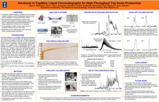
- 1. INTRODUCTION Top Down proteomics has benefited greatly from advances in mass spectrometry instrumentation and database searching, yet it has been hindered by the lack of robust separation platforms for intact proteins. Recently, the use of Gel-Eluted Liquid Fraction Entrapment Electrophoresis (GELFrEE)1,2 followed by capillary liquid chromatography-MS/MS has enabled hundreds of Top Down identifications from a single proteome run.3 However, most of the identifications occur in the low-molecular weight proteome (<40 kDa). Proteins of higher molecular weight, which comprise a majority of the human proteome, have been difficult to identify in great numbers4 largely due to poor chromatographic performance. In this work, we utilize column heating and modifications to the mobile phase to increase the peak capacity of LC separations. 1.Tran, J.C., Doucette, A.A. Anal. Chem. 2008, 80, 1568-73. 2. Tran, J.C., Doucette, A.A. Anal. Chem. 2009, 81, 6201-09. 3. Lee, J.E., Kellie, J.F., Tran, J.C., Tipton, J.D., Catherman, A.D., Thomas, H.M., Ahlf, D.R., Durbin, K.R., Vellaichamy, A., Ntai, I., Marshall, A.G., Kelleher, N.L. J. Am. Soc. Mass Spectrom., 2009, 20, 2183-91. 4. Vellaichamy, A., Tran, J.C., Catherman, A.D.; Lee, J.E., Kellie, J.F., Sweet, S.M., Zamdborg, L., Thomas, P.M., Ahlf, D.R., Durbin, K.R., Valaskovic, G.A., Kelleher, N.L. Anal. Chem. 2010, 82, 1234-44. HIGH RESOLUTION GELFREE OVERVIEW A separation platform utilizing a modified GELFrEE method coupled to capillary liquid chromatography reduces protein sample complexity for mass spectrometry analysis. Mass spectrometry data acquisition using ion trap precursor scans followed by nozzle-skimmer dissociation and FT fragmentation scans affords identification of proteins greater than 70 kDa on an LC timescale. A newly designed capillary column heater allows for decreased retention time and peak width. Modifications to the mobile phases show promise for further increasing protein separation. Advances in Capillary Liquid Chromatography for High-Throughput Top Down Proteomics Adam D. Catherman1, John C. Tran1, Lee Sawdey3, Kenneth R. Durbin1, Adaikkalam Vellaichamy2, Gary Valaskovic3, Neil L. Kelleher1,2 1Department of Chemistry, 2The Institute for Genomic Biology, University of Illinois, 3New Objective, Inc 100 75 ACKNOWLEDGEMENTS The authors would like to thank the other members of the Top Down Proteomics Development Team at the University of Illinois and the National High Magnetic Field Laboratory. Funding was provided by National Institutes of Health GM 067193-07 and the University of Illinois. CONCLUSIONS High resolution GELFrEE allows for the separation of a complicated proteome into narrow size bins for Top Down analysis of higher molecular weight proteins. A newly designed capillary column heater allows for heating the chromatographic separation, decreasing retention time and peak width. TFA has shown to decrease peak width but has led to a decrease in sensitivity. 15 20 25 37 50 75 100 150 250 MolecularWeight(kDa) FUTURE WORK Future work will be focused on determining the concentration of TFA or other ion pairing agents such as hexafluoroisopropanol that consistently allow for increased peak capacity as well as an increase in protein identifications. The use of isopropanol will be further explored for increasing resolution and sensitivity of high molecular weight proteins. Hydrophilic interaction liquid chromatography (HILIC) will be coupled to the mass spectrometer for the analysis of GELFrEE fractions. METHODS All buffers and gels were prepared according to Laemmli protocol. The separation potential was 180 V. The gel tube consisted of a 1 cm 4% gel and a 3 cm 8% resolving gel. 150 µL fractions were collected and 10 µL were used for SDS-PAGE visualization. SDS was removed from the GELFrEE fractions using MeOH/CHCl3/H2O precipitation and resuspended in 30 to 40 µL of H2O with 5% ACN and 0.2% formic acid. Capillary-LC separations were performed using a 10 cm long, 75 µm inner diameter columns packed with 5-µm PLRPS particles with 1000 Å pore size. Typical capillary LC solvents were A: H2O with 0.2% formic acid and B: 95% ACN, 5% H2O with 0.2% formic acid. In some experiments TFA was added at 0.05% or 0.1% or IPA with 0.2% formic acid was used as solvent B. Fragmentation was performed using nozzle-skimmer dissociation set to 75V. Protein Sample Solution IEF (Optional) GELFrEE Protein Precipitation and Resuspension for SDS Removal Nano-LC MS/MS on 12 T LTQ-FT Ultra Database Search SDS-PAGE Gel for Visualization ANALYSIS PLATFORM CAPILLARY COLUMN HEATER Easily couples to New Objective’s PicoView® nanospray source via a magnetic stage. Allows for entire packed bed of an analytical column to be heated, spraying sample from the pulled tip extending from the heater block. Conductive transfer block allows for fast temperature stabilization and has been used to heat the column to 60°C. USE OF TRIFLUOROACETIC ACID Use of TFA as an ion paring agent has shown significant decreases in peak width across the molecular weight range. Signal suppression has been observed using as low as 0.05%, leading to a decrease in identifications. Prosight ID: 78 kDa glucose-regulated protein w/ signal peptide cleaved 8 matching fragments (10 ppm tolerance) Best E-value: 2.5 x 10⁻⁹ Intact Mass ~ 71 kDa Ion Trap Intact Spectrum CAPILLARY COLUMN HEATING 25°C 45°C Figure 3: Selected ion chromatogram using two high-resolution fragments of glyceraldehyde-3-phosphate dehydrogenase (36 kDa) identified in an IEF-GELFrEE fraction. The peak width and retention time decrease when the capillary temperature is increase to 45°C. The separation used isopropanol as solvent B. Figure 4: Selected ion chromatogram using two high-resolution fragments of an identified 65 kDa protein from a high-resolution GELFrEE fraction. Again, the peak width and retention time both decrease significantly when the capillary temperature is increase to 45°C. The separation used 0.1% TFA. Figure 1: SDS-PAGE (12% T) visualization of the GELFrEE separation of 400 µg of HeLa cytosolic proteins. The visualization shows the ability of the GELFrEE platform to fractionate higher molecular weight proteins into narrow size bins. Figure 2: Base peak chromatogram from GELFrEE fraction 2 using 0.1% TFA. 25°C Base Peak Chromatogram GELFrEE Fraction 15 Nozzle-Skimmer Dissociation Ion Trap Precursor Scan FT Fragmentation Scan PROTEIN DETECTION AND IDENTIFICATION Additional IDs in fraction 15 Moesin 68 kDa Eukaryotic translation initiation factor 4B 69 kDa 4F2 cell-surface antigen heavy chain 62 kDa Heat shock protein HSP 90-beta 83 kDa Uncharacterized protein C19orf21 76 kDa m/z m/z 45°C