1. Motivation
Metalloproteins
Preparation
Conclusions and Future Work Acknowledgements
Approximately one-third of all proteins contain metal cofactors or bind to various metals. Determining the absolute number of metals contained in these proteins has proven to
be difficult. We hope to accurately characterize a protein’s stoichiometry with two common ion beam analysis methods along with a standard preparation technique. A
combination of Particle Induced X-ray Emission spectroscopy (PIXE) and Nuclear Reaction Analysis (NRA) is being developed to determine the areal density of a thin protein
target along with the target’s concentration of heavy elements. The results of this analytical method yield a stoichiometric ratio, along with the absolute numbers of the metal ions
contained in a certain metalloprotein. Current work focuses refining the present method of protein preparation to yield the most consistent results while reducing uncertainty in
the metallic ratios and absolute values.
Results
Cytochrome C
• The Nuclear Group
• Hope College Department of Physics
• Matthew Weiss
• Andrew McCubbin
• Megan Sibley
• Dave Daugherty
• This work is supported by the Nation Science
Foundation under grant No. PHY-1306074 and
grant No. PHY/DMR-1004811.
Ion Beam Analysis
Particle Induced X-ray Emission (PIXE) Nuclear Reaction Analysis (NRA)
Nuclear Reaction Analysis (NRA) is a non-destructive ion
beam analysis technique which is capable of measuring
the areal density of a thin target. The scattering of protons
is measured for a piece of Mylar® of a known thickness.
These counts are then compared to the counts of protons
scattering off a protein spot on the Mylar® substrate.
This allows for a determination of Mylar® correction
factor, so that counts of scattering off just the protein can
be calculated. To do this, the correction factor is applied
to the normalized Mylar® counts. These counts are then
subtracted from the total counts of both the protein spot
and the Mylar®. Once the counts of just the protein spot
are known, the absolute number of proteins and the
thickness of the protein spot can be derived.
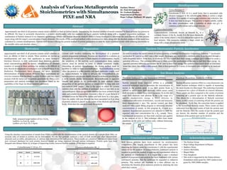
Particle Induced X-ray Emission (PIXE) is a powerful
analytical technique used to determine the concentration of
metals in the protein spots. A 3.4 MeV proton beam is
utilized to ionize inner shell electrons, which causes outer
shell electrons to replace these inner vacancies. To do this the
outer shell electrons emit photons in the x ray range of a
specific characteristic energy. A Si(Li) detector, in
combination with series of electronics, records and measures
these characteristic x rays. The spectra created, are then
analyzed with a peak fitting program to determine absolute
concentrations of metals. In this program B12 is used as a
standard. Experimental parameters are adjusted to yield the
expected ratio and concentration for a B12 sample. These
experimental parameters are then held constant and applied
to the analysis of Cyt. C. This technique offers trace metal
sensitivity on the parts per million scale, without destroying
the sample.
Using the absolute concentration of metals from PIXE coupled with the determination of the protein density in a sample from NRA, an
accurate ratio of metals to protein can be determined. For the five proteins analyzed a ratio of both metals and other characteristic
elements such as sulfur, phosphorous, or calcium were determined for each protein. These results are consistent with known values from
peer reviewed databases for each protein however results are being improved through various techniques. Protein spots have been
dropped onto thinner Mylar ® in hopes of improving results, however further analysis of this data is required.
Cytochrome C (Cyt. C) Cyanocobalamin (B12)
Cyanocobalamin commonly known as Vitamin B12 is a
common vitamer of the B12 family. It is the most chemically
stable of the family which is why it is utilized throughout the
food industry. This coenzyme contains a single cobalt ion
along with a single phosphorous atom.
Cytochrome C (Cyt. C) is a small heme that is associated with
electron transport in the mitochondria, where it carries a single
electron. It is capable of undergoing oxidation and reduction, but
it does not bind to oxygen. This protein is highly soluble, unlike
the other cytochromes, with a solubility of about 100 g/L. It
contains 4 sulfur atoms and a single iron ion.
Well known proteins have been analyzed for stoichiometric
composition. The largest impediment to this project has been
obtaining the correct ratios for Cytochrome C with the experimental
values set by the analysis of Cyanocobalamin. Continued analysis is
required to perfect the method of applying the experimental values to
the analysis of Cytochrome C. It is hoped that a once a standard
method of preparation and analysis has been confirmed with various
well-known proteins, that the technique be expanded to unknown
proteins. This method will hopefully be a time efficient way to
determine the function of an unknown protein by analyzing the ions
which they contain.
Tandem Electrostatic Particle Accelerator
In order to analyze the metal content of various proteins, a National Electrostatics Corporation Pelletron ™ Accelerator
accelerated H+ ions to 3.4 MeV onto a thin target . Hydrogen gas, excited by a rubidium oven, is accelerated through a
radio-frequency source. From here, to accelerate the ions to half their final energy, a stripper gas is utilized to create a
potential difference. This potential difference is what causes the acceleration of the ions to half their final energy. An
opposite potential difference accelerates the H+ ions to their final energy. The particles then travel down an evacuated
beam line where they strike a thin sample. Data is then gathered by a series of electronics and detectors to be analyzed
later.
Abstract
Cyanocobalamin
Cytochrome C PIXE spectrum
Fe Peak
S peak
Current work involves continuing the development of a standard
protein preparation technique. Proteins are dissolved in a solution of
ultra-pure-de-ionized water to limit chances of contamination. Due to
the sensitivity of the analysis used, contamination from various
sources must be limited in order to obtain consistent results.
Depending on protein characteristics the drying method must be
modified in order to limit recrystallization. Recrystallization occurs
when the protein is a fine powder and small molecular weight, such
as, Cyanocobalamin. In order to mitigate the recrystallization, the
aqueous protein solvent was slightly diluted in methanol providing for
a more volatile drying environment. Recrystallization is apparent
visually and proves to be problematic in data analysis where
integration of the particle spectra is complicated by long tails and
non-uniform peak shapes. Due to the speed in which the protein
solution dries with the addition of methanol, there is very little to no
recrystallization. Once an agreeable solution has been created it is set
aside and a ladder is assembled. Currently a film of 1.5 µm Mylar® is
stretched across the face of the ladder and held down by a face plate.
Once the ladder with taut Mylar® has been assembled an 8 µL drop
of protein solution is placed on the center of the Mylar® and allowed
to dry. From here the sample is ready for analysis.
Approximately one-third of all proteins contain metal cofactors or
have an affinity to bind to metals. These proteins are known as
metalloproteins and have vital significance to many biological
functions However, to fully understand these functions absolute
metal concentrations must be known. Identification of absolute
numbers of metals in these proteins has proven to be difficult at
best. Recent developments in techniques such as Synchrotron X-ray
Radiation Fluorescence have allowed for more accurate
determinations of metal content. However, these instruments are
even less common than Ion Beam Analysis facilities, and they don’t
measure both the metal and the protein quantitatively. Thus, a new
preparation and analysis technique was developed based on the
capabilities of the Hope Ion Beam Analysis Laboratory.
Fully prepared target ladders of Cyt. C & B12
Ladders 1-3: 8 µL B12 spots
Ladders 4-6: 8 µL Cyt. C Spots
Zachary Diener
Dr. Paul DeYoung and
Dr. Graham Peaslee
Department of Physics
Hope College, Holland, MI 49423
0
1000
2000
3000
4000
5000
6000
1 26 51 76 101 126 151 176 201 226 251 276
Methanol Added
Problem Region
Recrystallized 8 µL B12 Spot Standard 8 µL B12 Spot
NRA Cytochrome C spectrum
Element Expected Experimental
S 4 3.7 ± 0.15
Fe 1 0.98 ± 0.04
Element Expected Experimental
P 1 0.99 ± 0.04
Co 1 1.00 ± 0.04