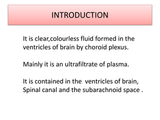
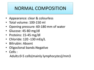
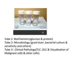
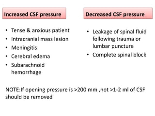
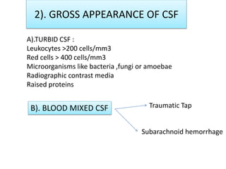
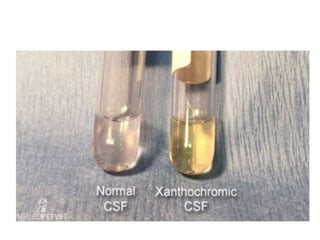
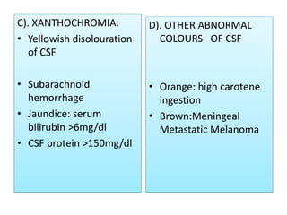
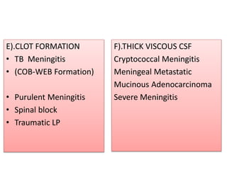
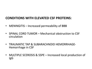
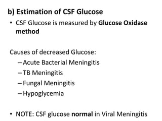
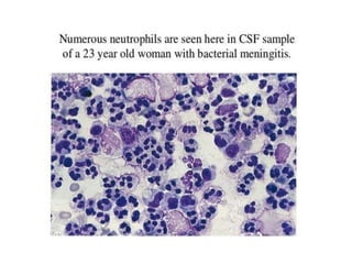
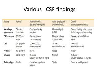
CSF ANALYSIS
INTRODUCTION
It is clear,colourless fluid formed in the
ventricles of brain by choroid plexus.
Mainly it is an ultrafiltrate of plasma.
It is contained in the ventricles of brain,
Spinal canal and the subarachnoid space .
NORMAL COMPOSITION
• Appearance: clear & colourless
• Total volume: 100-150 ml
• Opening pressure: 60-180 mm of water
• Glucose: 45-80 mg/dl
• Proteins: 15-45 mg/dl
• Chloride: 120 -130 mEq/L
• Bilirubin: Absent
• Oligoclonal bands:Negative
• Cells :
Adults:0-5 cells(mainly lymphocytes)/mm3
Functions of CSF
1).Protection of brain & spinal cord thereby acting as
shock absorber.
2).Serve as medium between blood and brain for supply of
nutrients to and removal of waste products from the brain.
Collection of CSF
Collected by Lumbar puncture.
• Patient in lateral recumbent
position.
• Under aseptic precautions,
22g LP needle is introduced .
• Collected in 3 sterile tubes.
Collection of CSF
• LUMBAR PUNCTURE-
btwn L3-L4 vertebrae in adults
btwn L4-L5 vertebrae in children
• CISTERNAL PUNCTURE- done in both
• VENTRICULAR PUNCTURE
• SHUNT DRAINAGE
Tube 1: BioChemistry(glucose & protein)
Tube 2: Microbiology (gram’stain ,bacterial culture &
sensitivity and others)
Tube 3 : Clinical Pathology(TLC ,DLC & Visualisation of
Malignant cells & other cells)
INDICATIONS OF LUMBAR
PUNCTURE
Detection and diagnosis of Suspected Meningitis,Subarachnoid
Haemorrhage , Spinal cord tumor ,CNS Syphilis
Differential diagnosis of Cerebral Infarction vs Intracerebral
Haemorrhage
Introduction of Anaesthetics ,Radiographic contrast media or Drugs
To reduce CSF pressure in Benign Intracranial Hypertension
Removal of exudate or blood from Subarachnoid space.
COMPLICATIONS OF LUMBAR PUNCTURE
1).Post-puncture headache : most common side effect.
So small bore needle(22G) is used.
2).Introduction of infection in spinal canal
3).Subdural hematoma
4).Dry tap
5).Herniation of Brain
CONTRAINDICATIONS TO LUMBAR
PUNCTURE
A). Raised Intracranial pressure
B). Cardiorespiratory compromise
C). Bleeding diathesis
D).Local infection at the site.
EXAMINATION OF CSF
•CSF fluid is examined within 1 hour of collection
because A). Cells degenerate rapidly
B). Reduction of glucose due to Spontaneous
Glycolysis and usage by bacteria
1). Opening pressure
During Lumbar Puncture,when CSF appears, manomete
Is attached to the spinal needle.
It is recorded with patient in lateral recumbent position
Increased CSF pressure
• Tense & anxious patient
• Intracranial mass lesion
• Meningitis
• Cerebral edema
• Subarachnoid
hemorrhage
Decreased CSF pressure
• Leakage of spinal fluid
following trauma or
lumbar puncture
• Complete spinal block
NOTE:If opening pressure is >200 mm ,not >1-2 ml of CSF
should be removed
2). GROSS APPEARANCE OF CSF
A).TURBID CSF :
Leukocytes >200 cells/mm3
Red cells > 400 cells/mm3
Microorganisms like bacteria ,fungi or amoebae
Radiographic contrast media
Raised proteins
B). BLOOD MIXED CSF Traumatic Tap
Subarachnoid hemorrhage
CSF Finding Traumatic LP Subarachnoid hemorrhage
a).Gross appearance Blood more in initial tubes
as compared to later tubes
Blood clots on standing.
Blood uniform in all tubes
Blood does not clot on standing
b).Supernatant after
centrifugation
clear Pink or yellow
(Xanthochromia)
e).Microscopy Progressive decrease of
red cell counts in later tubes
Red cell counts uniform
In all tubes
c).CSF Pressure Normal Increased
d).CSF protein Normal Increased
C). XANTHOCHROMIA:
• Yellowish disolouration
of CSF
• Subarachnoid
hemorrhage
• Jaundice: serum
bilirubin >6mg/dl
• CSF protein >150mg/dl
D). OTHER ABNORMAL
COLOURS OF CSF
• Orange: high carotene
ingestion
• Brown:Meningeal
Metastatic Melanoma
E).CLOT FORMATION
• TB Meningitis
• (COB-WEB Formation)
• Purulent Meningitis
• Spinal block
• Traumatic LP
F).THICK VISCOUS CSF
Cryptococcal Meningitis
Meningeal Metastatic
Mucinous Adenocarcinoma
Severe Meningitis
3). CHEMICAL EXAMINATION
a) Estimation of proteins :
• Normal levels : 15-45 mg/dl
Methods of estimation of proteins:-
•TURBIDIMETRIC METHOD
•PANDY’S TEST
CONDITIONS WITH ELEVATED CSF PROTEINS:
• MENINGITIS – Increased permeability of BBB
• SPINAL CORD TUMOR – Mechanical obstruction to CSF
circulation
• TRAUMATIC TAP & SUBARACHNOID HEMORRHAGE-
Hemorrhage in CSF
• MULTIPLE SCEROSIS & SSPE – Increased local production of
IgG
b) Estimation of CSF Glucose
• CSF Glucose is measured by Glucose Oxidase
method
Causes of decreased Glucose:
–Acute Bacterial Meningitis
–TB Meningitis
–Fungal Meningitis
–Hypoglycemia
• NOTE: CSF glucose normal in Viral Meningitis
c).Estimation of CSF ADA level
• Tuberculous : ADA >/- 10U/L
• Non-Tuberculous : ADA < 10U/L
d).Estimation of CSF Chloride
Normal levels –
Viral Meningitis
Viral Encephalitis
Poliomyelitis
Moderately Depressed level –Pyogenic Meningitis
Pronounced depression - TB Meningitis
Fungal Meningitis
4).CELL COUNTS IN CSF
NOTE: It’s essential to do microscopic examination of all CSF samples
since WBC COUNT upto 200 cells/mm3 & RBC count upto 400 cells/mm3
are associated with clear appearance of CSF.
a).METHODS OF TLC:
Manual Method &
Automated Method
Manual Method
If CSF Clear – NO DILUENT used
If CSF turbid /cloudy –DILUENT used with 1:20 dilution
Diluting fluid used – TURK’S FLUID
WBC Pipette used
• For counting cells in CSF ,FUCHS-ROSENTHAL
chamber >> NEUBAUER chamber
In Fuchs –Rosenthal chamber
• Cells are counted in 5 large squares
• If CSF IS DILUTED – number of cells counted x
dilution factor(20)
• If CSF UNDILUTED – total cells counted in 5
squares(total WBC count)
In Neubauer’s chamber
• Cells are counted in 4 WBC squares
• If CSF IS DILUTED – number of cells counted x 50
• If CSF UNDILUTED – total cells counted in 4
squares(total WBC count)
b)DLC
• If CSF CLEAR – Few drops of sediment taken on the slide and it
is spread over it.
• If CSF TURBID – smear is made directly from the
uncentrifuged sample.
• Now smears are stained with Romanowsky & examined under
Microscope.
• In NORMAL ADULTS -DLC shows 70% Lymphocytes
30% Monocytes
• In CHILDREN shows -70% Monocytes
Causes of Pleocytosis
• Meningitis & other CNS
infections
• Intracranial Haemorrhage
• Meningeal Infiltration by
Malignancy
• Repeated Lumbar punctures
• Injection of foreign substances
into subarachnoid space.
Differential count
`
REACTIVE LYMPHOCYTES IN CSF
5).Microbiological examination
a).Direct WET MOUNT OF CSF : Suspected cases of cryptococcal
infection,trypanosomiasis
and others.
OTHER TESTS are
b).GRAM’S STAIN
c).ZIEHL-NEELSEN STAIN
d).CULTURE for bacteria and Mycobacterium
e).PCR for Mycobacterium Tuberculosis and Viruses
Various CSF findings
SUMMARY
CSF Fluid which is collected in suspected CNS Pathology
via LUMBAR PUNCTURE, Processed and derived reports
from different departments thereby reaching the
appropriate Diagnosis and helping in the Management
of the CNS Disease.
CONCLUSION
Thus Analysis of CSF Fluid play a very important role in the
Diagnosis and Management of INFECTIOUS and
MALIGNANT Conditions of CNS
CSF ANALYSIS.pptx

CSF ANALYSIS.pptx

  • 1.
  • 2.
    INTRODUCTION It is clear,colourlessfluid formed in the ventricles of brain by choroid plexus. Mainly it is an ultrafiltrate of plasma. It is contained in the ventricles of brain, Spinal canal and the subarachnoid space .
  • 3.
    NORMAL COMPOSITION • Appearance:clear & colourless • Total volume: 100-150 ml • Opening pressure: 60-180 mm of water • Glucose: 45-80 mg/dl • Proteins: 15-45 mg/dl • Chloride: 120 -130 mEq/L • Bilirubin: Absent • Oligoclonal bands:Negative • Cells : Adults:0-5 cells(mainly lymphocytes)/mm3
  • 4.
    Functions of CSF 1).Protectionof brain & spinal cord thereby acting as shock absorber. 2).Serve as medium between blood and brain for supply of nutrients to and removal of waste products from the brain. Collection of CSF Collected by Lumbar puncture.
  • 5.
    • Patient inlateral recumbent position. • Under aseptic precautions, 22g LP needle is introduced . • Collected in 3 sterile tubes.
  • 6.
    Collection of CSF •LUMBAR PUNCTURE- btwn L3-L4 vertebrae in adults btwn L4-L5 vertebrae in children • CISTERNAL PUNCTURE- done in both • VENTRICULAR PUNCTURE • SHUNT DRAINAGE
  • 7.
    Tube 1: BioChemistry(glucose& protein) Tube 2: Microbiology (gram’stain ,bacterial culture & sensitivity and others) Tube 3 : Clinical Pathology(TLC ,DLC & Visualisation of Malignant cells & other cells)
  • 8.
    INDICATIONS OF LUMBAR PUNCTURE Detectionand diagnosis of Suspected Meningitis,Subarachnoid Haemorrhage , Spinal cord tumor ,CNS Syphilis Differential diagnosis of Cerebral Infarction vs Intracerebral Haemorrhage Introduction of Anaesthetics ,Radiographic contrast media or Drugs To reduce CSF pressure in Benign Intracranial Hypertension Removal of exudate or blood from Subarachnoid space.
  • 9.
    COMPLICATIONS OF LUMBARPUNCTURE 1).Post-puncture headache : most common side effect. So small bore needle(22G) is used. 2).Introduction of infection in spinal canal 3).Subdural hematoma 4).Dry tap 5).Herniation of Brain
  • 10.
    CONTRAINDICATIONS TO LUMBAR PUNCTURE A).Raised Intracranial pressure B). Cardiorespiratory compromise C). Bleeding diathesis D).Local infection at the site.
  • 11.
    EXAMINATION OF CSF •CSFfluid is examined within 1 hour of collection because A). Cells degenerate rapidly B). Reduction of glucose due to Spontaneous Glycolysis and usage by bacteria
  • 12.
    1). Opening pressure DuringLumbar Puncture,when CSF appears, manomete Is attached to the spinal needle. It is recorded with patient in lateral recumbent position
  • 13.
    Increased CSF pressure •Tense & anxious patient • Intracranial mass lesion • Meningitis • Cerebral edema • Subarachnoid hemorrhage Decreased CSF pressure • Leakage of spinal fluid following trauma or lumbar puncture • Complete spinal block NOTE:If opening pressure is >200 mm ,not >1-2 ml of CSF should be removed
  • 14.
    2). GROSS APPEARANCEOF CSF A).TURBID CSF : Leukocytes >200 cells/mm3 Red cells > 400 cells/mm3 Microorganisms like bacteria ,fungi or amoebae Radiographic contrast media Raised proteins B). BLOOD MIXED CSF Traumatic Tap Subarachnoid hemorrhage
  • 15.
    CSF Finding TraumaticLP Subarachnoid hemorrhage a).Gross appearance Blood more in initial tubes as compared to later tubes Blood clots on standing. Blood uniform in all tubes Blood does not clot on standing b).Supernatant after centrifugation clear Pink or yellow (Xanthochromia) e).Microscopy Progressive decrease of red cell counts in later tubes Red cell counts uniform In all tubes c).CSF Pressure Normal Increased d).CSF protein Normal Increased
  • 17.
    C). XANTHOCHROMIA: • Yellowishdisolouration of CSF • Subarachnoid hemorrhage • Jaundice: serum bilirubin >6mg/dl • CSF protein >150mg/dl D). OTHER ABNORMAL COLOURS OF CSF • Orange: high carotene ingestion • Brown:Meningeal Metastatic Melanoma
  • 18.
    E).CLOT FORMATION • TBMeningitis • (COB-WEB Formation) • Purulent Meningitis • Spinal block • Traumatic LP F).THICK VISCOUS CSF Cryptococcal Meningitis Meningeal Metastatic Mucinous Adenocarcinoma Severe Meningitis
  • 19.
    3). CHEMICAL EXAMINATION a)Estimation of proteins : • Normal levels : 15-45 mg/dl Methods of estimation of proteins:- •TURBIDIMETRIC METHOD •PANDY’S TEST
  • 20.
    CONDITIONS WITH ELEVATEDCSF PROTEINS: • MENINGITIS – Increased permeability of BBB • SPINAL CORD TUMOR – Mechanical obstruction to CSF circulation • TRAUMATIC TAP & SUBARACHNOID HEMORRHAGE- Hemorrhage in CSF • MULTIPLE SCEROSIS & SSPE – Increased local production of IgG
  • 21.
    b) Estimation ofCSF Glucose • CSF Glucose is measured by Glucose Oxidase method Causes of decreased Glucose: –Acute Bacterial Meningitis –TB Meningitis –Fungal Meningitis –Hypoglycemia • NOTE: CSF glucose normal in Viral Meningitis
  • 22.
    c).Estimation of CSFADA level • Tuberculous : ADA >/- 10U/L • Non-Tuberculous : ADA < 10U/L
  • 23.
    d).Estimation of CSFChloride Normal levels – Viral Meningitis Viral Encephalitis Poliomyelitis Moderately Depressed level –Pyogenic Meningitis Pronounced depression - TB Meningitis Fungal Meningitis
  • 24.
    4).CELL COUNTS INCSF NOTE: It’s essential to do microscopic examination of all CSF samples since WBC COUNT upto 200 cells/mm3 & RBC count upto 400 cells/mm3 are associated with clear appearance of CSF. a).METHODS OF TLC: Manual Method & Automated Method Manual Method If CSF Clear – NO DILUENT used If CSF turbid /cloudy –DILUENT used with 1:20 dilution Diluting fluid used – TURK’S FLUID WBC Pipette used
  • 25.
    • For countingcells in CSF ,FUCHS-ROSENTHAL chamber >> NEUBAUER chamber In Fuchs –Rosenthal chamber • Cells are counted in 5 large squares • If CSF IS DILUTED – number of cells counted x dilution factor(20) • If CSF UNDILUTED – total cells counted in 5 squares(total WBC count)
  • 26.
    In Neubauer’s chamber •Cells are counted in 4 WBC squares • If CSF IS DILUTED – number of cells counted x 50 • If CSF UNDILUTED – total cells counted in 4 squares(total WBC count)
  • 27.
    b)DLC • If CSFCLEAR – Few drops of sediment taken on the slide and it is spread over it. • If CSF TURBID – smear is made directly from the uncentrifuged sample. • Now smears are stained with Romanowsky & examined under Microscope. • In NORMAL ADULTS -DLC shows 70% Lymphocytes 30% Monocytes • In CHILDREN shows -70% Monocytes
  • 28.
    Causes of Pleocytosis •Meningitis & other CNS infections • Intracranial Haemorrhage • Meningeal Infiltration by Malignancy • Repeated Lumbar punctures • Injection of foreign substances into subarachnoid space.
  • 30.
  • 40.
  • 42.
    5).Microbiological examination a).Direct WETMOUNT OF CSF : Suspected cases of cryptococcal infection,trypanosomiasis and others. OTHER TESTS are b).GRAM’S STAIN c).ZIEHL-NEELSEN STAIN d).CULTURE for bacteria and Mycobacterium e).PCR for Mycobacterium Tuberculosis and Viruses
  • 43.
  • 44.
    SUMMARY CSF Fluid whichis collected in suspected CNS Pathology via LUMBAR PUNCTURE, Processed and derived reports from different departments thereby reaching the appropriate Diagnosis and helping in the Management of the CNS Disease.
  • 45.
    CONCLUSION Thus Analysis ofCSF Fluid play a very important role in the Diagnosis and Management of INFECTIOUS and MALIGNANT Conditions of CNS