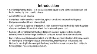
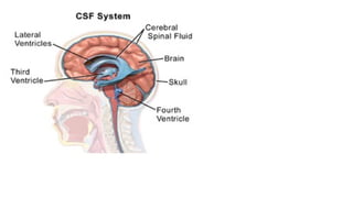
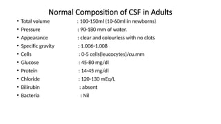
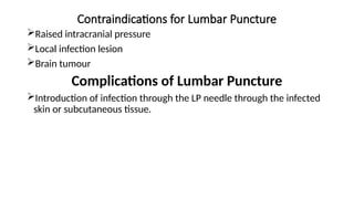
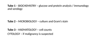
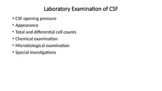
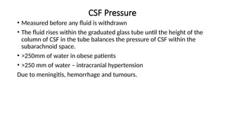
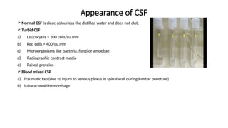
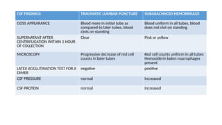
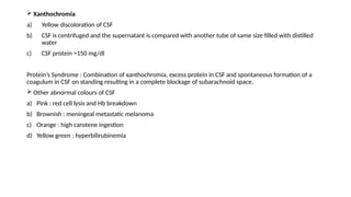
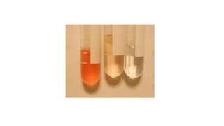
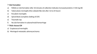
EXAMINATION OF
CEREBROSPINAL FLUID
GATLUAK JAMES KEDOK JIEK JANY (BBLT, MUK)
Introduction
• Cerebrospinal fluid (CSF) is a clear, colorless liquid found in the ventricles of the
brain mainly by the choroid plexus.
• An ultrafiltrate of plasma.
• Contained in the cerebral ventricles, spinal canal and subarachnoid space
(between arachnoid and pia matter).
• A CSF analysis is a group of tests that look at cerebrospinal fluid to help diagnose
diseases and conditions that affect the brain and spinal cord.
• Samples of cerebrospinal fluid are taken in cases of suspected meningitis,
subarachnoid haemorrhage and brain tumours as well as other conditions.
• Bacterial meningitis is an important and life threatening infection. Primary causes
of meningitis are Streptococcus pneumoniae, Haemophilus influenzae and
Neisseria meningitidis amongst the fungi and in immunocompromised patients,
Cryptococcus neoformans is common.
Functions of CSF
• Protection of the brain and spinal cord from injury by acting as shock
absorber or cushion.
• To serve as a medium between blood and brain for supply of nutrients
to and removal of waste products from the brain.
Normal Composition of CSF in Adults
• Total volume : 100-150ml (10-60ml in newborns)
• Pressure : 90-180 mm of water.
• Appearance : clear and colourless with no clots
• Specific gravity : 1.006-1.008
• Cells : 0-5 cells(leucocytes)/cu.mm
• Glucose : 45-80 mg/dl
• Protein : 14-45 mg/dl
• Chloride : 120-130 mEq/L
• Bilirubin : absent
• Bacteria : Nil
Collection of CSF
• Cerebrospinal fluid must be collected by an experienced medical officer or health worker. It is
usually obtained through a lumbar puncture (spinal tap). CSF must be collected aseptically to
prevent organisms being introduced into the central nervous system.
1. Lumbar puncture
2. Cisternal puncture : done in spinal cord block, vertebral deformity or infections in the tissue
where lumbar puncture is usually done.
3. Ventricular puncture : performed in infants who have an open fontanelle.
• Note: A delay in examining CSF reduces the chances of isolating a pathogen. It will also result in a
lower cell count due to WBCs being lyzed, and to a falsely low glucose value due to glycolysis.
When trypanosomes are present, they will be difficult to find because they are rapidly lyzed once
the CSF has been withdrawn.
Lumbar Puncture
Also called spinal tap where a sample of CSF is taken for examination.
Using a special needle called LP needle, length of 10 cm and 1-1.5mm with an
internal stylet.
The stylet has a pin which fits into the slot of the head of the needle and helps to
keep the needle patent.
Lumbar Puncture Procedure
• Patient usually lied on a bed on side (lateral recumbent position) with knees pulled up against the chest.
• Sterilize the area, a local anaesthetic is usually applied.
• The needle is introduced into the sunarachnoid space carefully, when the tip of the needle has reached the
correct depth, the stylet is removed and CSF will flow into the needle.
• Between 3rd
and 4th
lumbar vertebrae in adults.
• Between 4th
and 5th
lumbar vertebrae in children.
• CSF pressure is measured by using a glass manometer attached to a needle.
• 6-8 ml of CSF is collected into 3 sterile containers.
Indications for Lumbar Puncture
Diagnostic Indications
Infection of meninges
a) Bacterial – pyogenic, tuberculosis, syphilitic
b) Viral
c) Fungal
Subarachnoid hemorrhage
Primary or metastatic malignancy
Spinal cord blockage
Injecting a radio-opaque dye for myelography
Therapeutic Indications
Spinal anaesthesia
Injection of chemotherapeutic drugs
Contraindications for Lumbar Puncture
Raised intracranial pressure
Local infection lesion
Brain tumour
Complications of Lumbar Puncture
Introduction of infection through the LP needle through the infected
skin or subcutaneous tissue.
Tube 1 – BIOCHEMISTRY – glucose and protein analysis / immunology
and serology
Tube 2 – MICROBIOLOGY – culture and Gram’s stain
Tube 3 – HAEMATOLOGY – cell counts
CYTOLOGY – if malignancy is suspected
Points to Note
• Glass tubes avoided since cell adhesion to glass affects the cell count
and differential.
• Specimens delivered quickly to the laboratory to minimize cellular
degradation
• Refrigeration not indicated for culture specimens : because fastidious
organisms like H. influenzae and N. meningitidis will not survive.
Laboratory Examination of CSF
• CSF opening pressure
• Appearance
• Total and differential cell counts
• Chemical examination
• Microbiological examination
• Special investigations
CSF Pressure
• Measured before any fluid is withdrawn
• The fluid rises within the graduated glass tube until the height of the
column of CSF in the tube balances the pressure of CSF within the
subarachnoid space.
• >250mm of water in obese patients
• >250 mm of water – intracranial hypertension
Due to meningitis, hemorrhage and tumours.
Appearance of CSF
 Normal CSF is clear, colourless like distilled water and does not clot.
 Turbid CSF
a) Leucocytes > 200 cells/cu.mm
b) Red cells > 400/cu.mm
c) Microorganisms like bacteria, fungi or amoebae
d) Radiographic contrast media
e) Raised proteins
 Blood mixed CSF
a) Traumatic tap (due to injury to venous plexus in spinal wall during lumbar puncture)
b) Subarachnoid hemorrhage
CSF FINDINGS TRAUMATIC LUMBAR PUNCTURE SUBARACHNOID HEMORRHAGE
GOSS APPEARANCE Blood more in initial tube as
compared to later tubes, blood
clots on standing
Blood uniform in all tubes, blood
does not clot on standing
SUPERNATANT AFTER
CENTRIFUGATION WITHIN 1 HOUR
OF COLLECTION
Clear Pink or yellow
MICROSCOPY Progressive decrease of red cell
counts in later tubes
Red cell counts uniform in all tubes
Hemosiderin laden macrophages
present
LATEX AGGLUTINATION TEST FOR A
DIMER
negative positive
CSF PRESSURE normal Increased
CSF PROTEIN normal Increased
 Xanthochromia
a) Yellow discoloration of CSF
b) CSF is centrifuged and the supernatant is compared with another tube of same size filled with distilled
water
c) CSF protein >150 mg/dl
Protein’s Syndrome : Combination of xanthochromia, excess protein in CSF and spontaneous formation of a
coagulum in CSF on standing resulting in a complete blockage of subarachnoid space.
 Other abnormal colours of CSF
a) Pink : red cell lysis and Hb breakdown
b) Brownish : meningeal metastatic melanoma
c) Orange : high carotene ingestion
d) Yellow green : hyperbilirubinemia
 Clot Formation
a) Pellicle or clot formation after 10 minutes of collection indicates increased proteins (>150 mg/dl)
b) Tuberculosis meningitis (fine cobweb-like clot after 12 to 24 hours)
c) Purudent meningitis
d) Spinal block (complete clotting of CSF)
e) Traumatic tap
f) No clot formation in subarachnoid hemorrhage
 Thick viscous CSF
a) Cryptococcal meningitis
b) Meningeal metastatic adrenocarcinoma

DIAGNOSTIC_TESTS_ON_CEREBROSPINAL_FLUID.pptx

  • 1.
    EXAMINATION OF CEREBROSPINAL FLUID GATLUAKJAMES KEDOK JIEK JANY (BBLT, MUK)
  • 2.
    Introduction • Cerebrospinal fluid(CSF) is a clear, colorless liquid found in the ventricles of the brain mainly by the choroid plexus. • An ultrafiltrate of plasma. • Contained in the cerebral ventricles, spinal canal and subarachnoid space (between arachnoid and pia matter). • A CSF analysis is a group of tests that look at cerebrospinal fluid to help diagnose diseases and conditions that affect the brain and spinal cord. • Samples of cerebrospinal fluid are taken in cases of suspected meningitis, subarachnoid haemorrhage and brain tumours as well as other conditions. • Bacterial meningitis is an important and life threatening infection. Primary causes of meningitis are Streptococcus pneumoniae, Haemophilus influenzae and Neisseria meningitidis amongst the fungi and in immunocompromised patients, Cryptococcus neoformans is common.
  • 4.
    Functions of CSF •Protection of the brain and spinal cord from injury by acting as shock absorber or cushion. • To serve as a medium between blood and brain for supply of nutrients to and removal of waste products from the brain.
  • 5.
    Normal Composition ofCSF in Adults • Total volume : 100-150ml (10-60ml in newborns) • Pressure : 90-180 mm of water. • Appearance : clear and colourless with no clots • Specific gravity : 1.006-1.008 • Cells : 0-5 cells(leucocytes)/cu.mm • Glucose : 45-80 mg/dl • Protein : 14-45 mg/dl • Chloride : 120-130 mEq/L • Bilirubin : absent • Bacteria : Nil
  • 6.
    Collection of CSF •Cerebrospinal fluid must be collected by an experienced medical officer or health worker. It is usually obtained through a lumbar puncture (spinal tap). CSF must be collected aseptically to prevent organisms being introduced into the central nervous system. 1. Lumbar puncture 2. Cisternal puncture : done in spinal cord block, vertebral deformity or infections in the tissue where lumbar puncture is usually done. 3. Ventricular puncture : performed in infants who have an open fontanelle. • Note: A delay in examining CSF reduces the chances of isolating a pathogen. It will also result in a lower cell count due to WBCs being lyzed, and to a falsely low glucose value due to glycolysis. When trypanosomes are present, they will be difficult to find because they are rapidly lyzed once the CSF has been withdrawn.
  • 7.
    Lumbar Puncture Also calledspinal tap where a sample of CSF is taken for examination. Using a special needle called LP needle, length of 10 cm and 1-1.5mm with an internal stylet. The stylet has a pin which fits into the slot of the head of the needle and helps to keep the needle patent.
  • 8.
    Lumbar Puncture Procedure •Patient usually lied on a bed on side (lateral recumbent position) with knees pulled up against the chest. • Sterilize the area, a local anaesthetic is usually applied. • The needle is introduced into the sunarachnoid space carefully, when the tip of the needle has reached the correct depth, the stylet is removed and CSF will flow into the needle. • Between 3rd and 4th lumbar vertebrae in adults. • Between 4th and 5th lumbar vertebrae in children. • CSF pressure is measured by using a glass manometer attached to a needle. • 6-8 ml of CSF is collected into 3 sterile containers.
  • 9.
    Indications for LumbarPuncture Diagnostic Indications Infection of meninges a) Bacterial – pyogenic, tuberculosis, syphilitic b) Viral c) Fungal Subarachnoid hemorrhage Primary or metastatic malignancy Spinal cord blockage Injecting a radio-opaque dye for myelography Therapeutic Indications Spinal anaesthesia Injection of chemotherapeutic drugs
  • 10.
    Contraindications for LumbarPuncture Raised intracranial pressure Local infection lesion Brain tumour Complications of Lumbar Puncture Introduction of infection through the LP needle through the infected skin or subcutaneous tissue.
  • 11.
    Tube 1 –BIOCHEMISTRY – glucose and protein analysis / immunology and serology Tube 2 – MICROBIOLOGY – culture and Gram’s stain Tube 3 – HAEMATOLOGY – cell counts CYTOLOGY – if malignancy is suspected
  • 12.
    Points to Note •Glass tubes avoided since cell adhesion to glass affects the cell count and differential. • Specimens delivered quickly to the laboratory to minimize cellular degradation • Refrigeration not indicated for culture specimens : because fastidious organisms like H. influenzae and N. meningitidis will not survive.
  • 13.
    Laboratory Examination ofCSF • CSF opening pressure • Appearance • Total and differential cell counts • Chemical examination • Microbiological examination • Special investigations
  • 14.
    CSF Pressure • Measuredbefore any fluid is withdrawn • The fluid rises within the graduated glass tube until the height of the column of CSF in the tube balances the pressure of CSF within the subarachnoid space. • >250mm of water in obese patients • >250 mm of water – intracranial hypertension Due to meningitis, hemorrhage and tumours.
  • 15.
    Appearance of CSF Normal CSF is clear, colourless like distilled water and does not clot.  Turbid CSF a) Leucocytes > 200 cells/cu.mm b) Red cells > 400/cu.mm c) Microorganisms like bacteria, fungi or amoebae d) Radiographic contrast media e) Raised proteins  Blood mixed CSF a) Traumatic tap (due to injury to venous plexus in spinal wall during lumbar puncture) b) Subarachnoid hemorrhage
  • 16.
    CSF FINDINGS TRAUMATICLUMBAR PUNCTURE SUBARACHNOID HEMORRHAGE GOSS APPEARANCE Blood more in initial tube as compared to later tubes, blood clots on standing Blood uniform in all tubes, blood does not clot on standing SUPERNATANT AFTER CENTRIFUGATION WITHIN 1 HOUR OF COLLECTION Clear Pink or yellow MICROSCOPY Progressive decrease of red cell counts in later tubes Red cell counts uniform in all tubes Hemosiderin laden macrophages present LATEX AGGLUTINATION TEST FOR A DIMER negative positive CSF PRESSURE normal Increased CSF PROTEIN normal Increased
  • 17.
     Xanthochromia a) Yellowdiscoloration of CSF b) CSF is centrifuged and the supernatant is compared with another tube of same size filled with distilled water c) CSF protein >150 mg/dl Protein’s Syndrome : Combination of xanthochromia, excess protein in CSF and spontaneous formation of a coagulum in CSF on standing resulting in a complete blockage of subarachnoid space.  Other abnormal colours of CSF a) Pink : red cell lysis and Hb breakdown b) Brownish : meningeal metastatic melanoma c) Orange : high carotene ingestion d) Yellow green : hyperbilirubinemia
  • 19.
     Clot Formation a)Pellicle or clot formation after 10 minutes of collection indicates increased proteins (>150 mg/dl) b) Tuberculosis meningitis (fine cobweb-like clot after 12 to 24 hours) c) Purudent meningitis d) Spinal block (complete clotting of CSF) e) Traumatic tap f) No clot formation in subarachnoid hemorrhage  Thick viscous CSF a) Cryptococcal meningitis b) Meningeal metastatic adrenocarcinoma