Gnathostoma spinigerum
•Download as PPTX, PDF•
7 likes•4,149 views

Parasitology - Gnathostoma spinigerum

Recommended
More Related Content
What's hot
What's hot (20)
Viewers also liked
Viewers also liked (11)
Similar to Gnathostoma spinigerum
Similar to Gnathostoma spinigerum (20)
More from Hazel Barcela
More from Hazel Barcela (15)
Recently uploaded
Recently uploaded (20)
Gnathostoma spinigerum
- 2. Gnathostoma spinegerum • Several species of the genus Gnathostoma are responsible for the zoonotic infections of man. • The most common being the species spinegerum commonly found in dogs, cats and several other carnivores • Human infections have been reported from Japan, China, Thailand, The Far East and The Philippines, mostly acquired from consumption of infected fresh water fish
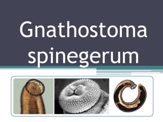
- 3. • Stout, reddish, slightly transparent with sub lobose cephalic swelling separated from the remainder of the worm by a cervical constriction • Curved ventrad at both ends • Posterior half is aspinous except for a few small terminal spines’ • Cephalic portion is covered with 4-8 rows of sharp, recurve hooks
- 4. Adult worm • Female ▫ 25-54µm long ▫ More curved tails than males ▫ larger • Male ▫ 11-25µm long ▫ Males have red tails
- 5. Eggs • 65-70 by 38-40µm • Ovoid, transparent, mucoid plug on one end, unembryonated
- 6. Life cycle • Natural DH : Domestic and wild felines, dogs, and foxes • Unatural DH : Man • Habitat : Tightly-coiled within tumors of the intestinal walls of the definitive hosts • Intermediate host ▫ 1st: Cyclops ▫ 2nd: Fresh water fish, snakes, crabs, crayfish and amphibians
- 7. Life Cycle
- 8. Local Epidemiology • The human cases of Gnathosomiasis (G.hispidum) are attributed to the consumtion of the fresh water fish Misgurnusangillicaudatus • In the Philippines the larvae of G. Dolorosi are found in “dalag” (Ophicephalustriatus in Laguna
- 9. Clinical Diagnosis • Gnathostomiasisinterna ▫ Adult worms are coiled inside tumors of the definitive hosts • Gnathostomiasisexterna ▫ Due to 3rd stage larva in humans ▫ Deep cutaneous and subcutaneous tunnels – visceral larva migrans • Migration to other tissue: results in cough, hematuria, ocular involvement, in serious manifestations eosinophilic meningitis with myeloencephalitis
- 10. Diagnosis • Presumptive diagnosis may be made on the basis on clinical symptoms • Definitive diagnosis is the removal and identification of the worm: presence of spines on the cuticle specially on the anterior end of worm, presence of large lateral chords, pigmented granular material in the intestinal cells
- 11. • Intradermal test : antigen extracted from adult or larva of the worm • Precipitin reaction – specific • Leukocytes with marked eosinophilia – highly suggestive END