SlideShare a Scribd company logo
1 of 29
Intravenous Cannulation
Group Members
Nageena
Shakeela
Shaista
Sonia
Kawish
7:21:12 AM
intravenous cannulation
1
Objectives
By the end of this session learner will be able to
• Define IV cannulation
• Explain the indication of IV cannulation
• Explain the advantages of IV cannulation
• Enlist the equipment's that are used for IV
• List the sites for iv placement
• Explain the IV cannulation procedure
• Explain the complication of IV cannulation
7:21:12 AM
intravenous cannulation
2
Cannula
A cannula (/ˈkænjʊlə/; from Latin "little reed"; plural
cannulae or cannulas)[1] is a tube that can be inserted
into the body, often for the delivery or removal of fluid
or for the gathering of data.
Origion of cannula
1675–85; < New Latin, Latin: small reed, equivalent to
cann(a) cane + -ula -ule
a metal tube for insertion into the body to draw off
fluid or to introduce medication
7:21:12 AM
intravenous cannulation
3
Cannulation
“The aim of intravenous management is safe, effective
delivery of treatment without discomfort or tissue
damage and without compromising venous access,
especially if long term therapy is proposed”
The Royal Marsden
NHS Trust Manual of Clinical Nursing Procedures
Fourth Addition
7:21:12 AM
intravenous cannulation
4
Anatomy and physiology
• Approximately 2/3 of the total blood volumeis in the
vein which is transport deoxygenated blood to the heart
from the tissues
• Veins are thin valled ,fibrous ,have large lumen and low
pressure
• Superificial and deep veins
• Some veins have valves to regulate the one way flow to
the heart especially lower limbs
7:21:12 AM
intravenous cannulation
5
Blood vessel walls have three layers
• Tunic Intima innermost, epithelial lining
• Tunic Media Elastic and Smooth muscle fibers
• Tunic Externa Outer coat
Major veins of the arm
• Dorsal • Cephalic • Basilic • Cubital Fossa
7:21:12 AM
intravenous cannulation
6
7:21:12 AM intravenous cannulation 7
• Fluid and electrolyte replacement
• Administration of medicines
• Administration of blood/blood products
• Administration of Total Parenteral Nutrition
• Haemodynamic monitoring
• Blood sampling
7:21:12 AM
intravenous cannulation
Indications
Advantages
• Immediate effect
• Control over the rate of administration
• Patient cannot tolerate drugs / fluids orally
• Some drugs cannot be absorbed by any other route
• Pain and irritation is avoided compared to some
substances when given SC/IM
7:21:12 AM
intravenous cannulation
9
7:21:12 AM
intravenous cannulation
10
7:21:12 AM
intravenous cannulation
11
7:21:12 AM intravenous cannulation 12
7:21:12 AM intravenous cannulation 13
What equipment do you need
• Dressing Tray - ANTT
• Non Sterile Gloves / Apron
• Cleaning Wipes
• Gauze swab
• IV cannula (separate slide)
• Tourniquet
• Dressing to secure cannula
• Alcohol wipes
• Saline flush and sterile syringe or fluid to be
administered
• Sharps bin
7:21:12 AM
intravenous cannulation
14
Procedure
• Wash hands prepare equipment ANTT
• Remove the cannula from the packaging and check
all parts are operational
• Loosen the white cap and gently replace it
• Apply tourniquet
• Identify vein
• Clean the site over the vein with alcohol wipe, allow
to dry
7:21:12 AM
intravenous cannulation
15
• Insert the needle (bevel side up) at an angle of 10-
30o to the skin (this will depend on vein depth.)
• Observe for blood in the flashback chamber
7:21:12 AM
intravenous cannulation
16
• Apply gentle pressure over the vein (beyond the
cannula tip) remove the white cap from the needle
7:21:12 AM
intravenous cannulation
17
• Remove the needle from the cannula and dispose of
it into a sharps container
• Attach the white lock cap
• Secure the cannula with an appropriate dressing
7:21:12 AM
intravenous cannulation
18
• Flush the cannula with 2-5 mls 0.9% Sodium
Chloride or attach an IV giving set and fluid
7:21:12 AM
intravenous cannulation
19
Document the procedure including:
• Date and time
• Site and size of cannula ,any problems
encountered
• Review date (cannula should be in situ no longer
than 72 hours without appropriate risk assessment.)
• Thanks to the patient
• Clean up, dispose of rubbish
7:21:12 AM
intravenous cannulation
20
Possible Complications:
The intravenous (IV) cannula offers direct access to a
patient's vascular system and provides a potential route
for entry of micro organisms into that system. These
organisms can cause serious infection if they are
allowed to enter and proliferate in the IV cannula,
insertion site, or IV fluid.
7:21:12 AM
intravenous cannulation
21
IV-Site Infection:
Does not produce much (if any) pus or inflammation at
the IV site. This is the most common cannula-related
infection, may be the most difficult to identify
7:21:12 AM
intravenous cannulation
22
Cellulites:
Warm, red and often tender skin surrounding the site
of cannula insertion; pus is rarely detectable.
7:21:12 AM
intravenous cannulation
23
Infiltration
• Infiltration or tissuing occurs when the infusion (fluid) leaks
into the surrounding tissue. It is important to detect early as
tissue necrosis could occur – re-site cannula immediately
7:21:12 AM
intravenous cannulation
24
Bruising
Bruising commonly results from failed IV placement -
particularly in the elderly and those on anticoagulant
therapy.
7:21:12 AM
intravenous cannulation
25
Haematoma:
Haematoma occurs when blood leaks out of the infusion
site. The common cause of this is using cannula that are
not tapered at the distal end. It will also occur if on
insertion the cannula has penetrated through the other
side of the vessel wall – apply pressure to the site for
approximately 4 minutes and elevate the limb
7:21:12 AM
intravenous cannulation
26
Phlebitis:
Phlebitiss is common in IV therapy and can be cause in many
ways. It is inflammation of a vein (redness and pain at the
infusion site) – prevention can be using aseptic insertion
techniques, choosing the smallest gauge cannula possible for the
prescribed treatment, secure the cannula properly to prevent
movement and carry out regular checks of the infusion site.
7:21:12 AM
intravenous cannulation
27
References:
• Clinical Skills Education Centre
http://www.qub.ac.uk/cskills/index.htm
• Standards for Infusion Therapy RCN
http://www.rcn.org.uk/publications/pdf/standardsi
nfusiontherapy.pdf
7:21:12 AM
intravenous cannulation
28
7:21:12 AM
intravenous cannulation
29

More Related Content

Similar to iv cannula.pptx

INTRAVENOUS INJECTION.pptx
INTRAVENOUS INJECTION.pptxINTRAVENOUS INJECTION.pptx
INTRAVENOUS INJECTION.pptx
ArpitPartil1
 
Iv Therapy
Iv TherapyIv Therapy
Iv Therapy
washinca
 

Similar to iv cannula.pptx (20)

INTRAVENOUS INJECTION.pptx
INTRAVENOUS INJECTION.pptxINTRAVENOUS INJECTION.pptx
INTRAVENOUS INJECTION.pptx
 
IV Cannulation
IV CannulationIV Cannulation
IV Cannulation
 
Pediatric lumbar puncture
Pediatric lumbar puncturePediatric lumbar puncture
Pediatric lumbar puncture
 
Blood collection.pptx
Blood collection.pptxBlood collection.pptx
Blood collection.pptx
 
cannulation and introduction, sizes and site of cannula
cannulation and introduction, sizes and site of cannulacannulation and introduction, sizes and site of cannula
cannulation and introduction, sizes and site of cannula
 
Cannulation
CannulationCannulation
Cannulation
 
Iv Therapy
Iv TherapyIv Therapy
Iv Therapy
 
Iv Therapy
Iv TherapyIv Therapy
Iv Therapy
 
9-Vascular-Access-updated.pdf
9-Vascular-Access-updated.pdf9-Vascular-Access-updated.pdf
9-Vascular-Access-updated.pdf
 
Blood Specimen Collection FY17.pptx
Blood Specimen Collection FY17.pptxBlood Specimen Collection FY17.pptx
Blood Specimen Collection FY17.pptx
 
Central venous catheterization
Central venous catheterizationCentral venous catheterization
Central venous catheterization
 
External Ventricular Drain
External Ventricular DrainExternal Ventricular Drain
External Ventricular Drain
 
Echocardiography, CSF study, IV and IM injections
Echocardiography, CSF study, IV and IM injectionsEchocardiography, CSF study, IV and IM injections
Echocardiography, CSF study, IV and IM injections
 
Central Venous Catheter Care- A Nursing skill
Central Venous Catheter Care- A Nursing skill Central Venous Catheter Care- A Nursing skill
Central Venous Catheter Care- A Nursing skill
 
Phlebotomy -2[1]
Phlebotomy -2[1]Phlebotomy -2[1]
Phlebotomy -2[1]
 
External Ventricular Drain (EVD)
External Ventricular Drain (EVD)External Ventricular Drain (EVD)
External Ventricular Drain (EVD)
 
Venesection
VenesectionVenesection
Venesection
 
IM,IV,IA.pptx
IM,IV,IA.pptxIM,IV,IA.pptx
IM,IV,IA.pptx
 
COMPLICATIONS OF PCNL.pptx
COMPLICATIONS OF PCNL.pptxCOMPLICATIONS OF PCNL.pptx
COMPLICATIONS OF PCNL.pptx
 
INTRAVENOUS-FLUID-INSERTION-2.ppt
INTRAVENOUS-FLUID-INSERTION-2.pptINTRAVENOUS-FLUID-INSERTION-2.ppt
INTRAVENOUS-FLUID-INSERTION-2.ppt
 

More from GhaffarAhmed3

More from GhaffarAhmed3 (6)

Spinal Cord Injuries.pptx
Spinal Cord Injuries.pptxSpinal Cord Injuries.pptx
Spinal Cord Injuries.pptx
 
CVA.ppt
CVA.pptCVA.ppt
CVA.ppt
 
Time Management.pptx
Time Management.pptxTime Management.pptx
Time Management.pptx
 
Concept of Elimination- GA.pptx
Concept of Elimination- GA.pptxConcept of Elimination- GA.pptx
Concept of Elimination- GA.pptx
 
Patricia Banner Theory.pptx
Patricia Banner Theory.pptxPatricia Banner Theory.pptx
Patricia Banner Theory.pptx
 
Medication Error.pptx
Medication Error.pptxMedication Error.pptx
Medication Error.pptx
 

Recently uploaded

(May 9, 2024) Enhanced Ultrafast Vector Flow Imaging (VFI) Using Multi-Angle ...
(May 9, 2024) Enhanced Ultrafast Vector Flow Imaging (VFI) Using Multi-Angle ...(May 9, 2024) Enhanced Ultrafast Vector Flow Imaging (VFI) Using Multi-Angle ...
(May 9, 2024) Enhanced Ultrafast Vector Flow Imaging (VFI) Using Multi-Angle ...
Scintica Instrumentation
 
CYTOGENETIC MAP................ ppt.pptx
CYTOGENETIC MAP................ ppt.pptxCYTOGENETIC MAP................ ppt.pptx
CYTOGENETIC MAP................ ppt.pptx
Cherry
 
Pteris : features, anatomy, morphology and lifecycle
Pteris : features, anatomy, morphology and lifecyclePteris : features, anatomy, morphology and lifecycle
Pteris : features, anatomy, morphology and lifecycle
Cherry
 
Cyathodium bryophyte: morphology, anatomy, reproduction etc.
Cyathodium bryophyte: morphology, anatomy, reproduction etc.Cyathodium bryophyte: morphology, anatomy, reproduction etc.
Cyathodium bryophyte: morphology, anatomy, reproduction etc.
Cherry
 
LUNULARIA -features, morphology, anatomy ,reproduction etc.
LUNULARIA -features, morphology, anatomy ,reproduction etc.LUNULARIA -features, morphology, anatomy ,reproduction etc.
LUNULARIA -features, morphology, anatomy ,reproduction etc.
Cherry
 
POGONATUM : morphology, anatomy, reproduction etc.
POGONATUM : morphology, anatomy, reproduction etc.POGONATUM : morphology, anatomy, reproduction etc.
POGONATUM : morphology, anatomy, reproduction etc.
Cherry
 

Recently uploaded (20)

Genome Projects : Human, Rice,Wheat,E coli and Arabidopsis.
Genome Projects : Human, Rice,Wheat,E coli and Arabidopsis.Genome Projects : Human, Rice,Wheat,E coli and Arabidopsis.
Genome Projects : Human, Rice,Wheat,E coli and Arabidopsis.
 
Human & Veterinary Respiratory Physilogy_DR.E.Muralinath_Associate Professor....
Human & Veterinary Respiratory Physilogy_DR.E.Muralinath_Associate Professor....Human & Veterinary Respiratory Physilogy_DR.E.Muralinath_Associate Professor....
Human & Veterinary Respiratory Physilogy_DR.E.Muralinath_Associate Professor....
 
Cot curve, melting temperature, unique and repetitive DNA
Cot curve, melting temperature, unique and repetitive DNACot curve, melting temperature, unique and repetitive DNA
Cot curve, melting temperature, unique and repetitive DNA
 
(May 9, 2024) Enhanced Ultrafast Vector Flow Imaging (VFI) Using Multi-Angle ...
(May 9, 2024) Enhanced Ultrafast Vector Flow Imaging (VFI) Using Multi-Angle ...(May 9, 2024) Enhanced Ultrafast Vector Flow Imaging (VFI) Using Multi-Angle ...
(May 9, 2024) Enhanced Ultrafast Vector Flow Imaging (VFI) Using Multi-Angle ...
 
TransientOffsetin14CAftertheCarringtonEventRecordedbyPolarTreeRings
TransientOffsetin14CAftertheCarringtonEventRecordedbyPolarTreeRingsTransientOffsetin14CAftertheCarringtonEventRecordedbyPolarTreeRings
TransientOffsetin14CAftertheCarringtonEventRecordedbyPolarTreeRings
 
GBSN - Microbiology (Unit 3)Defense Mechanism of the body
GBSN - Microbiology (Unit 3)Defense Mechanism of the body GBSN - Microbiology (Unit 3)Defense Mechanism of the body
GBSN - Microbiology (Unit 3)Defense Mechanism of the body
 
CYTOGENETIC MAP................ ppt.pptx
CYTOGENETIC MAP................ ppt.pptxCYTOGENETIC MAP................ ppt.pptx
CYTOGENETIC MAP................ ppt.pptx
 
Use of mutants in understanding seedling development.pptx
Use of mutants in understanding seedling development.pptxUse of mutants in understanding seedling development.pptx
Use of mutants in understanding seedling development.pptx
 
GBSN - Biochemistry (Unit 2) Basic concept of organic chemistry
GBSN - Biochemistry (Unit 2) Basic concept of organic chemistry GBSN - Biochemistry (Unit 2) Basic concept of organic chemistry
GBSN - Biochemistry (Unit 2) Basic concept of organic chemistry
 
Pteris : features, anatomy, morphology and lifecycle
Pteris : features, anatomy, morphology and lifecyclePteris : features, anatomy, morphology and lifecycle
Pteris : features, anatomy, morphology and lifecycle
 
Cyathodium bryophyte: morphology, anatomy, reproduction etc.
Cyathodium bryophyte: morphology, anatomy, reproduction etc.Cyathodium bryophyte: morphology, anatomy, reproduction etc.
Cyathodium bryophyte: morphology, anatomy, reproduction etc.
 
GBSN - Microbiology (Unit 5) Concept of isolation
GBSN - Microbiology (Unit 5) Concept of isolationGBSN - Microbiology (Unit 5) Concept of isolation
GBSN - Microbiology (Unit 5) Concept of isolation
 
Daily Lesson Log in Science 9 Fourth Quarter Physics
Daily Lesson Log in Science 9 Fourth Quarter PhysicsDaily Lesson Log in Science 9 Fourth Quarter Physics
Daily Lesson Log in Science 9 Fourth Quarter Physics
 
LUNULARIA -features, morphology, anatomy ,reproduction etc.
LUNULARIA -features, morphology, anatomy ,reproduction etc.LUNULARIA -features, morphology, anatomy ,reproduction etc.
LUNULARIA -features, morphology, anatomy ,reproduction etc.
 
Understanding Partial Differential Equations: Types and Solution Methods
Understanding Partial Differential Equations: Types and Solution MethodsUnderstanding Partial Differential Equations: Types and Solution Methods
Understanding Partial Differential Equations: Types and Solution Methods
 
X-rays from a Central “Exhaust Vent” of the Galactic Center Chimney
X-rays from a Central “Exhaust Vent” of the Galactic Center ChimneyX-rays from a Central “Exhaust Vent” of the Galactic Center Chimney
X-rays from a Central “Exhaust Vent” of the Galactic Center Chimney
 
POGONATUM : morphology, anatomy, reproduction etc.
POGONATUM : morphology, anatomy, reproduction etc.POGONATUM : morphology, anatomy, reproduction etc.
POGONATUM : morphology, anatomy, reproduction etc.
 
Thyroid Physiology_Dr.E. Muralinath_ Associate Professor
Thyroid Physiology_Dr.E. Muralinath_ Associate ProfessorThyroid Physiology_Dr.E. Muralinath_ Associate Professor
Thyroid Physiology_Dr.E. Muralinath_ Associate Professor
 
module for grade 9 for distance learning
module for grade 9 for distance learningmodule for grade 9 for distance learning
module for grade 9 for distance learning
 
Energy is the beat of life irrespective of the domains. ATP- the energy curre...
Energy is the beat of life irrespective of the domains. ATP- the energy curre...Energy is the beat of life irrespective of the domains. ATP- the energy curre...
Energy is the beat of life irrespective of the domains. ATP- the energy curre...
 

iv cannula.pptx

  • 2. Objectives By the end of this session learner will be able to • Define IV cannulation • Explain the indication of IV cannulation • Explain the advantages of IV cannulation • Enlist the equipment's that are used for IV • List the sites for iv placement • Explain the IV cannulation procedure • Explain the complication of IV cannulation 7:21:12 AM intravenous cannulation 2
  • 3. Cannula A cannula (/ˈkænjʊlə/; from Latin "little reed"; plural cannulae or cannulas)[1] is a tube that can be inserted into the body, often for the delivery or removal of fluid or for the gathering of data. Origion of cannula 1675–85; < New Latin, Latin: small reed, equivalent to cann(a) cane + -ula -ule a metal tube for insertion into the body to draw off fluid or to introduce medication 7:21:12 AM intravenous cannulation 3
  • 4. Cannulation “The aim of intravenous management is safe, effective delivery of treatment without discomfort or tissue damage and without compromising venous access, especially if long term therapy is proposed” The Royal Marsden NHS Trust Manual of Clinical Nursing Procedures Fourth Addition 7:21:12 AM intravenous cannulation 4
  • 5. Anatomy and physiology • Approximately 2/3 of the total blood volumeis in the vein which is transport deoxygenated blood to the heart from the tissues • Veins are thin valled ,fibrous ,have large lumen and low pressure • Superificial and deep veins • Some veins have valves to regulate the one way flow to the heart especially lower limbs 7:21:12 AM intravenous cannulation 5
  • 6. Blood vessel walls have three layers • Tunic Intima innermost, epithelial lining • Tunic Media Elastic and Smooth muscle fibers • Tunic Externa Outer coat Major veins of the arm • Dorsal • Cephalic • Basilic • Cubital Fossa 7:21:12 AM intravenous cannulation 6
  • 7. 7:21:12 AM intravenous cannulation 7
  • 8. • Fluid and electrolyte replacement • Administration of medicines • Administration of blood/blood products • Administration of Total Parenteral Nutrition • Haemodynamic monitoring • Blood sampling 7:21:12 AM intravenous cannulation Indications
  • 9. Advantages • Immediate effect • Control over the rate of administration • Patient cannot tolerate drugs / fluids orally • Some drugs cannot be absorbed by any other route • Pain and irritation is avoided compared to some substances when given SC/IM 7:21:12 AM intravenous cannulation 9
  • 12. 7:21:12 AM intravenous cannulation 12
  • 13. 7:21:12 AM intravenous cannulation 13
  • 14. What equipment do you need • Dressing Tray - ANTT • Non Sterile Gloves / Apron • Cleaning Wipes • Gauze swab • IV cannula (separate slide) • Tourniquet • Dressing to secure cannula • Alcohol wipes • Saline flush and sterile syringe or fluid to be administered • Sharps bin 7:21:12 AM intravenous cannulation 14
  • 15. Procedure • Wash hands prepare equipment ANTT • Remove the cannula from the packaging and check all parts are operational • Loosen the white cap and gently replace it • Apply tourniquet • Identify vein • Clean the site over the vein with alcohol wipe, allow to dry 7:21:12 AM intravenous cannulation 15
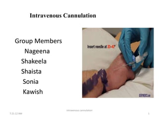
  • 16. • Insert the needle (bevel side up) at an angle of 10- 30o to the skin (this will depend on vein depth.) • Observe for blood in the flashback chamber 7:21:12 AM intravenous cannulation 16
  • 17. • Apply gentle pressure over the vein (beyond the cannula tip) remove the white cap from the needle 7:21:12 AM intravenous cannulation 17
  • 18. • Remove the needle from the cannula and dispose of it into a sharps container • Attach the white lock cap • Secure the cannula with an appropriate dressing 7:21:12 AM intravenous cannulation 18
  • 19. • Flush the cannula with 2-5 mls 0.9% Sodium Chloride or attach an IV giving set and fluid 7:21:12 AM intravenous cannulation 19
  • 20. Document the procedure including: • Date and time • Site and size of cannula ,any problems encountered • Review date (cannula should be in situ no longer than 72 hours without appropriate risk assessment.) • Thanks to the patient • Clean up, dispose of rubbish 7:21:12 AM intravenous cannulation 20
  • 21. Possible Complications: The intravenous (IV) cannula offers direct access to a patient's vascular system and provides a potential route for entry of micro organisms into that system. These organisms can cause serious infection if they are allowed to enter and proliferate in the IV cannula, insertion site, or IV fluid. 7:21:12 AM intravenous cannulation 21
  • 22. IV-Site Infection: Does not produce much (if any) pus or inflammation at the IV site. This is the most common cannula-related infection, may be the most difficult to identify 7:21:12 AM intravenous cannulation 22
  • 23. Cellulites: Warm, red and often tender skin surrounding the site of cannula insertion; pus is rarely detectable. 7:21:12 AM intravenous cannulation 23
  • 24. Infiltration • Infiltration or tissuing occurs when the infusion (fluid) leaks into the surrounding tissue. It is important to detect early as tissue necrosis could occur – re-site cannula immediately 7:21:12 AM intravenous cannulation 24
  • 25. Bruising Bruising commonly results from failed IV placement - particularly in the elderly and those on anticoagulant therapy. 7:21:12 AM intravenous cannulation 25
  • 26. Haematoma: Haematoma occurs when blood leaks out of the infusion site. The common cause of this is using cannula that are not tapered at the distal end. It will also occur if on insertion the cannula has penetrated through the other side of the vessel wall – apply pressure to the site for approximately 4 minutes and elevate the limb 7:21:12 AM intravenous cannulation 26
  • 27. Phlebitis: Phlebitiss is common in IV therapy and can be cause in many ways. It is inflammation of a vein (redness and pain at the infusion site) – prevention can be using aseptic insertion techniques, choosing the smallest gauge cannula possible for the prescribed treatment, secure the cannula properly to prevent movement and carry out regular checks of the infusion site. 7:21:12 AM intravenous cannulation 27
  • 28. References: • Clinical Skills Education Centre http://www.qub.ac.uk/cskills/index.htm • Standards for Infusion Therapy RCN http://www.rcn.org.uk/publications/pdf/standardsi nfusiontherapy.pdf 7:21:12 AM intravenous cannulation 28