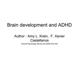
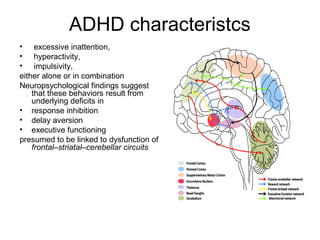
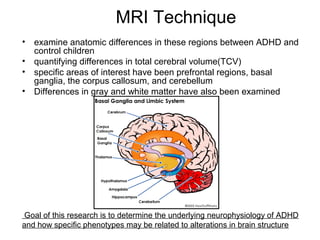
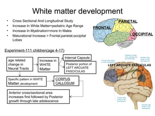
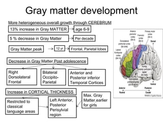
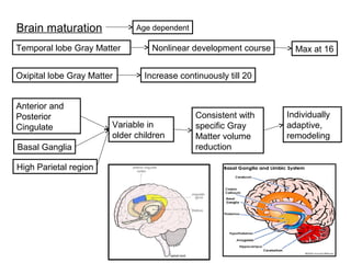
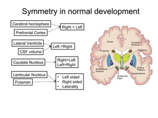
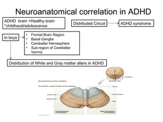
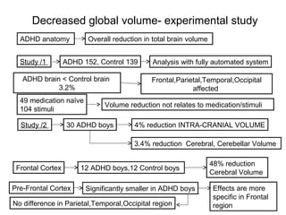
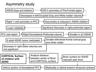
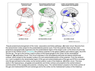
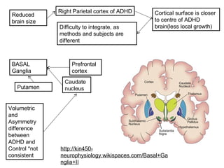
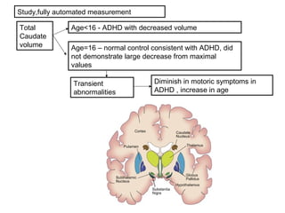
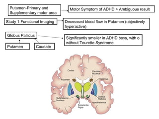
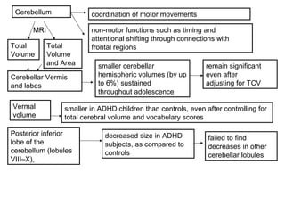
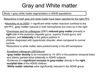
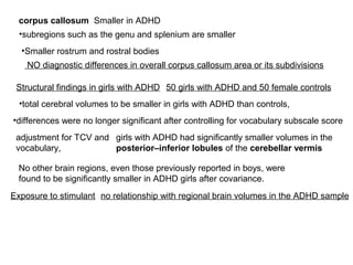
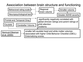
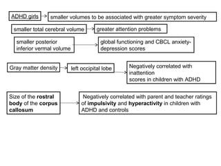
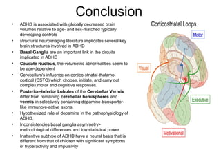
Brain development and ADHD
Author : Amy L. Krain, F. Xavier
Castellanos
Clinical Psychology Review 26 (2006) 433–444
ADHD characteristcs
• excessive inattention,
• hyperactivity,
• impulsivity,
either alone or in combination
Neuropsychological findings suggest
that these behaviors result from
underlying deficits in
• response inhibition
• delay aversion
• executive functioning
presumed to be linked to dysfunction of
frontal–striatal–cerebellar circuits
MRI Technique
• examine anatomic differences in these regions between ADHD and
control children
• quantifying differences in total cerebral volume(TCV)
• specific areas of interest have been prefrontal regions, basal
ganglia, the corpus callosum, and cerebellum
• Differences in gray and white matter have also been examined
Goal of this research is to determine the underlying neurophysiology of ADHD
and how specific phenotypes may be related to alterations in brain structure
Hypothesized pschychological deficits
• Dysfunction of frontal/striatal cerebellar circuits
Neural circuits
Prefrontal Cortex Basal Ganglia
Cerebellum
• MOTOR COORDINATION centre
• Closely linked to NONMOTOR region of
CEREBRAL CORTEX
• EXECUTIVE FUNCTION/Cognitive Planning
Module Response Inhibition
Anatomic MRI -Principle technology to study
Pediatric Brain
Advantage
1.Spacial resolution
2.No ionizing radiation
Disadvantage
1.Cost of MRI scan (small sample size)-> less statistical power-
2.Cost increases by loss of scans due to excessive motion (hyperactivity of
children)
3.ADHD characteristics vary with age, sex, clinical setting->heterogenous
dataset+small sample size->Difficulty in comparison
4.Stimulant medication->children already with medication, no medication ,
previously medicated.
Current methods
Hand-tracing of individual region of interest
•Decrease reliability
•Optimize validity
Fully-automated method
•Maximize test-retest reliability
•Best for large well/defined brain region
Semi-automated method
•Combined the two
Focus on Lateralization of language , indices and asymmetry
•Asymmetry measure , less reliable that volumetric measure
•Reliability inverses with degree of similarity between left and right side
Normal Brain Developement
• 90% of young adult’s brain volume attained by age 5
• Total Cerebral volume (TCV)->Max. early adolescence
• Experimental data: 1mm/yr in PFC
Gender difference
prominent
Cross-sectional analysis
Age related decrease-thalamus, lenticular neucleus
Increase>lentricular size, after controlling TCV
Sex difference in development
pattern
• Experiment-104 children(age 4-18)
• Decrease in CAUDATE and PUTAMEN in boys only
Cerebram
Cerebellum
Boy > Girl (7-10%)
Cortical Gray Matter Boy > Girl (10%) Even if
TCV
controlled
Subcortical
Region
Putamen
Globus
Pallidus
Boy > Girl
White matter development
• Cross Sectional And Longitudinal Study
• Increase In White Matter>pediatric Age Range
• Increase In Myelination>more In Males
• Maturational Increase > Frontal,parietal,occipital
Lobes
Experiment-111 children(age 4-17)
age related
change in
Neural Tracts
Increase in
WHITE
Matter
Internal Capsule
Posterior portion of
LEFT ARCUATE
FASCICULAS
Specific pattern in WHITE
Matter development
CORPUS
CALLOSUM
Anterior cross/sectional area
increases first followed by Posterior
growth through late adolescence
FRONTAL
PARIETAL
OCCIPITAL
LEFT ARCUATE FASCICULAS
Gray matter development
More heterogeneous overall growth through CEREBRUM
13% increase in Gray MATTER age 6-9
5 % decrease in Gray Matter Per decade
Gray Matter peak 12 yr Frontal, Parietal lobes
Decrease in Gray Matter Post adolescence
Right
Dorsolateral
Frontal
Bilateral
Occipito-
Parietal
Anterior and
Posterior inferior
Temporal Cortices
Increase in CORTICAL THICKNESS
Restricted to
classical
language areas
Left Anterior,
Posterior
Perisylvial
region
Max. Gray
Matter earlier
for girls
Brain maturation Age dependent
Temporal lobe Gray Matter Nonlinear development course Max at 16
Oxipital lobe Gray Matter Increase continuously till 20
Anterior and
Posterior
Cingulate
Basal Ganglia
High Parietal region
Variable in
older children
Consistent with
specific Gray
Matter volume
reduction
Individually
adaptive,
remodeling
Symmetry in normal development
Cerebral hemisphere
Prefrontal Cortex
Right > Left
Left >Right
Lateral Ventricle
CSF volume
Right>Left
Left>Right
Caudate Nucleus
Lenticular Nucleus
Putamen
• Left sided
• Right sided
• Laterality
Neuroanatomical correlation in ADHD
ADHD brain <Healthy brain
*childhood/adoloscence
Distributed Circuit ADHD syndrome
• Frontal Brain Region
• Basal Ganglia
• Cerebellar Hemesphere
• Sub-region of Cerebellar
Vermis
In boys
Distribution of White and Gray matter alters in ADHD
Decreased global volume- experimental study
ADHD anatomy Overall reduction in total brain volume
ADHD 152, Control 139Study /1 Analysis with fully automated system
ADHD brain < Control brain
3.2%
Frontal,Parietal,Temporal,Occipital
affected
Volume reduction not relates to medication/stimuli
49 medication naïve
104 stimuli
Study /2 30 ADHD boys 4% reduction INTRA-CRANIAL VOLUME
3.4% reduction Cerebral, Cerebellar Volume
Frontal Cortex 12 ADHD boys,12 Control boys
48% reduction
Cerebral Volume
Pre-Frontal Cortex Significantly smaller in ADHD boys Effects are more
specific in Frontal
regionNo difference in Parietal,Temporal,Occipital region
Asymmetry study
ADHD boys and bothers Diff in symmetry of Pre-Frontal region
Decreased in left/Occipital Gray and White matter volume
Right > Left symmetry in PFC Asymmetry is reduced in ADHD children
Significant Decrease in right-prefrontal regionLower reliability
PFC sub-region Right-Dorsolateral Prefrontal volume Smaller in 23 ADHD
8 adult ADHD never medicated Smaller left/orbital frontal cortical gray
and white matter
Decrease in right-sided volumes are
not significant
CORTICAL Surface
of children with
ADHD
Analyze distance
between center of Brain
and CORTICAL surface
Brain surface for ADHD
reduced upto 4mm
Bilaterality in lateral anterior temporal corticesInferior portion of dorsolateral PFC
Pseudo-anatomical arrangement of the motor, associative and limbic pathways. (A) motor circuit. Neurons from
the sensorimotor cortex project to the posterolateral putamen (put). From the putamen there are two main
projections topographically organized onto the posterolateral region of the target nuclei: (i) the direct circuit to the
gpi and (ii) the indirect circuit connecting the posterior putamen to the globus pallidus pars externa (gpe), the STN
and the gpi. The gpi is the primary output nucleus of the basal ganglia to the cortex via the ventrolateral
thalamus. (B) associative circuit. This circuit originates in the dorsolateral prefrontal and lateral orbitofrontal
cortices, which project to the caudate nucleus (cn) and anteromedial portion of the putamen. From the striatum
(cn + put) it projects to the dorsomedial region of the gpi and anteromedial parts of the gpe and STN to converge
onto the gpi and back to the cortex via the ventral anterior nuclei of the thalamus. (C) limbic circuit. This loop
starts in the hippocampus, amygdala and paralimbic and limbic cortices and projects to the ventral striatum
(ventral portion of the caudate and putamen, including nacc). The ventral striatum projects to the limbic portion of
the gpe and medioventral STN and ventral gpi and to the cortex via the mediodorsal nucleus of the thalamus
Reduced
brain size
Right Parietal cortex of ADHD
Difficulty to integrate, as
methods and subjects are
different
Cortical surface is closer
to centre of ADHD
brain(less local growth)
BASAL
Ganglia
Prefrontal
cortex
Caudate
nucleusPutamen
Volumetric
and
Asymmetry
difference
between
ADHD and
Control *not
consistent
http://kin450-
neurophysiology.wikispaces.com/Basal+Ga
nglia+II
Total
Caudate
volume
Study,fully automated measurement
Age<16 - ADHD with decreased volume
Age=16 – normal control consistent with ADHD, did
not demonstrate large decrease from maximal
values
Transient
abnormalities
Diminish in motoric symptoms in
ADHD , increase in age
Study 1-Functional Imaging
Putamen-Primary and
Supplementary motor area
Decreased blood flow in Putamen (objectively
hyperactive)
Motor Symptom of ADHD > Ambiguous result
CaudatePutamen
Significantly smaller in ADHD boys, with o
without Tourette Syndrome
Globus Pallidus
Effect of head trauma , damage to Basal Ganglia->
Secondary development of ADHD
Complete elimination of
basal ganglia
Case 1-traumatic amniocentesis
at 17 weeks of gestation
Lesions of Right Putamen
Posterior Ventral Putamen
Higher in SADHD
Higher in ADHD
Case 2-
99 children(age4 -19)
Closed head injury Chance of SADHD
basal ganglia-3.2% Thalamus-3.6%
Cerebellum coordination of motor movements
non-motor functions such as timing and
attentional shifting through connections with
frontal regionsTotal
Volume
smaller cerebellar
hemispheric volumes (by up
to 6%) sustained
throughout adolescence
Total
Volume
and Area
Cerebellar Vermis
and lobes
remain significant
even after
adjusting for TCV
Vermal
volume
smaller in ADHD children than controls, even after controlling for
total cerebral volume and vocabulary scores
decreased size in ADHD
subjects, as compared to
controls
failed to find
decreases in other
cerebellar lobules
Posterior inferior
lobe of the
cerebellum (lobules
VIII–X)
MRI
Gray and White matter
Study > gray–white matter segmentation in ADHD populations
Reductions in both gray and white matter have been reported for the right PFC
•Mostofsky et al.(2002) -> significant white matter reduction confined to the
left PFC, gray matter reduced in both hemispheres but more so in the right.
•Overmeyer and his colleagues (2001) reduced gray matter primarily in
right side in the posterior cingulate gyrus, superior frontal gyrus, and
putamen, and bilaterally in the globus pallidus in
children diagnosed with hyperkinetic disorder, when compared to normal
controls.
•Reductions in white matter were predominantly in the left hemisphere
Sowelland colleagues (2003)found
•gray matter density to be increased by 15–30% in the posterior temporal lobes
and inferior parietal lobes bilaterally in ADHD subjects.
•Evidence of a significant increase in gray-matter density in the right
occipital lobe of the ADHD children.
•White matter volumes were significantly reduced in the ADHD group
corpus callosum Smaller in ADHD
•subregions such as the genu and splenium are smaller
•Smaller rostrum and rostral bodies
NO diagnostic differences in overall corpus callosum area or its subdivisions
Structural findings in girls with ADHD 50 girls with ADHD and 50 female controls
•total cerebral volumes to be smaller in girls with ADHD than controls,
•differences were no longer significant after controlling for vocabulary subscale score
adjustment for TCV and
vocabulary,
girls with ADHD had significantly smaller volumes in the
posterior–inferior lobules of the cerebellar vermis
No other brain regions, even those previously reported in boys, were
found to be significantly smaller in ADHD girls after covariance.
Exposure to stimulant no relationship with regional brain volumes in the ADHD sample
Association between brain structure and functioning
Behavioral rating scale
Neuropsychological test
Regional
brain volume
Smaller volume
Greater ADHD severity
Caudate
Frontal and Temporal Gray
Cerebellar Volume
significantly negatively correlated with
global clinician ratings and parent ratings of
child attention
problems
Semrud-Clikeman
et al. (2000)
smaller left caudate head and white matter volumes
associated with higher Child Behavior Checklist (CBCL)
Externalizing scores.
ADHD girls smaller volumes to be associated with greater symptom severity
smaller total cerebral volume greater attention problems
smaller posterior
inferior vermal volume
global functioning and CBCL anxiety-
depression scores
Gray matter density left occipital lobe Negatively correlated with
inattention
scores in children with ADHD
Size of the rostral
body of the corpus
callosum
Negatively correlated with parent and teacher ratings
of impulsivity and hyperactivity in children with
ADHD and controls
Executive function deficits in ADHD
children
study of 26 ADHD and 26 control boys
• ADHD task performance was positively correlated with prefrontal cortex,
caudate, and globus pallidus volumes
• Correlations between sensory selection task performance and prefrontal
and caudate volumes were predominantly localized to the right
• Response selection and response execution tasks were correlated with
caudate symmetry and left globus pallidus size
• Prefrontal volumes were correlated with performance on the inhibitory
conditions, while basal ganglia volumes related to both control and
inhibitory conditions
study of 23 ADHD children and 24 normal controls
larger volumes in total superior prefrontal cortex and right superior
prefrontal cortex were correlated with worse performance on a test of
attention (Conners' Continuous Performance Test; CPT)
Proton magnetic resonance spectroscopy study
Conners' CPT Right dorsolateral volumes
Larger volumes poorer performance on the CPT composite,
variability, and reaction time standard error scores
not found in healthy control
Right dorsolateral region may be dysfunctional in
ADHD
More tissue in right dorsolateral region leads to greater disruption in attention
Study comparing anatomic MRI measures with the performance of children with
ADHD and normal controls on Executive function tests
Reversed normal
asymmetry of the caudate
Poorer performance on the Stroop color–word
test and Wisconsin Card Sorting Test (WCST)
Reversal of normal left-greater-
than-right asymmetry
Greater disinhibition on the stroop and a
higher incidence of loss of set on the WCST
Ability to name colors quickly compromised in the ADHD group
smaller volumes of white
matter of the anterior–
superior region
worse
performance on
rapid naming
Role in maintaining attention
Conclusion
• ADHD is associated with globally decreased brain
volumes relative to age- and sex-matched typically
developing controls
• structural neuroimaging literature implicates several key
brain structures involved in ADHD
• Basal Ganglia are an important link in the circuits
implicated in ADHD
• Caudate Nucleus, the volumetric abnormalities seem to
be age-dependent
• Cerebellum's influence on cortico-striatal-thalamo-
cortical (CSTC) which choose, initiate, and carry out
complex motor and cognitive responses.
• Posterior–inferior Lobules of the Cerebellar Vermis
differ from remaining cerebellar hemispheres and
vermis in selectively containing dopamine-transporter-
like immunore-active axons.
• Hypothesized role of dopamine in the pathophysiology of
ADHD,
• Inconsistencies basal ganglia asymmetry>
methodological differences and low statistical power
• Inattentive subtype of ADHD have a neural basis that is
different from that of children with significant symptoms
of hyperactivity and impulsivity

Brain developm adhd2

  • 1.
    Brain development andADHD Author : Amy L. Krain, F. Xavier Castellanos Clinical Psychology Review 26 (2006) 433–444
  • 2.
    ADHD characteristcs • excessiveinattention, • hyperactivity, • impulsivity, either alone or in combination Neuropsychological findings suggest that these behaviors result from underlying deficits in • response inhibition • delay aversion • executive functioning presumed to be linked to dysfunction of frontal–striatal–cerebellar circuits
  • 3.
    MRI Technique • examineanatomic differences in these regions between ADHD and control children • quantifying differences in total cerebral volume(TCV) • specific areas of interest have been prefrontal regions, basal ganglia, the corpus callosum, and cerebellum • Differences in gray and white matter have also been examined Goal of this research is to determine the underlying neurophysiology of ADHD and how specific phenotypes may be related to alterations in brain structure
  • 4.
    Hypothesized pschychological deficits •Dysfunction of frontal/striatal cerebellar circuits Neural circuits Prefrontal Cortex Basal Ganglia Cerebellum • MOTOR COORDINATION centre • Closely linked to NONMOTOR region of CEREBRAL CORTEX • EXECUTIVE FUNCTION/Cognitive Planning Module Response Inhibition
  • 5.
    Anatomic MRI -Principletechnology to study Pediatric Brain Advantage 1.Spacial resolution 2.No ionizing radiation Disadvantage 1.Cost of MRI scan (small sample size)-> less statistical power- 2.Cost increases by loss of scans due to excessive motion (hyperactivity of children) 3.ADHD characteristics vary with age, sex, clinical setting->heterogenous dataset+small sample size->Difficulty in comparison 4.Stimulant medication->children already with medication, no medication , previously medicated.
  • 6.
    Current methods Hand-tracing ofindividual region of interest •Decrease reliability •Optimize validity Fully-automated method •Maximize test-retest reliability •Best for large well/defined brain region Semi-automated method •Combined the two Focus on Lateralization of language , indices and asymmetry •Asymmetry measure , less reliable that volumetric measure •Reliability inverses with degree of similarity between left and right side
  • 7.
    Normal Brain Developement •90% of young adult’s brain volume attained by age 5 • Total Cerebral volume (TCV)->Max. early adolescence • Experimental data: 1mm/yr in PFC Gender difference prominent Cross-sectional analysis Age related decrease-thalamus, lenticular neucleus Increase>lentricular size, after controlling TCV
  • 8.
    Sex difference indevelopment pattern • Experiment-104 children(age 4-18) • Decrease in CAUDATE and PUTAMEN in boys only Cerebram Cerebellum Boy > Girl (7-10%) Cortical Gray Matter Boy > Girl (10%) Even if TCV controlled Subcortical Region Putamen Globus Pallidus Boy > Girl
  • 9.
    White matter development •Cross Sectional And Longitudinal Study • Increase In White Matter>pediatric Age Range • Increase In Myelination>more In Males • Maturational Increase > Frontal,parietal,occipital Lobes Experiment-111 children(age 4-17) age related change in Neural Tracts Increase in WHITE Matter Internal Capsule Posterior portion of LEFT ARCUATE FASCICULAS Specific pattern in WHITE Matter development CORPUS CALLOSUM Anterior cross/sectional area increases first followed by Posterior growth through late adolescence FRONTAL PARIETAL OCCIPITAL LEFT ARCUATE FASCICULAS
  • 10.
    Gray matter development Moreheterogeneous overall growth through CEREBRUM 13% increase in Gray MATTER age 6-9 5 % decrease in Gray Matter Per decade Gray Matter peak 12 yr Frontal, Parietal lobes Decrease in Gray Matter Post adolescence Right Dorsolateral Frontal Bilateral Occipito- Parietal Anterior and Posterior inferior Temporal Cortices Increase in CORTICAL THICKNESS Restricted to classical language areas Left Anterior, Posterior Perisylvial region Max. Gray Matter earlier for girls
  • 11.
    Brain maturation Agedependent Temporal lobe Gray Matter Nonlinear development course Max at 16 Oxipital lobe Gray Matter Increase continuously till 20 Anterior and Posterior Cingulate Basal Ganglia High Parietal region Variable in older children Consistent with specific Gray Matter volume reduction Individually adaptive, remodeling
  • 12.
    Symmetry in normaldevelopment Cerebral hemisphere Prefrontal Cortex Right > Left Left >Right Lateral Ventricle CSF volume Right>Left Left>Right Caudate Nucleus Lenticular Nucleus Putamen • Left sided • Right sided • Laterality
  • 13.
    Neuroanatomical correlation inADHD ADHD brain <Healthy brain *childhood/adoloscence Distributed Circuit ADHD syndrome • Frontal Brain Region • Basal Ganglia • Cerebellar Hemesphere • Sub-region of Cerebellar Vermis In boys Distribution of White and Gray matter alters in ADHD
  • 14.
    Decreased global volume-experimental study ADHD anatomy Overall reduction in total brain volume ADHD 152, Control 139Study /1 Analysis with fully automated system ADHD brain < Control brain 3.2% Frontal,Parietal,Temporal,Occipital affected Volume reduction not relates to medication/stimuli 49 medication naïve 104 stimuli Study /2 30 ADHD boys 4% reduction INTRA-CRANIAL VOLUME 3.4% reduction Cerebral, Cerebellar Volume Frontal Cortex 12 ADHD boys,12 Control boys 48% reduction Cerebral Volume Pre-Frontal Cortex Significantly smaller in ADHD boys Effects are more specific in Frontal regionNo difference in Parietal,Temporal,Occipital region
  • 15.
    Asymmetry study ADHD boysand bothers Diff in symmetry of Pre-Frontal region Decreased in left/Occipital Gray and White matter volume Right > Left symmetry in PFC Asymmetry is reduced in ADHD children Significant Decrease in right-prefrontal regionLower reliability PFC sub-region Right-Dorsolateral Prefrontal volume Smaller in 23 ADHD 8 adult ADHD never medicated Smaller left/orbital frontal cortical gray and white matter Decrease in right-sided volumes are not significant CORTICAL Surface of children with ADHD Analyze distance between center of Brain and CORTICAL surface Brain surface for ADHD reduced upto 4mm Bilaterality in lateral anterior temporal corticesInferior portion of dorsolateral PFC
  • 16.
    Pseudo-anatomical arrangement ofthe motor, associative and limbic pathways. (A) motor circuit. Neurons from the sensorimotor cortex project to the posterolateral putamen (put). From the putamen there are two main projections topographically organized onto the posterolateral region of the target nuclei: (i) the direct circuit to the gpi and (ii) the indirect circuit connecting the posterior putamen to the globus pallidus pars externa (gpe), the STN and the gpi. The gpi is the primary output nucleus of the basal ganglia to the cortex via the ventrolateral thalamus. (B) associative circuit. This circuit originates in the dorsolateral prefrontal and lateral orbitofrontal cortices, which project to the caudate nucleus (cn) and anteromedial portion of the putamen. From the striatum (cn + put) it projects to the dorsomedial region of the gpi and anteromedial parts of the gpe and STN to converge onto the gpi and back to the cortex via the ventral anterior nuclei of the thalamus. (C) limbic circuit. This loop starts in the hippocampus, amygdala and paralimbic and limbic cortices and projects to the ventral striatum (ventral portion of the caudate and putamen, including nacc). The ventral striatum projects to the limbic portion of the gpe and medioventral STN and ventral gpi and to the cortex via the mediodorsal nucleus of the thalamus
  • 17.
    Reduced brain size Right Parietalcortex of ADHD Difficulty to integrate, as methods and subjects are different Cortical surface is closer to centre of ADHD brain(less local growth) BASAL Ganglia Prefrontal cortex Caudate nucleusPutamen Volumetric and Asymmetry difference between ADHD and Control *not consistent http://kin450- neurophysiology.wikispaces.com/Basal+Ga nglia+II
  • 18.
    Total Caudate volume Study,fully automated measurement Age<16- ADHD with decreased volume Age=16 – normal control consistent with ADHD, did not demonstrate large decrease from maximal values Transient abnormalities Diminish in motoric symptoms in ADHD , increase in age
  • 19.
    Study 1-Functional Imaging Putamen-Primaryand Supplementary motor area Decreased blood flow in Putamen (objectively hyperactive) Motor Symptom of ADHD > Ambiguous result CaudatePutamen Significantly smaller in ADHD boys, with o without Tourette Syndrome Globus Pallidus
  • 20.
    Effect of headtrauma , damage to Basal Ganglia-> Secondary development of ADHD Complete elimination of basal ganglia Case 1-traumatic amniocentesis at 17 weeks of gestation Lesions of Right Putamen Posterior Ventral Putamen Higher in SADHD Higher in ADHD Case 2- 99 children(age4 -19) Closed head injury Chance of SADHD basal ganglia-3.2% Thalamus-3.6%
  • 21.
    Cerebellum coordination ofmotor movements non-motor functions such as timing and attentional shifting through connections with frontal regionsTotal Volume smaller cerebellar hemispheric volumes (by up to 6%) sustained throughout adolescence Total Volume and Area Cerebellar Vermis and lobes remain significant even after adjusting for TCV Vermal volume smaller in ADHD children than controls, even after controlling for total cerebral volume and vocabulary scores decreased size in ADHD subjects, as compared to controls failed to find decreases in other cerebellar lobules Posterior inferior lobe of the cerebellum (lobules VIII–X) MRI
  • 22.
    Gray and Whitematter Study > gray–white matter segmentation in ADHD populations Reductions in both gray and white matter have been reported for the right PFC •Mostofsky et al.(2002) -> significant white matter reduction confined to the left PFC, gray matter reduced in both hemispheres but more so in the right. •Overmeyer and his colleagues (2001) reduced gray matter primarily in right side in the posterior cingulate gyrus, superior frontal gyrus, and putamen, and bilaterally in the globus pallidus in children diagnosed with hyperkinetic disorder, when compared to normal controls. •Reductions in white matter were predominantly in the left hemisphere Sowelland colleagues (2003)found •gray matter density to be increased by 15–30% in the posterior temporal lobes and inferior parietal lobes bilaterally in ADHD subjects. •Evidence of a significant increase in gray-matter density in the right occipital lobe of the ADHD children. •White matter volumes were significantly reduced in the ADHD group
  • 23.
    corpus callosum Smallerin ADHD •subregions such as the genu and splenium are smaller •Smaller rostrum and rostral bodies NO diagnostic differences in overall corpus callosum area or its subdivisions Structural findings in girls with ADHD 50 girls with ADHD and 50 female controls •total cerebral volumes to be smaller in girls with ADHD than controls, •differences were no longer significant after controlling for vocabulary subscale score adjustment for TCV and vocabulary, girls with ADHD had significantly smaller volumes in the posterior–inferior lobules of the cerebellar vermis No other brain regions, even those previously reported in boys, were found to be significantly smaller in ADHD girls after covariance. Exposure to stimulant no relationship with regional brain volumes in the ADHD sample
  • 24.
    Association between brainstructure and functioning Behavioral rating scale Neuropsychological test Regional brain volume Smaller volume Greater ADHD severity Caudate Frontal and Temporal Gray Cerebellar Volume significantly negatively correlated with global clinician ratings and parent ratings of child attention problems Semrud-Clikeman et al. (2000) smaller left caudate head and white matter volumes associated with higher Child Behavior Checklist (CBCL) Externalizing scores.
  • 25.
    ADHD girls smallervolumes to be associated with greater symptom severity smaller total cerebral volume greater attention problems smaller posterior inferior vermal volume global functioning and CBCL anxiety- depression scores Gray matter density left occipital lobe Negatively correlated with inattention scores in children with ADHD Size of the rostral body of the corpus callosum Negatively correlated with parent and teacher ratings of impulsivity and hyperactivity in children with ADHD and controls
  • 26.
    Executive function deficitsin ADHD children study of 26 ADHD and 26 control boys • ADHD task performance was positively correlated with prefrontal cortex, caudate, and globus pallidus volumes • Correlations between sensory selection task performance and prefrontal and caudate volumes were predominantly localized to the right • Response selection and response execution tasks were correlated with caudate symmetry and left globus pallidus size • Prefrontal volumes were correlated with performance on the inhibitory conditions, while basal ganglia volumes related to both control and inhibitory conditions study of 23 ADHD children and 24 normal controls larger volumes in total superior prefrontal cortex and right superior prefrontal cortex were correlated with worse performance on a test of attention (Conners' Continuous Performance Test; CPT)
  • 27.
    Proton magnetic resonancespectroscopy study Conners' CPT Right dorsolateral volumes Larger volumes poorer performance on the CPT composite, variability, and reaction time standard error scores not found in healthy control Right dorsolateral region may be dysfunctional in ADHD More tissue in right dorsolateral region leads to greater disruption in attention Study comparing anatomic MRI measures with the performance of children with ADHD and normal controls on Executive function tests Reversed normal asymmetry of the caudate Poorer performance on the Stroop color–word test and Wisconsin Card Sorting Test (WCST) Reversal of normal left-greater- than-right asymmetry Greater disinhibition on the stroop and a higher incidence of loss of set on the WCST Ability to name colors quickly compromised in the ADHD group smaller volumes of white matter of the anterior– superior region worse performance on rapid naming Role in maintaining attention
  • 28.
    Conclusion • ADHD isassociated with globally decreased brain volumes relative to age- and sex-matched typically developing controls • structural neuroimaging literature implicates several key brain structures involved in ADHD • Basal Ganglia are an important link in the circuits implicated in ADHD • Caudate Nucleus, the volumetric abnormalities seem to be age-dependent • Cerebellum's influence on cortico-striatal-thalamo- cortical (CSTC) which choose, initiate, and carry out complex motor and cognitive responses. • Posterior–inferior Lobules of the Cerebellar Vermis differ from remaining cerebellar hemispheres and vermis in selectively containing dopamine-transporter- like immunore-active axons. • Hypothesized role of dopamine in the pathophysiology of ADHD, • Inconsistencies basal ganglia asymmetry> methodological differences and low statistical power • Inattentive subtype of ADHD have a neural basis that is different from that of children with significant symptoms of hyperactivity and impulsivity