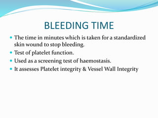
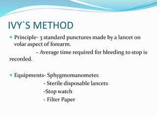
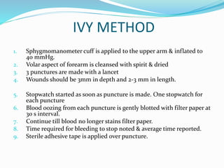
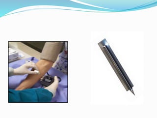
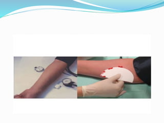
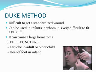
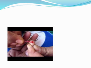
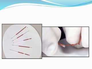
DR.SAMIYAH SYEED
BLEEDING TIME
 The time in minutes which is taken for a standardized
skin wound to stop bleeding.
 Test of platelet function.
 Used as a screening test of haemostasis.
 It assesses Platelet integrity & Vessel Wall Integrity
METHODS
1) IVY METHOD
2) DUKE METHOD
3) TEMPLATE METHOD
IVY`S METHOD
 Principle- 3 standard punctures made by a lancet on
volar aspect of forearm.
- Average time required for bleeding to stop is
recorded.
 Equipments- Sphygmomanometer.
- Sterile disposable lancets
-Stop watch
- Filter Paper
IVY METHOD
1. Sphygmomanometer cuff is applied to the upper arm & inflated to
40 mmHg.
2. Volar aspect of forearm is cleansed with spirit & dried
3. 3 punctures are made with a lancet
4. Wounds should be 3mm in depth and 2-3 mm in length.
5. Stopwatch started as soon as puncture is made. One stopwatch for
each puncture
6. Blood oozing from each puncture is gently blotted with filter paper at
30 s interval.
7. Continue till blood no longer stains filter paper.
8. Time required for bleeding to stop noted & average time reported.
9. Sterile adhesive tape is applied over puncture.
DUKE METHOD
 Difficult to get a standardized wound
 Can be used in infants in whom it is very difficult to fit
a BP cuff.
 It can cause a large hematoma
SITE OF PUNCTURE:
- Ear lobe in adult or older child
- Heel of foot in infant
 Clean the area
 Make a wound 3mm deep as in Ivy method
 Time is begun soon after this
 Blot drop of blood with filter paper every 30 seconds
and report the time when blood no longer stains the
filter paper.
TEMPLATE METHOD
 Newer method
 Use a glass or plastic template with a transverse slit,
which allows a lancet to make a cut on the volar aspect
of forearm – Exactly 11 mm long and 1 mm deep
 Very sensitive and reproducible test
REFERENCE RANGES
 Normal Bleeding Time- 2-7 minutes
 Usually, BT <4 mins
 When bleeding continues beyond 20 minutes- report
at >20 minutes
- Test discontinued.
PROLONGED BT
 1. Disorders of Platelets
- Thrombocytopenia
- Increased Destruction of platelets
- ITP
- Infection- malaria, dengue, septicemia
- Drugs- Quinine, Heparin
- DIC
- Increased Pooling of platelets
- Hypersplenism
- Decreased production of platelets in bone marrow
- Aplastic anemia
- Bone marrow infiltration- leukemia, lymphomas
- Megaloblastic anemia
- Drugs, radiation, infections
PROLONGED BT
- Disorders of platelet function
- Acquired- Drugs- Aspirin, NSAID
- Myeloproliferative disorders
- Hereditary- Bernard Soulier Syndrome
- Glanzmanns Thrombasthenia
2. Disorders of coagulation
- Hereditary- Von Willebrands Disease
- Hemophilia A, B
- Acquired – DIC, Liver disease, Vit. K Deficiency,
Heparin therapy.
PROLONGED BT
3. Disorders of Blood Vessels
Acquired- Allergic Purpura
Infections- Viral Septicemia
Scurvy, Senile Purpura
Inherited- Hereditary hemorrhagic telangiectasia
CLOTTING TIME
 Time required for blood to form a clot
 It is a rough measure to assess clotting factors in blood
 It is a crude test, has been replaced by aPTT
 PROCEDURE- Clean finger with spirit
- Prick with lancet, note the time of prick
- Take a capillary tube & hold
horizontally over the blood
- Place in water bath after sealing tube.
 After 2 minutes of prick, take the capillary tube &
break it , separate the 2 ends slowly.
 Repeat the process at 30 seconds interval
 When a thin thread-like clot is formed, note the time.
 Time from finger prick to appearance of clot is
Clotting Time.
 Normal Referance Range- 5-15 mins
 Prolonged in deficiency of intrinsic coagulation
Pathway. Eg- Hemophila due to Factor VIII deficiency
BLEEDING TIME & CLOTTING TIME.pptx

BLEEDING TIME & CLOTTING TIME.pptx

  • 1.
  • 2.
    BLEEDING TIME  Thetime in minutes which is taken for a standardized skin wound to stop bleeding.  Test of platelet function.  Used as a screening test of haemostasis.  It assesses Platelet integrity & Vessel Wall Integrity
  • 3.
    METHODS 1) IVY METHOD 2)DUKE METHOD 3) TEMPLATE METHOD
  • 4.
    IVY`S METHOD  Principle-3 standard punctures made by a lancet on volar aspect of forearm. - Average time required for bleeding to stop is recorded.  Equipments- Sphygmomanometer. - Sterile disposable lancets -Stop watch - Filter Paper
  • 5.
    IVY METHOD 1. Sphygmomanometercuff is applied to the upper arm & inflated to 40 mmHg. 2. Volar aspect of forearm is cleansed with spirit & dried 3. 3 punctures are made with a lancet 4. Wounds should be 3mm in depth and 2-3 mm in length. 5. Stopwatch started as soon as puncture is made. One stopwatch for each puncture 6. Blood oozing from each puncture is gently blotted with filter paper at 30 s interval. 7. Continue till blood no longer stains filter paper. 8. Time required for bleeding to stop noted & average time reported. 9. Sterile adhesive tape is applied over puncture.
  • 8.
    DUKE METHOD  Difficultto get a standardized wound  Can be used in infants in whom it is very difficult to fit a BP cuff.  It can cause a large hematoma SITE OF PUNCTURE: - Ear lobe in adult or older child - Heel of foot in infant
  • 9.
     Clean thearea  Make a wound 3mm deep as in Ivy method  Time is begun soon after this  Blot drop of blood with filter paper every 30 seconds and report the time when blood no longer stains the filter paper.
  • 10.
    TEMPLATE METHOD  Newermethod  Use a glass or plastic template with a transverse slit, which allows a lancet to make a cut on the volar aspect of forearm – Exactly 11 mm long and 1 mm deep  Very sensitive and reproducible test
  • 11.
    REFERENCE RANGES  NormalBleeding Time- 2-7 minutes  Usually, BT <4 mins  When bleeding continues beyond 20 minutes- report at >20 minutes - Test discontinued.
  • 12.
    PROLONGED BT  1.Disorders of Platelets - Thrombocytopenia - Increased Destruction of platelets - ITP - Infection- malaria, dengue, septicemia - Drugs- Quinine, Heparin - DIC - Increased Pooling of platelets - Hypersplenism - Decreased production of platelets in bone marrow - Aplastic anemia - Bone marrow infiltration- leukemia, lymphomas - Megaloblastic anemia - Drugs, radiation, infections
  • 13.
    PROLONGED BT - Disordersof platelet function - Acquired- Drugs- Aspirin, NSAID - Myeloproliferative disorders - Hereditary- Bernard Soulier Syndrome - Glanzmanns Thrombasthenia 2. Disorders of coagulation - Hereditary- Von Willebrands Disease - Hemophilia A, B - Acquired – DIC, Liver disease, Vit. K Deficiency, Heparin therapy.
  • 14.
    PROLONGED BT 3. Disordersof Blood Vessels Acquired- Allergic Purpura Infections- Viral Septicemia Scurvy, Senile Purpura Inherited- Hereditary hemorrhagic telangiectasia
  • 15.
    CLOTTING TIME  Timerequired for blood to form a clot  It is a rough measure to assess clotting factors in blood  It is a crude test, has been replaced by aPTT  PROCEDURE- Clean finger with spirit - Prick with lancet, note the time of prick - Take a capillary tube & hold horizontally over the blood - Place in water bath after sealing tube.  After 2 minutes of prick, take the capillary tube & break it , separate the 2 ends slowly.
  • 16.
     Repeat theprocess at 30 seconds interval  When a thin thread-like clot is formed, note the time.  Time from finger prick to appearance of clot is Clotting Time.  Normal Referance Range- 5-15 mins  Prolonged in deficiency of intrinsic coagulation Pathway. Eg- Hemophila due to Factor VIII deficiency