
Recommended
More Related Content
What's hot
What's hot (20)
Similar to Deoxyribozymes
Similar to Deoxyribozymes (20)
More from Ayush Jain
More from Ayush Jain (12)
Recently uploaded
Recently uploaded (20)
Deoxyribozymes
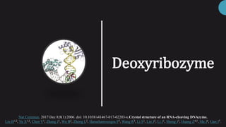
- 1. Deoxyribozyme Nat Commun. 2017 Dec 8;8(1):2006. doi: 10.1038/s41467-017-02203-x.Crystal structure of an RNA-cleaving DNAzyme. Liu H1,2, Yu X1,2, Chen Y1, Zhang J1, Wu B2, Zheng L2, Haruehanroengra P3, Wang R3, Li S1, Lin J2, Li J1, Sheng J3, Huang Z4,5, Ma J6, Gan J7.
- 2. Overview 1.What are Deoxyribozymes?? 2. Historical perspective 3.Structure of 8-17 RNA Cleaving DNA enzyme. a) Purpose of the Research b) What they did ?? c) What did they Find ?? d) Interpretations 4. Summary 2 03/11/2018,IDC 451 Ronald Breaker Gerald Joyce
- 3. What are Deoxyribozyme s ?? Natural duplex of DNA is unlikely to be Catalytic It obeys Michaelis-Menten kinetics. Existence of DNA World is debateable, but seems mostly unlikely. 1 2 3 • A DNA sequence with catalytic activity is called Deoxyribozyme. • They are single SS DNA, identified by in vitro selection, so are called synthetic catalyst. 3 03/11/2018,IDC 451 Catalytic DNA: Scope, Applications, and Biochemistry of Deoxyribozymes, Scott K. Silverman, Trends in Biochemical Sciences, July 2016, Vol. 41, No. 7
- 4. Historical perspective • They used an in-vitro selection strategy to isolate RNA cleaving Deoxyribozyme. • It Binds to its substrate using Watson-crick base pairing. • RNA cleaving Deoxyribozyme has activity similar to RNAse. • It requires metal ions (Pb2+,Mg2+ ). 4 03/11/2018,IDC 451
- 5. Crystal structure of 8-17 RNA Cleaving DNA Enzyme 5 03/11/2018,IDC 451 Image Source: Protein Data Bank
- 6. 1. To find the Structure of 8-17 RNA cleaving DNAzyme. 2. To study the Mechanism of substrate recognition and the catalysis of this DNAzyme. 3. To elucidate the structural and catalytic role of Pb2+ in this DNA enzyme. 03/11/2018,IDC 451 6 What was the purpose this research ???
- 7. What they did ? The Experiment • They carried out crystallographic studies of 8–17 DNAzyme. • They used these 3 substrate enzyme combinations • To confirm the functional importance of some conserved residues of DNAzymes, they performed activity studies using the native and modified DNAzymes. 7 03/11/2018,IDC 451 Short hand Notation Substrate/Dz36 Complex Type Length of substrate Presence or Absence of Pb2+ D1 Native DNA/ Dz36 23nt Absent D2 Native DNA/Dz36 23nt Present D3 2′-OMe-G modified DNA/Dz36 23nt Absent
- 8. What did they Find ! The Results • Dz36 paired with the native DNA substrate (D1 and D2) in two structures and it paired with the 2′-OMe-G (D3) modified DNA in the third structure. 8 03/11/2018,IDC 451 Sequence of 8-17 DNAzyme
- 9. Pb2+/DNAzyme (D2) complex in high resolution at 2.55 Å 03/11/2018,IDC 451 9 • The D1 complex adopts the same space group as D2 complex (C2221), indicated by the very low rmsd value (0.307 Å). • While the D3 structure when compared to D2 show different space group (P43212). But the central catalytic regions where found to be identical. (As shown in Fig.) Super position of D2(White) and D3 (Magenta) Complex, Black box show identical central catalytic region
- 10. • The Dz36/substrate(All) complexes assemble into a “V” shape with two arms (P1 and P2) orientated ~70° with respect to each other. • P1 and P2 are connected through the catalytic core (15 nt) and substrate binds to DNAzyme by forming a dinucleotide kink at the junction (G-1 and G+1). 10 03/11/2018,IDC 451 Dinucleotide kink DZ36/substrate complex showing P1,P2 arm and catalytic core Fig. showing Dinucleotide kink
- 11. • The catalytic core coincides with pseudoknot consisting of two short duplexes (P3 and P4) perpendicular to each other. (which shows Conserved regions) 11 03/11/2018,IDC 451 Pseudoknot DZ36/substrate-complex showing Conserved Regions
- 12. • Conformations of D2 Catalytic core remain same as D1 Even after Pb2+ is used with D2. • In D2 structure, Pb2+ ion was captured in the catalytic core. • Compared with the native Dz36, Methylation at the N1 position (for Dz36-1mG13) caused reduction on the DNA enzyme’s activity. 12 03/11/2018,IDC 451 Effect of methylation at N1 position for Dz36-1mG13 on DNAzyme activity Surface representation showing the preformed cation binding cage. The Pb2+ is shown as a black sphere.
- 13. . 13 03/11/2018,IDC 451 • The Pb2+ ion coordinates with the O6 atom of G6, which forms G6:C12 pair identified structurally and also with a water molecule interacting with the O5′ atom of G+1. • Structure factors and coordinates have been deposited in the Protein Data Bank under accession codes 5XM8, 5XM9, and 5XMA for the DNAzyme-Pb2+ complex, DNAzyme, and DNAzyme-(2′-OMe-G) structures respectively. Coordination of Pb2+ and the catalytic water observed in the DNAzyme- Pb2+ structure. URL rcsb.org: The Protein Data Bank H.M. Berman, J. Westbrook, Z. Feng, G. Gilliland, T.N. Bhat, H. Weissig, I.N. Shindyalov, P.E. Bourne (2000) Nucleic Acids Research, 28: 235-242. doi:10.1093/nar/28.1.235
- 14. 14 03/11/2018,IDC 451 • 8–17 DNAzyme may follow an In-line attack mechanism. • Conserved G13 residue may play the key role in the catalysis, functioning as the base to deprotonate the 2′-OH of G-1 for attacking the 3′-phosphate of G-1. • Observations suggest that the N1 atom of G13 plays critical role in the catalytic process of the DNAzyme. • 8–17 DNAzyme may catalysis indirectly via Pb2+, as it activates the coordinated H2O Molecule, which serve as Acid for Acid-Base Catalysis, by provide a proton for displacing O5′ atom of G+1. • Low pKa of hydrated Pb2+ may contribute to the catalytic efficiency of 8–17 DNAzyme. In-line attack mechanism, Red box shows part of substrate Interpretation s Phosphodiester bond which will be broken
- 15. Summary 15 03/11/2018,IDC 451 The 8–17 DNAzyme adopts a V-shape fold consisting of two substrate-recognizing arms (P1&P2) and one twisted DNA pseudoknot having a Central catalytic core(15nt). Substrate form a GG Kink at Recognition junction. Catalysis is performed by In-Line Acid-base Reaction. The Pb2+ is bound at the pre organized pocket and activates the coordinated H2O Molecule, which serve as Acid for Acid-Base Catalysis.
- 16. Thankyou Presented by: Ayush Jain Int-PhD IISER, Mohali 16 03/11/2018,IDC 451