1. The study examined the incidence of junctional ectopic tachycardia (JET) in 343 patients after surgery for congenital heart defects.
2. JET occurred in 37 patients (10.8%), most frequently after repair of tetralogy of Fallot. JET significantly increased ventilation time and intensive care unit stay.
3. Treatment for JET, including surface cooling and amiodarone, was associated with further increases in ventilation time and intensive care unit stay, though it successfully converted the arrhythmia in most patients.
1. Impact of junctional ectopic tachycardia on postoperative morbidity
following repair of congenital heart defectsq
A. Dodge-Khatamia
, O.I. Millera
, R.H. Andersona,1
, J.M. Gil-Jaurenab
,
A.P. Goldmana
, M.R. de Levala,*
a
Cardiothoracic Unit, Great Ormond Street Hospital for Children NHS Trust, Great Ormond Street, London WC1N 3JH, UK
b
Institute of Child Health, University of London, 30 Guilford Street, London WC1N 1EH, UK
Received 6 April 2001; received in revised form 23 October 2001; accepted 12 November 2001
Abstract
Objective: To determine the incidence of postoperative junctional ectopic tachycardia (JET), we reviewed 343 consecutive patients
undergoing surgery between 1997 and 1999. The impact of this arrhythmia on inhospital morbidity and our protocol for treatment were
assessed. Methods: We reviewed the postoperative course of patients undergoing surgery for ventricular septal defect (VSD; n ¼ 161),
tetralogy of Fallot (TOF; n ¼ 114), atrioventricular septal defect (AVSD; n ¼ 58) and common arterial trunk (n ¼ 10). All patients with JET
received treatment, in a stepwise manner, beginning with surface cooling, continuous intravenous amiodarone, and/or atrial pacing if the
haemodynamics proved unstable. A linear regression model assessed the effect of these treatments upon hours of mechanical ventilation, and
stay on the cardiac intensive care unit (CICU). Results: Overall mortality was 2.9% (n ¼ 10), with three of these patients having JET and
TOF. JET occurred in 37 patients (10.8%), most frequently after TOF repair (21.9%), followed by AVSD (10.3%), VSD (3.7%), and with no
occurrence after repair of common arterial trunk. Mean ventilation time increased from 83 to 187 h amongst patients without and with JET
patients (P , 0:0001). Accordingly, CICU stay increased from 107 to 210 h when JET occurred (P , 0:0001). Surface cooling was
associated with a prolongation of ventilation and CICU stay, by 74 and 81 h, respectively (P , 0:02; P , 0:02). Amiodarone prolonged
ventilation and CICU stay, respectively, by 274 and 275 h (P , 0:05; P , 0:06). Conclusions: Postoperative JET adds considerably to
morbidity after congenital cardiac surgery, and is particularly frequent after TOF repair. Aggressive treatment with cooling and/or amiodar-
one is mandatory, but correlates with increased mechanical ventilation time and CICU stay. Better understanding of the mechanism
underlying JET is required to achieve prevention, faster arrhythmic conversion, and reduction of associated inhospital morbidity. q 2002
Elsevier Science B.V. All rights reserved.
Keywords: Junctional ectopic tachycardia; Postoperative morbidity; Congenital heart defects
1. Background
Junctional ectopic tachycardia (JET) is a malignant
arrhythmia of unknown aetiology, and a growing source
of concern in the postoperative setting after repair of conge-
nital heart defects (CHD). It has been reported after every
type of surgical repair, but is most frequently observed after
complete repair of tetralogy of Fallot (TOF) and surgery in
the vicinity of the atrioventricular (av) node and the bundle
of His. It frequently creates haemodynamic instability and
requires aggressive management to allow survival. Despite
a relatively high success rate in achieving a resolution of the
arrhythmia with the current protocols for treatment, recov-
ery is often prolonged, and the potential remains for death to
occur. This study reviews the incidence of postoperative
JET after surgery for selected congenital heart defects and
its impact on inhospital morbidity. We present our current
protocol for treatment and analyse its effect on stay in inten-
sive care.
2. Methods
A retrospective analysis was performed on 343 consecu-
tive patients undergoing surgery for certain congenital heart
defects between January 1997 and December 1999 at the
Cardiothoracic Unit of Great Ormond Street Hospital for
Children NHS Trust, London, UK. Although JET has been
European Journal of Cardio-thoracic Surgery 21 (2002) 255–259
1010-7940/02/$ - see front matter q 2002 Elsevier Science B.V. All rights reserved.
PII: S1010-7940(01)01089-2
www.elsevier.com/locate/ejcts
q
Presented at the 14th Annual Meeting of the European Association for
Cardio-thoracic Surgery, Frankfurt, Germany, October 7–11, 2000.
* Corresponding author. Tel.: 144-20-7404-4383; fax: 144-20-7831-
4931.
E-mail address: delevm@gosh.nhs.uk (M.R. de Leval).
1
Professor Anderson is supported by the British Heart Foundation and
the Joseph Levy Foundation.
2. reported after all types of surgical repairs, it is most
commonly observed after surgical procedures which include
closure of a ventricular sepal defect, as well as after repair of
tetralogy of Fallot. Accordingly, this study focused on those
congenital heart anomalies requiring repair of a defect in the
vicinity of the conduction system. The lesions targeted for
analysis, therefore, included ventricular septal defect (VSD;
n ¼ 161), tetralogy of Fallot (TOF; n ¼ 114), atrioventricu-
lar septal defect (AVSD; n ¼ 58), and common arterial
trunk (n ¼ 10).
Surgery was performed by three surgeons using similar
surgical technique, approaches to VSD closure, and relief of
right ventricular outflow tract obstruction (RVOTO).
Accordingly, cardiopulmonary bypass technique was stan-
dardized, including routine continuous arterial filtration, and
modified ultrafiltration after discontinuation of bypass.
Data collection was based on patient charts, cardiac inten-
sive care unit (CICU) records, and 12-lead surface or atrial
electrocardiograms (ECG). Diagnostic ECG criteria for JET
included: (1) a narrow QRS tachycardia (unless surgically
induced right bundle branch block (RBBB) was evident)
with a QRS rate between 170 and 230 beats per min; (2)
av dissociation with a ventricular rate faster than the atrial
rate or retrograde ventriculo-atrial 1:1 conduction; and (3)
unresponsiveness to adenosine, direct current (DC) cardio-
version or overdrive pacing.
Upon diagnosis of JET, a stepwise treatment protocol was
initiated until tachycardia converted to a stable rhythm, or
else the patient died. Initial management commenced with
avoidance of fever, followed by active surface cooling to
32–358C, as assessed by a rectal temperature probe. This
was performed using cooling blankets and insulated ice
packs placed around the head and trunk. Patients remained
intubated and ventilated during treatment for the tachycar-
dia. To avoid any stress-induced release of catecholamine,
all patients were maximally sedated, using morphine (40
mg/kg per min) and midazolam (2–6 mg/kg per min). To
facilitate active cooling, all patients were relaxed with
vecuronium (2–4 mg/kg per min) infusions. Concomitant
postoperative electrolyte imbalance was aggressively mana-
ged with appropriate infusions of potassium, calcium, and
magnesium, to achieve levels above 4.0, 1.2, and 2.3 mmol/
l, respectively. In the frequent cases of atrioventricular (av)
dissociation with haemodynamic instability, atrial or
sequential pacing was initiated in an attempt to restore av
synchrony. This was achieved by using the temporary
epicardial pacing wires placed at surgery. If the accelerated
nodal rhythm further reduced cardiac output, with persistent
metabolic acidosis, rate-control therapy with amiodarone
was added. An initial intravenous loading dose of 25 mg/
kg per min during 4 h was followed by a continuous infusion
at 5–15 mg/kg per min. Monitoring of levels of amiodarone
in the serum was performed when therapy was prolonged
over several days.
Postoperative intensive care morbidity was considered in
terms of hours of mechanical ventilation until successful
extubation, and hours of total stay in the CICU until
discharge to a paediatric cardiac stepdown unit. Transfer
from the CICU to the stepdown unit may in some instances
be dependent on staffing levels and bed availability, rather
than patient acuity. For this reason, a Pearson’s correlations
test between hours of ventilation and CICU stay was
performed to assess the validity of this variable selection.
An analysis of variance was performed using hours of
mechanical ventilation (VENT) and hours spent in the
CICU as an end-point. Variables studied included the treat-
ment modalities cooling (COOL), pacing (PACE), amiodar-
one (AMIOD), or a combination thereof. A one-way
analysis of variance was performed to assess the effect of
JET on hours of VENT and CICU with a two-sample t-test.
3. Results
Median age was 5.9 months (range 0.1–368.8 months).
Per diagnosis, the youngest patients were those with
common arterial trunk (median age ¼ 0.4 months), followed
by patients with AVSD (median age ¼ 5.4 months), VSD
(median age ¼ 6.8 months), and TOF (median age ¼ 11.2
months). Operative mortality was 3% (n ¼ 10), including
four patients dying after repair of TOF, four after repair of
common arterial trunk, and two after AVSD repair. In
patients with JET, mortality was 8% (n ¼ 3), all of which
occurred after TOF repair.
JET occurred in 37 instances (10.8%), most frequently
after repair of TOF (n ¼ 25; 21.9%), followed by AVSD
(n ¼ 6; 10.3%), VSD (n ¼ 6; 3.7%), with no occurrences
after repair of common arterial trunk. Per diagnosis, patients
with common arterial trunk remained ventilated and in the
CICU the longest (median ¼ 220 and 270 h, respectively),
followed by patients with AVSD (median ¼ 90 and 118 h),
TOF (median ¼ 73 and 96 h), and VSD (median ¼ 17 and
37 h).
While standard criteria for extubation are universally
accepted, the indications for transfer from the intensive
care to a stepdown unit are less so. Despite this potential
variability, the Pearson’s correlations between hours of
VENT and CICU stay was 0.988 (P , 0:0001). The diag-
nosis of JET, and its associated strategies for treatment,
significantly increased the mean duration of postoperative
mechanical ventilation as compared to controls without JET
(187 ^ 25 versus 83 ^ 12 h, respectively, P , 0:0001).
Correspondingly, the mean duration of CICU stay was
prolonged in patients with JET, as compared to controls
without (210 ^ 25 versus 107 ^ 13 h, respectively,
P , 0:0001) (Table 1).
Cooling was performed in 47 patients, including 34
patients with TOF (29.8%), one with common arterial
trunk (10%), five with VSD (3.1%), and seven with
AVSD (12.1%). Atrial or sequential pacing was performed
in 21 patients, including 16 patients with TOF (14%), three
with VSD (1.9%) and two with AVSD (3.4%), but in none
A. Dodge-Khatami et al. / European Journal of Cardio-thoracic Surgery 21 (2002) 255–259256
3. after repair of common arterial trunk. Amiodarone was
initiated in 22 patients, including 15 patients with TOF
repair (13.2%), five with VSD (3.1%), and two with
AVSD (3.4%). Fourteen patients required all three modal-
ities, including nine with TOF, three with VSD, and two
with AVSD. Ten children needed cooling and pacing (five
with TOF and five with AVSD). Seven children needed
cooling and amiodarone (five TOF 1 two VSD). Cooling
alone resulted in a lower heart rate until spontaneous
arrhythmic conversion in 21 patients, and two patients bene-
fited from atrial pacing alone.
According to our protocol, cooling and antiarrhythmic
treatment with amiodarone were discontinued 24–48 h
after conversion to sinus rhythm with a heart rate in normal
limits. This was achieved in 34/37 patients, giving a rate of
success of 91.9%. The rate of mortality in patients with JET
was 8% (n ¼ 3). All three deaths were in patients after
repair of TOF, giving a JET-mortality, or protocol failure
rate, of 12% in patients with TOF.
Cooling was initiated at the slightest suspicion of JET.
Thus, 47 patients underwent cooling, of whom only 37 were
subsequently established to have JET. Similarly, five
patients received amiodarone, and three patients were atri-
ally paced, without surface or atrial-lead ECG confirmation
of JET. This somewhat aggressive early initiation of
management may be clarified in light of the finding that
21/37 (56.8%) of patients with a confirmed diagnosis of
JET responded to cooling alone, without further escalation
in the protocol for treatment.
Using a linear model with analysis of variance, cooling
was significantly associated with a prolongation of VENT
time by 74 ^ 30 h (P , 0:02) and CICU stay by 81 ^ 32 h
(P , 0:02). Treatment with amiodarone was significantly
associated with an increase of mechanical ventilation by
274 ^ 133 h (P , 0:05) and length of CICU stay by
275 ^ 142 h (P , 0:06) (Table 2).
4. Discussion
Junctional ectopic tachycardia is a potentially life-threa-
tening, although eventually self-limiting, arrhythmia. It
occurs rarely in a spontaneous congenital form, but most
commonly arises in the postoperative setting of surgery
for congenital heart diseases [1–14]. Villain et al noted a
familial variant in half of their patients with congenital JET
[4], and mortality in these patients has been as high as 35%
despite treatment.
The true incidence of postoperative JET is probably
underreported, and is estimated to be between 1 and 22%
after repair of CHD [7,14]. The precise aetiology of JET is
unknown, but it is believed to be the result of enhanced
automaticity in the bundle of His, either in its right atrial
or right ventricular portion, promoted by haemorrhage into
the conduction tissues [1]. Successful radiofrequency abla-
tion is sometimes difficult in refractory JET, leading some
authors to suggest a ‘left-sided’ bundle of His, or a JET
focus emerging from the left side of the ventricular crest,
before propagating to the His bundle [4,9]. Autopsy studies
of surgical specimens with JET have disclosed streaks of
haemorrhage penetrating the atrioventricular bundle and
node on the left side of the ventricular crest, in addition to
direct suture damage within the central fibrous body [1,12].
It is postulated that disruption of conduction tissue, either by
direct trauma or penetrating blood and interstitial inflamma-
tory cells, may result in an irritable focus leading to JET [1].
The incidence of postoperative JET after repair of TOF is
relatively high (21.9%) in our series. Speculating on causa-
tive surgical, anatomical or cardioplumonary bypass factors
in the genesis of JET, a critical analysis of the complete
patient population (n ¼ 343) was performed [15]. Specifi-
cally, variables looking at surgical techniques of VSD
closure, and approaches towards relief of RVOTO were
studied. After univariate analysis, followed by multivariate
analysis including 16 perioperative and operative variables
of surgical technique, approaches to repair, and cardiopul-
monary bypass data, only resection of RVOTO muscle
bundles (versus sharp division), higher temperatures on
cardiopulmonary bypass, and transatrial relief of RVOTO,
were significant and independent risk factors for JET
(P , 0:0001, P , 0:03, and P , 0:05, respectively) [15].
The diagnosis of JET is best established by a 12-lead
surface ECG at 50 mm per s, followed by an atrial-lead
ECG, performed during the tachycardia [3–8]. The tempor-
ary epicardial atrial pacing wires routinely placed during
surgery make an atrial ECG readily available. It is important
to exclude a sinus or supraventricular tachycardia by an
adenosine challenge, and atrial pacing to assess atrial
capture [3]. JET is characterised by a narrow QRS complex
(unless surgical right bundle branch block has occurred),
with atrioventricular dissociation, most often creating
haemodynamic instability from loss of atrial contraction
and its contribution to cardiac output [1,3,5,7,8]. The arter-
ial and venous pressure wave forms will correspondingly
A. Dodge-Khatami et al. / European Journal of Cardio-thoracic Surgery 21 (2002) 255–259 257
Table 2
Effect of treatment protocol on inhospital morbiditya
Vent (h) CICU (h)
Cooling 74 ^ 30 (P , 0.02) 81 ^ 32 (P , 0.02)
Amiodarone 274 ^ 133 (P , 0.05) 275 ^ 142 (P , 0.06)
a
Vent, ventilation; h, hours; and CICU, cardiac intensive care unit.
Table 1
Effect of JET on inhospital morbiditya
Vent (h) CICU (h)
Jet (n ¼ 37) 187 ^ 25 210 ^ 25
Controls (n ¼ 306) 83 ^ 12 107 ^ 13
P , 0.0001 P , 0.0001
a
Vent, ventilation; h, hours; and CICU, cardiac intensive care unit.
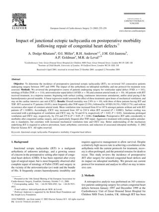
4. demonstrate a beat-to-beat variability (Fig. 1). In the great
majority of cases (90%), the atrial rate is slower than the
ventricular rate, but P waves may be retrograde with ventri-
culo-atrial 1:1 conduction in 10% of cases [1,7]. In common
with other automatic tachycardias, JET typically presents in
a ‘warm-up’ and ‘warm-down’ pattern, with progressive
acceleration into, and deceleration out of the tachycardia
[3,6–8]. The QRS rate is above the 98th percentile for the
age of the patient, typically between 170 and 230 beats per
min. JET is unresponsive to overdrive pacing or direct
current (DC) cardioversion [3,6–8]. Once the rate of the
tachycardia is slowed by cooling or medication, atrial
pacing becomes a useful adjunct to support cardiac output.
Atrial pacing conducts normally to the ventricle, thus yield-
ing a normal QRS morphology and av synchrony [3,6,8].
Aggressive management is required in the postoperative
setting, as the arrhythmia frequently induces haemodynamic
instability through atrioventricular dissociation. It typically
feeds on itself in a vicious circle pattern, as the lack of av
synchrony and the high ectopic rate lead to diminished
cardiac output, leading to a reflex increase in adrenergic
tone, which further accelerates the arrhythmia.
Concomitant postoperative electrolytic imbalance is
frequent, and requires appropriate treatment [14,16]. Hypo-
kalemia, hypocalcemia and hypomagnesemia must be
managed to keep levels of potassium above 3.5–4.0
mmol/l [13,14], levels of calcium above 0.8–1.2 mmol/l
[13,14], and levels of magnesium from 1.5 to 2.3 mg/dl
[13,14]. Recent evidence suggests that magnesium deple-
tion in the postoperative setting may significantly increase
the risk of developing JET [13]. Dorman et al presented data
from 28 patients undergoing surgical repair for CHD in
which magnesium supplementation was prophylactically
given in a double-blinded fashion, directly after disconti-
nuation of CPB. JET did not occur in those receiving
magnesium, as compared to an incidence of 27% in patients
given saline, before the study was discontinued for ethical
reasons [13]. Prophylactic supplementation with magne-
sium, therefore, may have a role in preventing JET, and is
performed in our unit. Acidosis and hypovolemia are
commonly associated and should be addressed. Endogenous
and exogenous catecholamines may worsen the tachycardia.
Accordingly, cardiac inotropic support should be reduced,
and even avoided, if possible [3]. Meticulous medical and
nursing care, including adequate sedation and avoidance of
vagolytic therapies, should reduce patient stress and any rise
of endogenous catecholamines [3]. Hyperthermia must be
avoided [7], and active surface cooling to 32–358C is a first-
line integral part of treatment during JET [3,5,8]. Aggres-
sive antiarrhythmic intervention usually achieves stabilisa-
tion, and subsequent spontaneous termination is the rule,
commonly occurring between 2 and 8 days [3,5].
Many antiarrhythmic therapies have been employed with
variable rates of success. Beta blockade with propranolol
may provide sufficient adrenergic block, but is limited by
its negative inotropic effect, which is undesirable in a post-
operative setting [3]. The class IC agents flecainide, encai-
nide, and propafenone, have been successfully used in
management [3,8,17]. Many centres, including our own,
favour the class III agent amiodarone as the agent of choice
[6,7,18]. Favourable pharmacokinetics allow various regi-
mens for administration, including intermittent bolus doses
or loading, plus continuous infusion protocols. Upon
achieving sinus rhythm, amiodarone is discontinued after
a minimum of 24 h of drug therapy, but the patient remains
continuously monitored for a further 24 h. As this is a self-
limiting disorder which usually abates in 2–8 days, long-
term therapy with amiodarone is unwarranted if no further
recurrence occurs during inhospital stay [7].
Intractable JET refractory to all antiarrhythmic medica-
tion or pacing methods may be aborted by transcatheter
ablation of the His bundle [3,9,17]. Although radiofre-
quency ablation has been successfully employed in infants
and children with the congenital form of JET [19,20], its use
in self-limiting postoperative JET should be restricted to
refractory cases, as there may be an unacceptably high
risk of complete heart block [17].
Extracorporeal membrane oxygenation (ECMO) as an
emergency procedure has been successfully used in patients
with JET refractory to all conventional treatments outlined
earlier [10]. The self-limited nature of JET makes ECMO
support a reasonable adjunct to conventional therapy that
allows circulatory assistance, and precise control of
hypothermia, until spontaneous recovery from the arrhyth-
mia occurs. The various risks and contraindications pertain-
A. Dodge-Khatami et al. / European Journal of Cardio-thoracic Surgery 21 (2002) 255–259258
Fig. 1. Beat-to-beat variability of arterial and venous pressure wave forms
in JET, before and after cooling.
5. ing to bleeding, exposure to additional blood products,
neurological damage, and infections, must be outweighed
by the life-saving nature of ECMO in this setting.
In conclusion, JET is an increasingly recognised malig-
nant arrhythmia arising in the postoperative setting after
surgery for congenital heart disease. Multiple protocols
exist for treatment, and most have demonstrated their effi-
ciency in sustaining patients until spontaneous arrhythmic
conversion. Nearly all protocols call for profound sedation,
paralysis, maintenance of mechanical ventilation, avoidance
of fever, and surface cooling in the first instance. We found
this initial step to be therapeutic in itself in a majority of
cases, without need for further escalation to pacing or anti-
arrhythmic therapy. In five patients, amiodarone may have
been required anyway to treat a refractory supraventricular
arrhythmia, despite overdiagnosis of JET. Accordingly,
temporary pacing in three patients without a confirmed diag-
nosis of JET achieved atrioventricular synchrony and
improved cardiac output. Nonetheless, the distinction
between true JET and other atrial arrhythmias is essential,
as excessive therapy may result in unnecessarily prolonged
ventilation time and CICU stay, both of which carry their
own potential morbidity, mortality, and cost considerations.
Strict interdisciplinary collaboration between surgeons,
cardiologists, and intensive care physicians is necessary to
assess this difficult arrhythmia, in terms of both diagnosis
and potential escalation in management. A better under-
standing of the underlying mechanisms of JET after surgery
for congenital heart disease may lead to a reduction or
avoidance of this malignant arrythmia, and to faster and
more efficient conversion in the remaining cases.
5. Study limitations
Although more frequently encountered after cardiac
surgery involving closure of a VSD, JET may occur after
the repair of virtually every congenital heart defect, even
extracardiac ones [17]. However, Walsh and colleagues [17]
reported a negative association of JET after surgery to close
defects within the oval fossa, after coarctation or aortic arch
repair, pacemaker implantation, and confection of systemic-
to-pulmonary arterial shunts. Accordingly, in this study,
patients with simpler intracardiac or extracardiac lesions
were not included. Also, it may be argued that many
complex congenital defects include closure of a VSD
amongst a long list of other anomalies to correct. However,
these were not analysed in the current study, in an attempt to
minimalize the potential effect of a wide spectrum of
morphology.
Acknowledgements
We thank Mr Thierry Murier of the University of
Neuchatel, Switzerland, for his statistical analysis. Research
at the Institute of Child Health and Great Ormond Street
Hospital for Children NHS Trust benefits from R&D fund-
ing received from the NHS Executive.
References
[1] Till JA, Ho SY, Rowland E. Histopathological findings in three chil-
dren with His bundle tachycardia occurring subsequent to cardiac
surgery. Eur Heart J 1992;13:709–712.
[2] Lupoglazoff JM, Denjoy I, Luton D, Magnier S, Azancot A. Prenatal
diagnosis of a familial form of junctional ectopic tachycardia. Prenat
Diagn 1999;19:767–770.
[3] Gillette PC. Diagnosis and management of postoperative junctional
ectopic tachycardia. Am Heart J 1989;118:192–194.
[4] Villain E, Vetter V, Garcia JM, Herre J, Cifarelli A, Garson Jr A.
Evolving concepts in the management of congenital junctional ecto-
pic tachycardia. Circulation 1990;81:1544–1549.
[5] Balaji S, Sullivan I, Deanfield J, James I. Moderate hypothermia in
the management of resistant automatic tachycardias in children. Br
Heart J 1991;66:221–224.
[6] Till JA, Rowland E. Atrial pacing as an adjunct to the management of
post-surgical His bundle tachycardia. Br Heart J 1991;66:225–229.
[7] Raja P, Hawker RE, Chaikitpinyo A, Cooper SG, Lau KC, Nunn GR,
Cartmill TB, Sholler GF. Amiodarone management of junctional
ectopic tachycardia after cardiac surgery in children. Br Heart J
1994;72:261–265.
[8] Pfammatter J-P, Paul T, Ziemer G, Kallfelz HC. Successful manage-
ment of junctional tachycardia by hypothermia after cardiac opera-
tions in infants. Ann Thorac Surg 1995;60:556–560.
[9] Bharati S, Moskowitz WB, Scheinman M, Estes NAM, Lev M. Junc-
tional tachycardias: anatomic substrate and its significance in ablative
procedures. J Am Coll Cardiol 1991;18:179–186.
[10] Azzam FJ, Fiore AC. Postoperative junctional ectopic tachycardia.
Can J Anaesth 1998;45(9):898–902.
[11] Garson A, Moak JP, Smith RT, Norton JB. Usefulness of intravenous
propafenone for control of postoperative junctional ectopic tachycar-
dia. Am J Cardiol 1987;59:1422–1424.
[12] Anderson RH, Ho SY. The morphologic substrate for pediatric
arrhythmias. Cardiol Young 1991;1:159–176.
[13] Dorman BH, Sade RM, Burnette JS, Wiles HB, Pinosky ML, Reeves
ST, Bond BR, Spinale FG. Magnesium supplementation in the
prevention of arrhythmias in pediatric patients undergoing surgery
for congenital heart defects. Am Heart J 2000;139:522–528.
[14] Braunstein PW, Sade RM, Gillette PC. Life-threatening postoperative
junctional ectopic tachycardia. Ann Thorac Surg 1992;53:726–728.
[15] Dodge-Khatami A, Miller OI, Anderson RH, Goldman AP, Gil-Jaur-
ena JM, Elliott MJ, Tsang VT, de Leval MR. Surgical substrates of
postoperative junctional ectopic tachycardia in congenital heart
defects. J Thorac Cardiovasc Surg (in press).
[16] Mandapati R, Byrum CJ, Kavey REW, Smith FC, Kveselis DA,
Hannan WP, Brandt III B, Gaum WE. Procainamide for rate control
of postsurgical junctional tachycardia. Pediatr Cardiol 2000;21:123–
128.
[17] Walsh EP, Saul JP, Sholler GF, Triedman JK, Jonas RA, Mayer JE,
Wessel DL. Evaluation of a staged protocol for rapid automatic junc-
tional tachycardia after operation for congenital heart disease. J Am
Coll Cardiol 1997;29:1046–1053.
[18] LuedtkeSA, Kuhn RJ, McCaffreyFM.Pharmacologicalmanagementof
supraventricular tachycardia. Part 2: Atrial flutter, atrial fibrillation, and
junctional ectopic tachycardia. Ann Pharmacother 1997;31:1347–1359.
[19] Wu MH, Lin JL, Chang YC. Catheter ablation of junctional ectopic
tachycardia by guarded low-dose radiofrequency energy application.
Pacing Clin Electrophysiol 1996;19:1655–1658.
[20] Wren C. Incessant tachycardias. Eur Heart J 1998;19:E32–E36.
A. Dodge-Khatami et al. / European Journal of Cardio-thoracic Surgery 21 (2002) 255–259 259