Bright field microscope .pptx

A **bright field microscope** is a type of compound light microscope that illuminates the background against a stained specimen ¹². It is commonly used in practical labs to study organisms' behavior and characteristics such as size, shape, and arrangement ². The microscope uses light rays to produce a dark image against a bright background ¹. It is specially designed with magnifying glasses known as lenses that modify the specimen to produce an image seen through the eyepiece ¹. The bright field microscope is made up of various parts, including the eyepiece, objective lenses, focusing knobs, and stage ¹. I hope this helps! Source: Conversation with Bing, 7/11/2023 (1) Brightfield Microscope (Compound Light Microscope)- Definition .... https://microbenotes.com/brightfield-microscope/. (2) Bright Field Microscopy - Biology Reader. https://biologyreader.com/bright-field-microscopy.html. (3) Bright-field microscopy - Wikipedia. https://en.wikipedia.org/wiki/Bright-field_microscopy. (4) Bright Field Microscope: Definition, Parts, Working Principle, Application. https://microbiologynote.com/bright-field-microscope-definition-parts-working-principle-application/.

Recommended
More Related Content
What's hot
What's hot (20)
Similar to Bright field microscope .pptx
Similar to Bright field microscope .pptx (20)
More from AjayDesouza V
More from AjayDesouza V (14)
Recently uploaded
Recently uploaded (20)
Bright field microscope .pptx
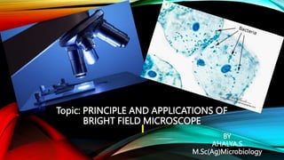
- 1. Topic: PRINCIPLE AND APPLICATIONS OF BRIGHT FIELD MICROSCOPE BY AHALYA.S M.Sc(Ag)Microbiology
- 2. MICROSCOPE MICROSCOPE : Microscope is an optical instrument that uses a lens or a combination of lenses to produce highly magnified images of small specimens especially which are too small to be seen by the naked eye. Light source is either mirror(natural light) or lamps. MICROSCOPY: Microscopy is the technical field of using microscope or investigation by a microscope.
- 3. HISTORY OF MICROSCOPE ● Hans and zacharias Janssen - 1590 First compound microscope (2 lenses),tube with lenses at each end. ● Anton van Leeuwenhoek -1670 Simple microscope(1 lenses) Father of microbiology ● Robert Hooke - 1667 Compound microscope improvement with 2 lenses - objective & ocular lenses.
- 4. CLASSIFICATION OF MICROSCOPE MICROSCOPE SIMPLE MICROSCOPE COMPOUND MICROSCOPE LIGHT MICROSCOPE ELECTRON MICROSCOPE BRIGHT FIELD DARK FIELD FLUORESCENCE PHASE CONFOCAL SCANNING TRANSMISSION MICROSCOPE MICROSCOPE MICROSCOPE CONTRAST MICROSCOPE ELECTRON ELECTRON MICROSCOPE MICROSCOPE MICROSCOPE
- 6. PARTS OF MICROSCOPE OPTICAL COMPONENTS : o Eyepiece or Ocular Lenses is what you look through at the top of the microscope. Typically, standard eyepieces have a magnifying power of 10x. Optional eyepieces of varying powers are available, typically from 5x-30x. o Objective Lenses are the primary optical lenses on a microscope. They range from 4x-100x and typically, include, three, four or five on lens on most microscopes. Objective lenses can be forward or rear-facing. Standard objectives include 10x, 40x and 100x although different power objectives are available. o Condenser Lens is used to collect and focus the light from the illuminator on to the specimen. It is located under the stage often in conjunction with an iris diaphragm.
- 7. PARTS OF MICROSCOPE MECHANICAL COMPONENTS: ● Head/Body - houses the optical parts in the upper part of the microscope. ● Base - supports the microscope and houses the illuminator. ● Arm - connects to the base and supports the microscope head. It is also used to carry the microscope. ● Eyepiece Tube holds the eyepieces in place above the objective lens. ● Nosepiece houses the objective lenses.. The objectives are exposed and are mounted on a rotating turret so that different objectives can be conveniently selected.
- 8. PARTS OF MICROSCOPE ● Coarse (wide focus) and Fine Focus knobs (sharp & fine focus) are used to focus the microscope. ● Specimen Stage is where the specimen to be viewed is placed. A mechanical stage is used when working at higher magnifications where delicate movements of the specimen slide are required. ● Stage Clips are used when there is no mechanical stage. The viewer is required to move the slide manually to view different sections of the specimen. ● Aperture is the hole in the stage through which the base (transmitted) light reaches the stage.
- 9. PARTS OF MICROSCOPE ● Illuminator is the light source for a microscope, typically located in the base of the microscope. Most light microscopes use low voltage, halogen bulbs with continuous variable lighting control located within the base. ● Iris Diaphragm controls the amount of light reaching the specimen. It is located above the condenser and below the stage. ● Light Switch present at bottom of right side of microscope to switch on/off the light source(illuminator). ● Brightness Adjustment helps to adjust the brightness of light that comes out from the illuminator.
- 10. SIMPLE MICROSCOPE. COMPOUND MICROSCOPE
- 11. SIMPLE MICROSCOPE. COMPOUND MICROSCOPE ● Invented by Anton van Leeuwenhoek in 1670. ● Structure : Stand is small, hollow cylindrical attached to base is used to hold the microscope. ● Contain single eyepiece. Total magnification is limited to ocular lens magnification. ● Light source is natural light. ● Mirror - concave which helps in reflection of light. ● No condenser lenses. ● Magnification power up to 300x. ● adjustment of magnification is not available. ● Used for study of microscopic fungi,algae. ● Invented by Hans and Zacharias Janssen in 1590. ● Structure : arm is curved used to hold the microscope. ● Contains either single or double eyepiece - ocular lens & objective lens. ● Total magnification is eyepiece*obj lens. ● Light source - natural light or illuminator. ● Mirror - plane at one side & concave at other side. ● Condenser lens present which adjust the intensity of light ● Magnification power up to 2000x. ● Adjustment of magnification by 3 obj lenses 10x,40x,100x. ● Used for study of morphology of bacteria & other microbes.
- 12. LIGHT MICROSCOPE ● A Llight microscope uses focused light and lenses to magnify a specimen, usually a CellS, microorganisms. ● Also known as OPTICAL MICROSCOPE. ● Highest practical magnification is 4000x. ● best resolution 0.2 micrometer. ● Illuminating source: visible light wavelength of 450nm - 750nm. ADVANTAGES: ● Easy to use. ● Low cost when compared to others. ● Live specimens can be studied. ● Shows true colours but sometimes stains required.
- 13. DISADVANTAGES : ● Low resolution due to shorter wavelength of light. ● Low magnification. TYPES OF LIGHT MICROSCOPE ● Bright field microscope ● Dark field microscope ● Phase contrast microscope ● Fluorescence microscope ● Confocal microscope
- 14. MAGNIFICATION ● Magnification is the enlargement of the image. ● Light microscope in schools & colleges can magnify 400 times than actual size. ● MMICROSCOPE = MOCULAR LENS * MOBJECTIVE LENS MAGNIFICATION OF OBJ LENS MAGNIFICATION OF OCULAR LENS TOTAL MAGNIFICATION SCANNING 4x 10x 40x LOW POWER 10x 10x 100x HIGH POWER 40x 10x 400x OIL IMMERSION 100x 10x 1000x
- 15. APPROPRIATE MAGNIFICATION SAMPLE Stained – bacteria Thick tissue sections Blood smears Negative stained bacteria Algae and other microscope plant material MAGNIFICATION 1000x 100x , 400x 400x , 1000x 400x , 1000x 40x , 100x , 400x
- 16. RESOLVING POWER RESOLUTION / RESOLVING POWER : ● Resolution is the ability of lenses to distinguish fine detail and structure of the specimens.(distance between two points as separate entities) ● Human eye resolution is 0.2 mm. ● Rmicroscope > Rhuman eye . ● d = wavelength of light(λ) 2×Numerical Aperture ● d = λ 2 n sinα Smaller the d, greater the Resolution. Shorter the wavelength of light(λ) used, greater the resolution. Lenses with higher numerical aperture(α) provide better resolving power. Greater the refractive index(n) , higher the resolution.
- 17. NUMERICAL APERTURE ● Number represents the angle of light produced by refraction & is the measure of quantity of light gathered by lens. ● Mathematical constant derived from Physical structure of lenses. ● Each objective lens has fixed numerical aperture reading ranging from 0.1 in lowest power lens(10x) to approximately 1.25 in highest power lens(100x). WAVELENTGH : ● Wavelength is the actual distance between the two successive crests or troughs of a wave. ● Visible light wavelength : VIBGYOR (300nm – 700nm). REFRACTIVE INDEX : ● Refractive index of air = 1.003 ● Refractive index of water = 1.33 ● Refractive index of oil = 1.51(higher magnification)
- 18. BRIGHT FIELD MICROSCOPE ● Bright field microscope are named because the microscope field is bright while the object being viewed is black (dark sample on a bright background). ● Used to observe various stained specimens, naturally pigmented specimens and to count microbes. ● In bright field microscopy, the sample illumination is transmitted white light and contrast in sample is caused by absorbance of some of the transmitted light in dense areas of the sample and this contrast allows us to see the specimens. ● High contrast sample.
- 19. PRINCIPLE
- 20. PRINCIPLE ● It has a series of two lenses: ● The OBJECTIVES LENS close to the object to be observed ● The OCULAR LENS or EYEPIECE, through which the image is viewed by eye. ● Light from the light source (electric lamp) passes through a thin transparent object. ● The objective lens produce a magnified “REAL IMAGE” (1st image) of the object. This image is again magnified by the ocular lens ( eyepiece) to obtain a magnified “VIRTUAL IMAGE” (final image), which can be seen by eye through the eyepiece. ● As light passes directly from the source to the eye through the two lenses, the field of vision is brightly illuminated. That is why : it is a BRIGHT – FIELD MICROSCOPE.
- 21. SPECIMENS
- 22. ADVANTAGES OF BRIGHT FIELD MICROSCOPE Shows the morphological as well as the internal structure of the specimen. Less expensive than dark field microscope. Stained, fixed and live specimens are observed. Specimen colour appears depending on the stain colour. Easy to use. DISADVANTAGES: Does not allow us to observe the metal and minerals. Specimen preparation or staining is a complex and lengthy process. Takes too much time. Opaque disc is absent. Does not resolve very small specimens, such as viruses. Requires a strong light source for high magnification applications and intense lighting can produce heat that will damage specimens or kill living microorganisms.
- 23. APPLICATIONS Widely use in pathology to view fixed tissue sections or cell films/smears. Important for hematology, microbiology, Tb and malaria testing. Microscope magnifies the blood samples, so, the doctor can see the malaria parasites attacking the red blood cells. Used in bacteriology, biology and medicine to examine minute objects such as bacteria, other unicellular organisms and plant cell , animal cells and tissues. Advances in fluorochrome stains and monoclonal antibody techniques caused grow in use of analysis and cell biology.
- 24. APPLICATIONS