Fluorescent Microscopy
•
2 likes•1,060 views

Fluorescence microscopes use fluorescence to generate an image by exciting a specimen with one wavelength of light and detecting emitted light of a different, longer wavelength. Fluorescence microscopes have various applications including staining biological molecules, labeling proteins or other targets within cells using fluorescent antibodies in immunofluorescence techniques, and genetically modifying proteins to directly carry fluorescent protein reporters to track protein location.

Recommended
More Related Content
What's hot
What's hot (20)
Similar to Fluorescent Microscopy
Similar to Fluorescent Microscopy (20)
More from Syed Muhammad Khan
More from Syed Muhammad Khan (20)
Recently uploaded
Recently uploaded (20)
Fluorescent Microscopy
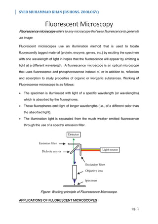
- 1. SYED MUHAMMAD KHAN (BS HONS. ZOOLOGY) pg. 1 Fluorescent Microscopy Fluorescence microscope refers to any microscope that uses fluorescence to generate an image. Fluorescent microscopes use an illumination method that is used to locate fluorescently tagged material (protein, enzyme, genes, etc.) by exciting the specimen with one wavelength of light in hopes that the fluorescence will appear by emitting a light at a different wavelength. A fluorescence microscope is an optical microscope that uses fluorescence and phosphorescence instead of, or in addition to, reflection and absorption to study properties of organic or inorganic substances. Working of Fluorescence microscope is as follows: The specimen is illuminated with light of a specific wavelength (or wavelengths) which is absorbed by the fluorophores. These fluorophores emit light of longer wavelengths (i.e., of a different color than the absorbed light). The illumination light is separated from the much weaker emitted fluorescence through the use of a spectral emission filter. Figure: Working principle of Fluorescence Microscope. APPLICATIONS OF FLUORESCENT MICROSCOPES
- 2. SYED MUHAMMAD KHAN (BS HONS. ZOOLOGY) pg. 2 The following are some of the applications of fluorescent microscopes: Biological Fluorescent Stains: Many fluorescent stains have been designed for a range of biological molecules. Some of these are small molecules that are intrinsically fluorescent and bind a biological molecule of interest. Many nucleic acid (fluorescent) stains bind the minor groove of DNA, thus labeling the nuclei of cells. Fluorophores / Fluorochromes: These are many fluorescent molecules that can be chemically linked to a different molecule which binds the target of interest within the sample. Immunofluorescence: Immunofluorescence is a technique that uses the highly specific binding of an antibody to its antigen to label specific proteins or other molecules within the cell. A sample is treated with a primary antibody specific for the molecule of interest. A fluorophore can be directly conjugated to the primary antibody. Alternatively, a secondary antibody, conjugated to a fluorophore, which binds specifically to the first antibody can be used. For example, a primary antibody raised in a mouse which recognizes tubulin combined with a secondary anti-mouse antibody derivatized with a fluorophore could be used to label microtubules in a cell. Figure: Basic mechanism of immunofluorescence Primary immunofluorescence involves an antibody with a fluorophore group bound to it directly binding to the epitope of the antigen for which it is specific. Once the antibody
- 3. SYED MUHAMMAD KHAN (BS HONS. ZOOLOGY) pg. 3 binds to the epitope, the sample can be viewed under a fluorescent microscope to confirm the presence of the antigen in the sample. Secondary immunofluorescence involves an untagged primary antibody that binds to the epitope of the antigen. However, after the primary antibodies have bound to their targets, a secondary antibody (tagged with a fluorophore) comes along which binds to the primary antibody. Fluorescent Proteins: The modern understanding of genetics and the techniques available for modifying DNA allow scientists to genetically modify proteins to also carry a fluorescent protein reporter. In biological samples this allows a scientist to directly make a protein of interest fluorescent. The protein location can then be directly tracked, including in live cells.