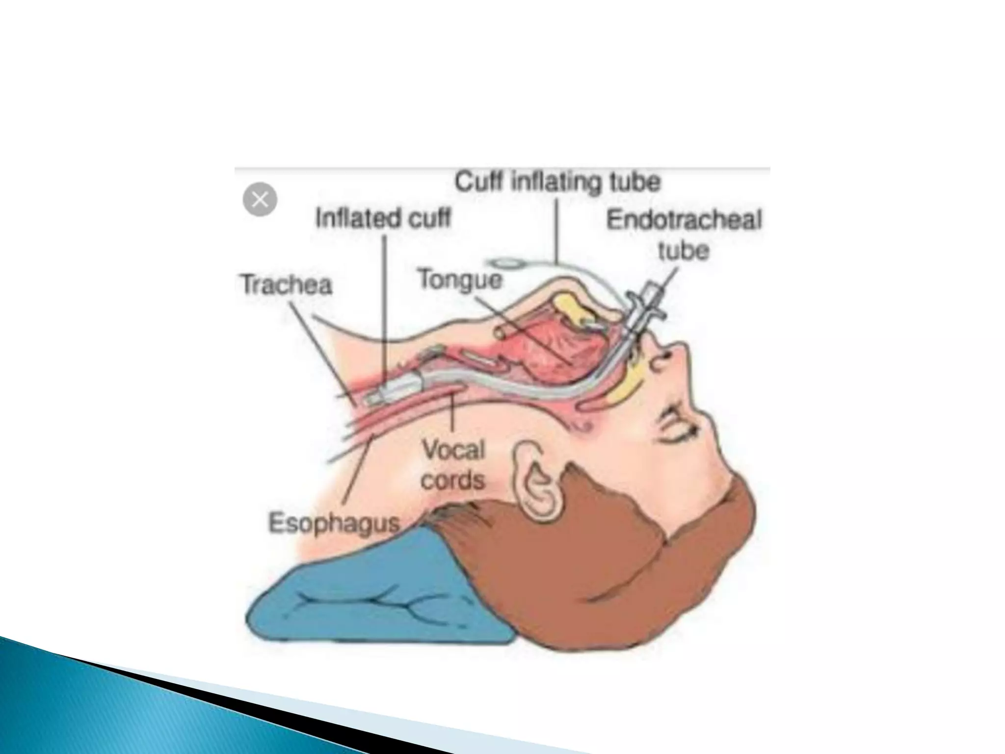
BY ADNAN QADIR
It is the procedure in which an
endotracheal tube is inserted
through the nose or mouth into
the trachea.
 To administer oxygen.
 To remove secretions.
 To promote airway patency.
 To assist when the patient has difficulty in
breathing.
 To administer anesthetics.
 Inability to maintain tone of the airway in
conditions such as
- trauma of the face and neck with
oropharyngeal bleeding.
- edema of upper airway secondary to
anaphylaxis.
 Inability to have spontaneous ventilation
despite prolonged respiratory effort in case of
COPD
Status asthmatics
 Poor oxygenation.
ARDs
Pneumonia
Carbon monoxide toxicity
Cyanide toxicity
 Clinically deteriorating conditions
CNS depression
Neuromuscular disease
Septic shock
 Complete obstruction of upper
airway in which surgical airway
is only remedy.
 Cervical spine injury
 A wide variety of endotracheal tubes are used
either oro-tracheal or naso-tracheal
intubations.
 Some of them are having cuffs while others
don’t.
 Oro-tracheal tubes arev larger than naso
tubes.
 Size is maked in mm on outer side of tube.
 New born 2.5mm – 4mm
 0-1 years 4mm – 4.5mm
 Upto 10 yrs 5mm- 7mm
 Above 10 yrs 7mm – 8mm
 Adults 8mm – 9.5mm
 Check the patient’s name and other
identification data.
 Check the vital signs
 Asses the level of consciousness
 Asses the anatomy of airway such as ability to
open the mouth,presence of dentures and
size of tongue.
 Check the oxygen saturation and ABG results.
 Findings of x ray chest.
An intubation tube of appropriate size
I. Endotracheal tube of appropriate size
II. Stylet –a wire
III. Laryngoscope with appropriate striaght/curved blade
IV. Oral airway
V. Magill’s forceps
VI. Suction catheter
VII. Suction apparatus oxygen source and tubing
VIII. Disposable syringe xiv. Cotton tape
IX. Xylocaine jelly xv. scissor
X. Towel xvi. Rolled towel
XI. Sterile gloves and mask xvii. Induction agents as per
XII. Ambu bag and mask physicians order .
XIII. Stethoscope
 Explain the procedure to the patients and family.
 Assemble and prepare all the equipments needed.
Assemble laryngoscope and ensure that light is bright.
Place the ET tube in a sterile field.
Lubricate the distal end of the tube with xylocaine jelly.
Insert the stylet into the ET tube
 Ensure that all the equipments are in working condition.
 Asses the loose teeth/dentures/foreign body in throat ;if so remove with
magill’s forceps.
 Position the patient with head and slightly elevated at 10 ;remove the
headboard if possible and needed . Place the rolled towel under the neck .
 Administer premedications (induction agents) as per order.
 Spray the anesthetic medicine in the throat if needed
 Apply suction to the oral cavity.
 Provide laryngoscope to doctor.
 Hold laryngoscope in the left hand and insert the blade along the right side of
the tongue ;with the right thumb and index finger ;pull lower lip away from
the patient’s lower teeth.
 Lift the laryngoscope upwards and forward at 45 .
 After visualizing the vocal cords ,insert the ET into the right side while
continuously visualizing the vocal cards .
 Insert the tube slowly ,gradually and downward till it reaches beyond the
cords.
 Once it reaches beyond the cords , remove laryngoscope and withdraw
stylet, the tube when inserted should have 22 mm marking at the incisor
teeth.
 Confirm the correct position of the tube by
BILATERAL CHEST MOVEMENTS
AUSCULTATION OF CHEST
 Attach the ambu bag with the oxygen
connection to ET tube and continue the
bagging to ventilate.
 Inflate the cuff with 10 ml of air.
 Insert the oral airway and apply suctioning if
necessary.
 Secure the tube in place with adhesive tube.
 Ensure the chest x ray is taken.
 Never leave the patient alone.
 Provide a comfortable position.
 Watch and maintain an open airway.
 Remove the secretions by effective suctioning.
 Prevent displacement of tube.
 Watch for complications such as laryngeal, oedema, tracheal
stenosis, heamorrhage.
 Provide for the humidification of air by boiling a kettle of
water in the patient’s unit.
 Prevent infection introduced into the lungs.
 Prevent contamination of inhaled air,
 Maintain oral hygiene.
 Maintain adequate nutrition of the patient by naso gastric
feeding or by giving IV fluids. They should never be fed an
oral feeds as long as tube is in the mouth.
 It includes the following
a. Date and time of intubation.
b. Pre procedure assessment.
c. Et tube type and size.
d. Cuff pressure
e. Condition during and post procedure.
f. Follow up.
LAUGH A LOT. IT CLEARS THE LUNGS …….

Endotracheal intubation

  • 1.
  • 2.
    It is theprocedure in which an endotracheal tube is inserted through the nose or mouth into the trachea.
  • 4.
     To administeroxygen.  To remove secretions.  To promote airway patency.  To assist when the patient has difficulty in breathing.  To administer anesthetics.
  • 5.
     Inability tomaintain tone of the airway in conditions such as - trauma of the face and neck with oropharyngeal bleeding. - edema of upper airway secondary to anaphylaxis.  Inability to have spontaneous ventilation despite prolonged respiratory effort in case of COPD Status asthmatics
  • 6.
     Poor oxygenation. ARDs Pneumonia Carbonmonoxide toxicity Cyanide toxicity  Clinically deteriorating conditions CNS depression Neuromuscular disease Septic shock
  • 7.
     Complete obstructionof upper airway in which surgical airway is only remedy.  Cervical spine injury
  • 8.
     A widevariety of endotracheal tubes are used either oro-tracheal or naso-tracheal intubations.  Some of them are having cuffs while others don’t.  Oro-tracheal tubes arev larger than naso tubes.  Size is maked in mm on outer side of tube.
  • 10.
     New born2.5mm – 4mm  0-1 years 4mm – 4.5mm  Upto 10 yrs 5mm- 7mm  Above 10 yrs 7mm – 8mm  Adults 8mm – 9.5mm
  • 11.
     Check thepatient’s name and other identification data.  Check the vital signs  Asses the level of consciousness  Asses the anatomy of airway such as ability to open the mouth,presence of dentures and size of tongue.  Check the oxygen saturation and ABG results.  Findings of x ray chest.
  • 12.
    An intubation tubeof appropriate size I. Endotracheal tube of appropriate size II. Stylet –a wire III. Laryngoscope with appropriate striaght/curved blade IV. Oral airway V. Magill’s forceps VI. Suction catheter VII. Suction apparatus oxygen source and tubing VIII. Disposable syringe xiv. Cotton tape IX. Xylocaine jelly xv. scissor X. Towel xvi. Rolled towel XI. Sterile gloves and mask xvii. Induction agents as per XII. Ambu bag and mask physicians order . XIII. Stethoscope
  • 14.
     Explain theprocedure to the patients and family.  Assemble and prepare all the equipments needed. Assemble laryngoscope and ensure that light is bright. Place the ET tube in a sterile field. Lubricate the distal end of the tube with xylocaine jelly. Insert the stylet into the ET tube  Ensure that all the equipments are in working condition.  Asses the loose teeth/dentures/foreign body in throat ;if so remove with magill’s forceps.  Position the patient with head and slightly elevated at 10 ;remove the headboard if possible and needed . Place the rolled towel under the neck .  Administer premedications (induction agents) as per order.  Spray the anesthetic medicine in the throat if needed
  • 15.
     Apply suctionto the oral cavity.  Provide laryngoscope to doctor.  Hold laryngoscope in the left hand and insert the blade along the right side of the tongue ;with the right thumb and index finger ;pull lower lip away from the patient’s lower teeth.  Lift the laryngoscope upwards and forward at 45 .  After visualizing the vocal cords ,insert the ET into the right side while continuously visualizing the vocal cards .  Insert the tube slowly ,gradually and downward till it reaches beyond the cords.  Once it reaches beyond the cords , remove laryngoscope and withdraw stylet, the tube when inserted should have 22 mm marking at the incisor teeth.  Confirm the correct position of the tube by BILATERAL CHEST MOVEMENTS AUSCULTATION OF CHEST
  • 16.
     Attach theambu bag with the oxygen connection to ET tube and continue the bagging to ventilate.  Inflate the cuff with 10 ml of air.  Insert the oral airway and apply suctioning if necessary.  Secure the tube in place with adhesive tube.  Ensure the chest x ray is taken.
  • 17.
     Never leavethe patient alone.  Provide a comfortable position.  Watch and maintain an open airway.  Remove the secretions by effective suctioning.  Prevent displacement of tube.  Watch for complications such as laryngeal, oedema, tracheal stenosis, heamorrhage.  Provide for the humidification of air by boiling a kettle of water in the patient’s unit.  Prevent infection introduced into the lungs.  Prevent contamination of inhaled air,  Maintain oral hygiene.  Maintain adequate nutrition of the patient by naso gastric feeding or by giving IV fluids. They should never be fed an oral feeds as long as tube is in the mouth.
  • 18.
     It includesthe following a. Date and time of intubation. b. Pre procedure assessment. c. Et tube type and size. d. Cuff pressure e. Condition during and post procedure. f. Follow up.
  • 19.
    LAUGH A LOT.IT CLEARS THE LUNGS …….