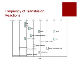
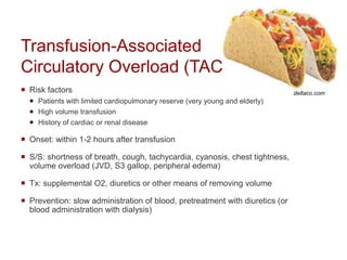
4. Case 1
Mr Red is a 17 year old male is brought to the ER
after a motor vehicle accident. He is in pain,
tachycardic to 100s, but normotensive.
Given his acute blood loss, transfusion of 2u PRBC is
initiated (after appropriate type and cross-matching
revealing no antibodies, and compatibility with donor
blood).
During transfusion, he develops a fever but otherwise
has no new signs or symptoms.
What is the diagnosis?
5. Febrile Nonhemolytic
Transfusion Reaction
Fevers are common during transfusion
Pathophysiology: likely involves recipient-derived
leukoreactive antibodies + donor-derived cytokines
Workup/Treatment: stop the transfusion!
Must r/o acute hemolytic transfusion reaction (AHTR)
Consider non-transfusion causes of fevers
Once AHTR is ruled out, may continue transfusion with
antipyretics
Prevention: antipyretics or leukoreduction of blood
products
6. Case 1 (continued)
Mr Red does well following discharge. Fifteen years later (age
32), however, he is unfortunately in a second MVA. He is brought
to the ER, again requiring blood products.
He is type and cross-matched, found to have no antibodies. He is
pre-treated with acetaminophen, and transfused 2 units PRBC
without issue.
The remainder of his hospital course is unremarkable and the pt
is discharged home.
Ten days after the accident he follows up at his PCP’s office with
a complaint of fatigue, fevers, and yellowing of his skin.
What is the diagnosis?
7. Delayed Hemolytic
Transfusion Reaction
Onset of symptoms: 5-10 days after
RBC transfusion
S/S: hemolytic anemia, jaundice,
fever (can also be asymptomatic)
Life-threatening complications are
rare
Confirmation: repeat type and screen
to detect alloantibody
Treatment: supportive
Abrupt onset of S/S
S/S: intravascular hemolysis,
hypotension, fevers, AKI, pain at the
infusion site, DIC, pink plasma or
urine
Treatment: stop the transfusion!
Send blood back to blood bank to check
for incompatibility, hemolysis
Supportive treatment with IVF, pressors,
diuresis
Acute Hemolytic
Transfusion Reaction
http://arimmuneresponseassignment.weebly.com/report.html
8. Case 1 (continued)
Mr Red is now 78 years old. Since we last saw
him, he has been diagnosed with diabetes,
complicated by ESRD 2/2 diabetic nephropathy
for which he is dialyzed three times per week.
He is admitted for a suspected GI bleed for
which he is transfused 2 units PRBC. An hour
after transfusion, he starts to complain of
shortness of breath and chest tightness. HR
120s, BP 180/90, an S3 gallop is noted, and
new bibasilar crackles are heard on pulmonary
exam. Post-transfusion CXR is shown (was
previously normal).
What is the diagnosis? https://www.med-ed.virginia.edu/courses/
rad/cxr/pathology2chest.html
9. Transfusion-Associated
Circulatory Overload (TACO)
Risk factors
Patients with limited cardiopulmonary reserve (very young and elderly)
High volume transfusion
History of cardiac or renal disease
Onset: within 1-2 hours after transfusion
S/S: shortness of breath, cough, tachycardia, cyanosis, chest tightness,
volume overload (JVD, S3 gallop, peripheral edema)
Tx: supplemental O2, diuretics or other means of removing volume
Prevention: slow administration of blood, pretreatment with diuretics (or
blood administration with dialysis)
deltaco.com
10. Case 2
Mr Red’s hospital roommate also
happens to be a 78 year old male
admitted for likely GI bleed. He also
underwent transfusion with 2 units
PRBC 1 hour ago and reports shortness
of breath.
He is febrile to 38.5C, HR 120s, BP
70/40, SpO2 is 85% on RA. New
bibasilar crackles are heard on
pulmonary exam. Post-transfusion CXR
is shown (was normal previously).
What is the diagnosis?
https://www.med-ed.virginia.edu/courses/
rad/cxr/pathology2chest.html
11. Transfusion-Related Acute
Lung Injury (TRALI)
Onset: during or within 6 hours of
transfusion
S/S: hypoxia, dyspnea, fevers,
hypotension, pulmonary edema
Treatment: stop the transfusion!
Supportive (may need intubation), O2
Prevention: notify blood bank of
reaction
thelancet.com
13. Back to Mr Red…
Mr Red is now 80 years old and is admitted after a fall
during which he sustained a left hip fracture.
Following surgery, he requires 1 unit PRBC. He is
appropriately type and crossmatched, pretreated with
acetaminophen, and a slow transfusion is initiated
during dialysis. During the transfusion, he develops
diffuse urticaria but is otherwise stable.
What is the diagnosis?
umm.edu
14. Allergic Reactions and
Anaphylaxis
Mild allergic reactions (urticaria) are common,
especially in pts who have undergone multiple
transfusions
Prevention: pretreat with anti histamines, or wash blood
products to remove plasma proteins
Severe anaphylaxis is rare
Mechanism: recipient who is IgA deficient and has anti-
IgA antibodies reacts to IgA in donor blood
Prevention: wash all subsequent blood products to
remove plasma proteins
If IgA deficient, then only give blood products from IgA
deficient donors
15. Summary
It is important to recognize the possible reactions that
can be associated with blood transfusions
If you suspect a reaction, stop the transfusion and
assess the patient’s vital signs, signs and symptoms
as some reactions may be life-threatening
Notify the blood bank if serious reactions are
suspected
16.
17. References
Kim J, Na S. Transfusion-related acute lung injury;
clinical perspectives. Korean J Anesthesiol. 2015
Apr;68(2):101-5.
MKSAP 16
UpToDate
Editor's Notes
We will be going over the more common transfusion reactions by discussing patient cases. The following will be covered:
Febrile nonhemolytic transfusion reaction
Delayed (and acute) hemolytic transfusion reactions
TACO
TRALI
Allergic and anaphylactic reactions
Note that we will not cover transfusion-associated graft versus host disease but should be aware of it, and who is at risk.
Just for reference, here’s some info on transfusion-associated GVHD
Rare, but often fatal transfusion reaction
Pathophysiology: donor lymphocytes recognize host HLA antigens as foreign, resulting in an immune response
Risk factors:
Hematopoietic stem cell transplant recipients
Transfusion of blood products from first-degree relatives
Immunosuppressed patient
Onset: 8-10 days after transfusion
S/S: severe pancytopenia, fever, diarrhea, rash, transaminitis
Treatment: none are effective
Prevention: for those at risk, pre-treat blood products with gamma-irradiation
Again, we’ll only cover the more common reactions. Note that a febrile nonhemolytic reaction is much more common than a hemolytic reaction. Also note that TACO is more common than TRALI.
A case to illustrate the first transfusion reaction
This patient is tachycardic, probably related to pain and/or blood loss. The only symptom he develops during transfusion is fever. This is getting at a nonhemolytic febrile reaction.
This is a common reaction
Likely related to antibodies, cytokines
In these instances the first thing to do is stop the transfusion (basically in ALL cases when a pt develops S/S during a transfusion, you’ll want to stop the transfusion and investigate further)
we’d need to rule out acute hemolytic transfusion reaction – check that the correct blood products were given to the right patient (we’ll discuss AHTR a little more later)
Make sure that there’s no infection going on (or if there is concern for one, for example, from the case patient’s trauma, draw cultures and start empiric antibiotics).
Otherwise, if it looks like a febrile nonhemolytic reaction, then we can continue the transfusion with close monitoring, give antipyretics.
We’ll continue looking at the same patient but fast-forward 15 years
He got tylenol to prevent the febrile nonhemolytic reaction that he had in the past
This time he develops symptoms days later
Last case: the patient’s history of prior transfusion, as well as timing of symptoms suggests delayed hemolytic transfusion reaction.
Pathophysiology: The patient likely developed alloantibodies (ie Kidd, duffy, kell etc) as a result of the initial transfusion (when he was 17 years old). Titers decrease over time with no transfusion so that pre-transfusion testing remains negative (as it did when the pt was cross-matched at 37 years old). When he is re-exposed to the same antigen during his second transfusion, the patient’s antibody levels skyrocket. This is known as an anamnestic response.
Risk: there is a 1% risk of developing alloantibodies after transfusion
For treatment/prevention: Also suggest minimizing transfusions however these should not be withheld if needed
Just for contrast, we’ll briefly go over acute hemolytic transfusion reactions, which are now quite uncommon.
Pathophys: due to ABO incompatibility (see figure) – ie giving type B blood to a type A recipient who has anti-B antibodies. This leads to hemolysis.
If this reaction happens, it’s usually due to a clerical error, ie specimens are mislabeled or blood given to the wrong patient
Go through S/S
Treatment
Blood bank should know about this and confirm that this is what happened
Treatment is supportive. AKI can be a big deal so per UpToDate, if there’s any suspicion that this reaction happened, then start aggressive fluid replacement to support UOP > 100-200cc/hr immediately to prevent development of acute oliguric renal failure
Our patient has become a dialysis pt – someone sensitive to volume overload
Last case: timing of symptoms and S/S of volume overload are consistent with TACO. His kidney disease put him at risk for this.
Now we’ll look a a similar scenario but make this patient less sensitive to volume to demonstrate another transfusion reaction.
CXR looks a lot like TACO… But presentation is slightly different – this patient has more systemic S/S – he is febrile, hypotensive, hypoxic. While he has pulmonary edema, he does not have any other signs of volume overload (no JVD, S3 gallop, LE edema, etc).
TRALI has an associated mortality of 5% and is the most common cause of transfusion-related death
The figure demonstrates pathophysiology of TRALI, which is basically as follows:
Recipient neutrophils are activated against an antigen in donor blood product
PMNs become sequestered in lung capillaries
Capillary leak
Pulmonary edema
Some info From UpToDate: TRALI presents as sudden onset of respiratory difficulties during or shortly after transfusion. Hypoxemia and lung infiltrates are detected on chest X-rays in almost all patients with TRALI, and half of patients show a pinkish, frothy sputum [25]. Tachypnea, tachycardia, and elevated airway pressure are frequently observed. Fever, hypotension, and cyanosis occur in less than one-third of patients with TRALI.
Confirming hypoxemia, obtaining a chest X-ray, and evaluating vital signs are required to diagnose TRALI.
No laboratory test is specific for diagnosing TRALI.
Blood bank should investigate associated donors for presence of anti-human leukocyte antigen (HLA) and possibly anti-hyman neutrophil antigen (HNA) antibodies. Goal is to find donors should should be deferred from future donations.
It may be difficult to differentiate between TRALI and TACO, so here are some things that point to one or the other.
Impaired myocardial function and rapid and aggressive fluid therapy are suggested risk factors for TACO. TRALI is more likely to be associated with signs and symptoms of inflammation, including fever, hypotension, and an exudative pulmonary infiltrate. In contrast, TACO is more likely to be associated with findings suggestive of cardiac dysfunction and/or volume overload
This is from UpToDate, just for reference
FNHTR = febrile nonhemolytic transfusion reaction