

Recommended
Recommended
More Related Content
What's hot
What's hot (20)
Similar to Nuclear medicine
Similar to Nuclear medicine (20)
More from cairo university
More from cairo university (20)
Recently uploaded
Recently uploaded (20)
Nuclear medicine
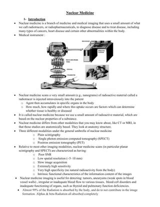
- 1. Nuclear Medicine 1- Introduction Nuclear medicine is a branch of medicine and medical imaging that uses a small amount of what we call radiotracers, or radiopharmaceuticals, to diagnose disease and to treat disease, including many types of cancers, heart disease and certain other abnormalities within the body. Medical instrument : Nuclear medicine scans a very small amount (e.g., nanograms) of radioactive material called a radiotracer is injected intravenously into the patient o Agent then accumulates in specific organs in the body o How much, how rapidly and where this uptake occurs are factors which can determine whether tissue is healthy or diseased It is called nuclear medicine because we use a small amount of radioactive material, which are based on the nuclear properties of a substance. Nuclear medicine differs from other modalities that you may know about, like CT or MRI, in that those studies are anatomically based. They look at anatomy structure. Three different modalities under the general umbrella of nuclear medicine o Plain scintigraphy o Single photon emission computed tomography (SPECT) o Positron emission tomography (PET) Relative to most other imaging modalities, nuclear medicine scans (in particular planar scintigraphy and SPECT) are characterized as having: o Poor SNR o Low spatial resolution (~5–10 mm) o Slow image acquisition o Extremely high sensitivity o Very high specificity (no natural radioactivity from the body) o Intrinsic functional characteristics of the information content of the images Nuclear medicine imaging is useful for detecting: tumors, aneurysms (weak spots in blood vessel walls) , irregular or inadequate blood flow to various tissues, blood cell disorders and inadequate functioning of organs, such as thyroid and pulmonary function deficiencies. Almost 99% of the Radiation is absorbed by the body, and do to not contribute in the image formation. Alphas & beta Radiation all absorbed completely
- 2. 2- Radioactivity The process of transmutation of an unstable element to another element by the emission of radiation termed disintegration. Radioactive Decay: For every radioactive element a probability of decay exists. The relationship between this probability and the number of atoms in a large sample that will decay in a given time is called radioactive decay “note: decay constant λ, It has a characteristic value for each nuclide. It also reflects the nuclide’s degree of instability; a larger decay constant connotes a more unstable nuclide (i.e., one that decays more rapidly) The activity of a sample is defined as the number of disintegrations per second (dps) and is given by Radioactivity is measured o Curies (Ci), where one curie equals 3.73*1010 disintegrations per second. Historically, this is the number of disintegrations per second from 1 gramme of radium (226 Ra) and is named after Pierre Curie. o Commercially, the most common units are millicuries (mCi), 1/1000 of a curie or 3.73*107 disintegrations per second. o Bequerel (Bq) : SI Unit of activity = 1 disintegrations per second Commonly expressed as KBq or MBq or GBq =λNo 𝑒−λt
- 3. 3- Decay Types A: Mass no. = no. of protons + neutrons Z: atomic no = no. of protons
- 4. Or Neutron decay 1 0 1 1 -1 0
- 5. 4- The technetium generator : A technetium generator is delivered to a nuclear medicine department at the beginning of the week, and is returned at the end of the week. It is a very convenient method for producing 99m Tc. Typically, the technetium is eluted every 24 hours, and the generator is replaced once a week. Most nuclear medicine scans are therefore scheduled for the beginning of a week. An on-site technetium generator comprises an alumina ceramic column with radioactive 99 Mo absorbed on to its surface in the form of ammonium molybdate, (NH4)2MoO4. The column is housed within a lead shield for safety. A schematic and photograph of a Tc-generator are shown, the 99Mo decays into 99mTc which in turn decays into 99gTc, the ground state of Tc . At any given time the generator column contains a mixture of 99 Mo, 99m Tc and 99g Tc. To remove the 99m Tc selectively a vial with physiological saline is placed at the input to the column and a needle and empty vial at the outlet. The saline is drawn through the column and washes out most of the 99mTc which does not bind strongly to the column and is eluted in the form of sodium pertechnetate: roughly 80% of the available 99mTc is eluted
- 6. Problems: 1- Two patients undergo nuclear medicine scans. One receives a dose of radiotracer A and the other radiotracer B. The half-life of A is 6 hours and of B is 24 hours. If the administered dose of radiotracer A is three times that of radiotracer B, and the biological half-lives of A and B are 6 and 12 hours respectively, at what time is the radioactivity in the body the same for the two patients? 2- (a) 𝑇𝐼81 201 has a decay constant of 9.49 × 10⁻³ hr−1. Find the activity in becquerels (Bq) of a sample containing 1010 atoms. (b) How many atoms of 𝐶6 11 with a decay constant of 2.08 hr−1 would be required to obtain the same activity as the sample in Part a? 3- (a) A sample of 113m In has a mass of 2 µg. How many 113m In atoms are present in the sample? (b) How many 113m In atoms remain after 4 hours have elapsed? (The physical half-life is 1.7 hours for 113m In) (c) What is the activity of the sample when t = 4.0 hours? (d) Enough 113m In must be obtained at 4 p.m. Thursday to provide 300 kBq at 1 p.m. Friday. How much 113m In should be obtained? 4- (a) In a sample of 20 000 atoms, if 400 decay in 8 seconds what is the radioactivity, measured in mCi, of the sample? (b) In order to produce a level of radioactivity of 1 mCi, how many nuclei of 99m Tc (λ = 3.223*10-5 s-1 ) must be present? What mass of the radiotracer does this correspond to? (Avogadro’s number is 6.0231023 ). (c) A radioactive sample of 99m Tc contains 10 mCi activity at 9 am. What is the radioactivity of the sample at 12 pm on the same day? 5- A dose of 1 mCi of 99mTc is administered to a patient. Calculate the total dose to the patient if the biological half-life of the radiotracer in the body is: (a) 2 years, (b) 6 hours, (c) 2 minutes. 6- In the technetium generator, show mathematically that if λ2 >> λ1 , the radioactivities of the parent and daughter nuclei become equal in value at long times . 7- For an 15 O PET scan, if an initial dose of 1 mCi is injected, calculate the total number of c- rays that are produced over a scan time of 4 minutes, assuming that scanning starts immediately after injection, and that clearance from the body is negligible. ( τ1/2=124.2 sec) References : Chapter 3 :Introduction to Medical Imaging Physics, Engineering and Clinical Applications , Chapter 3 : MEDICAL IMAGING PHYSICS , William R. Hendee,