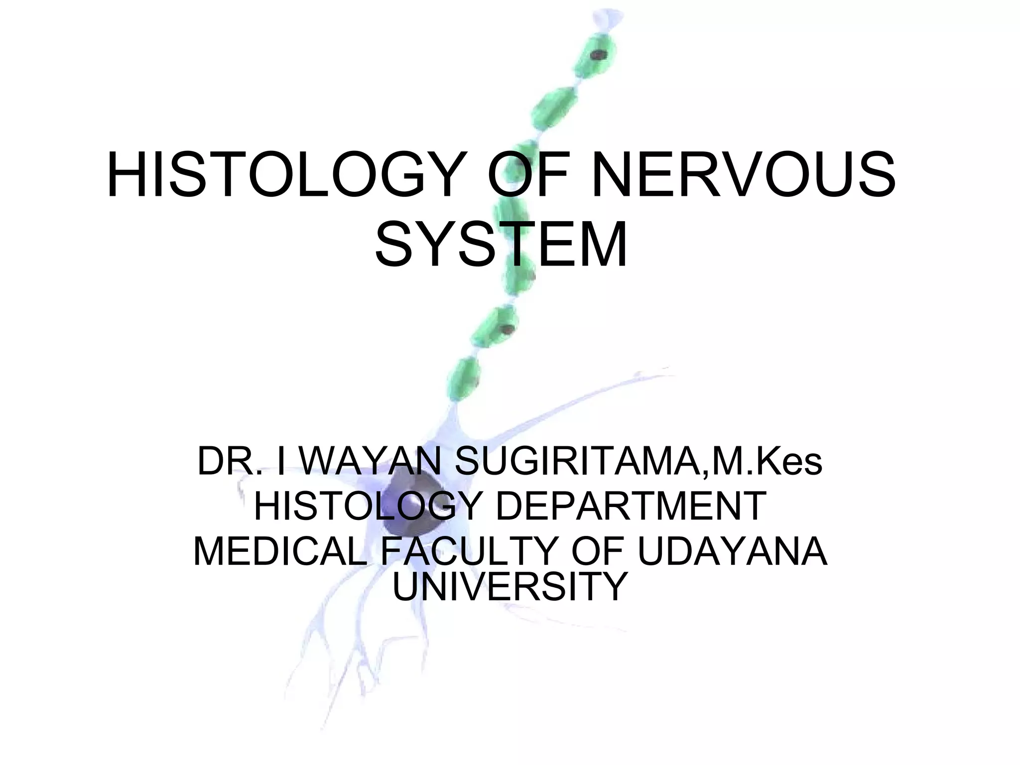
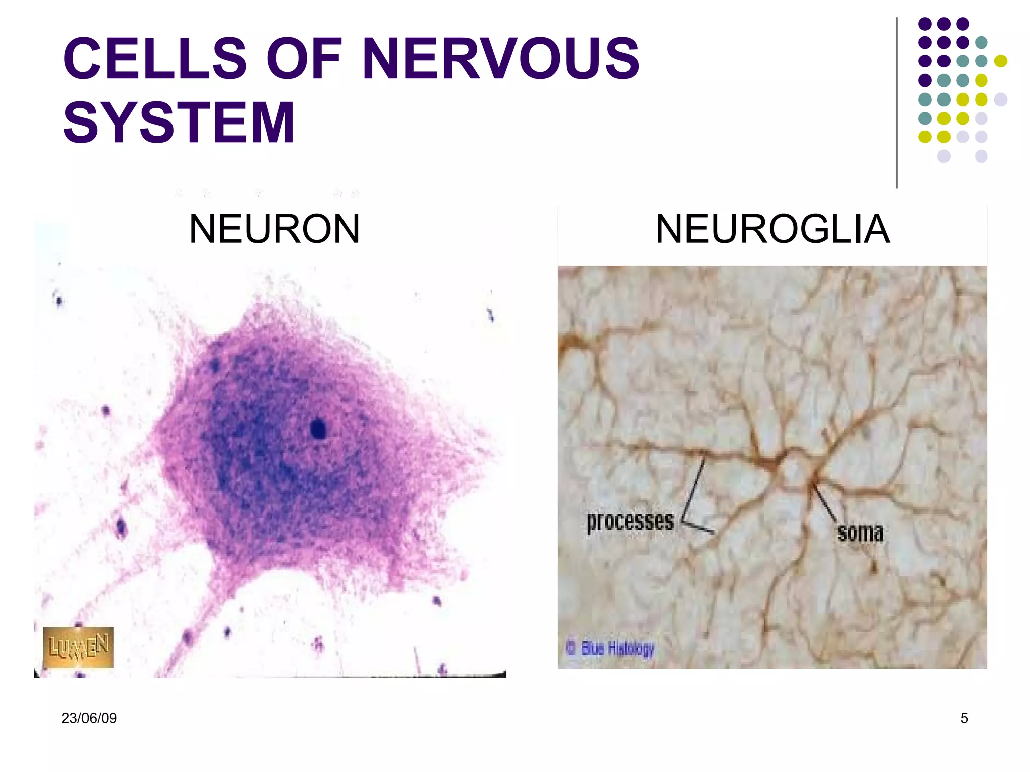
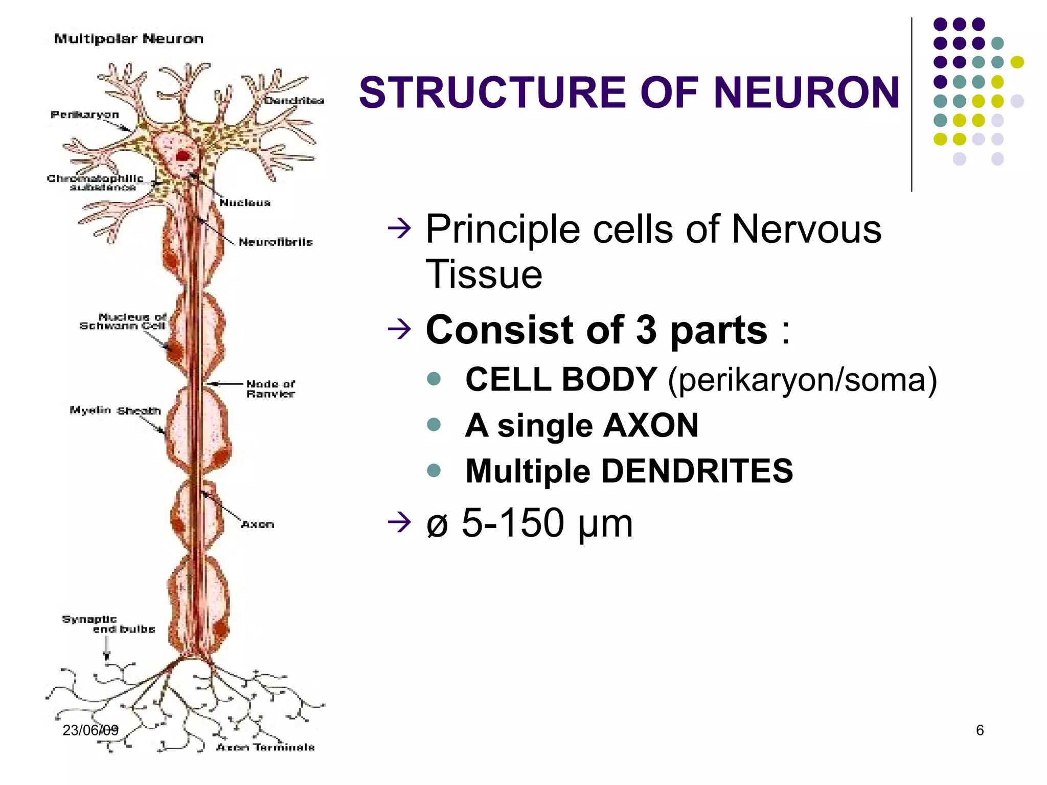
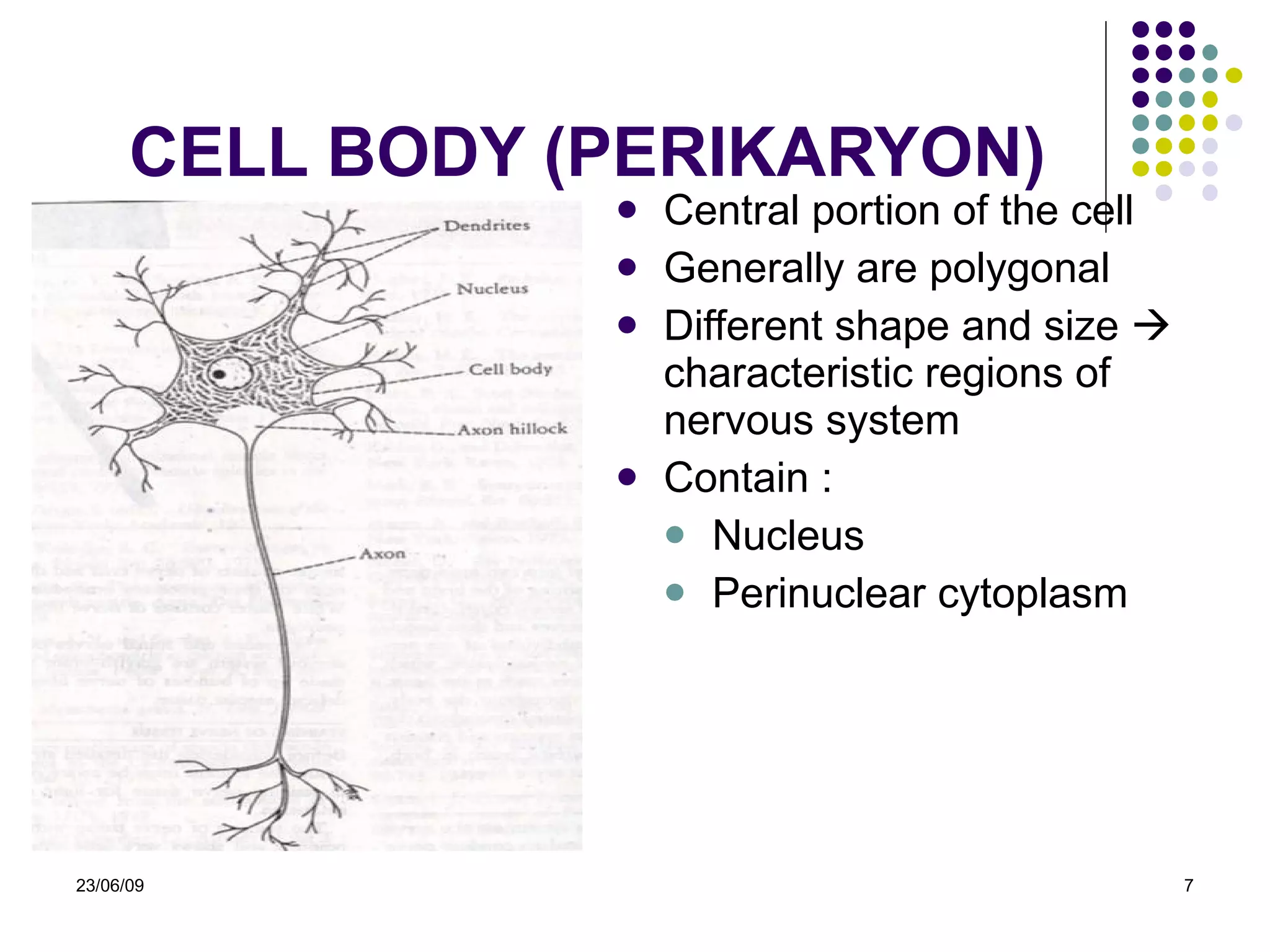
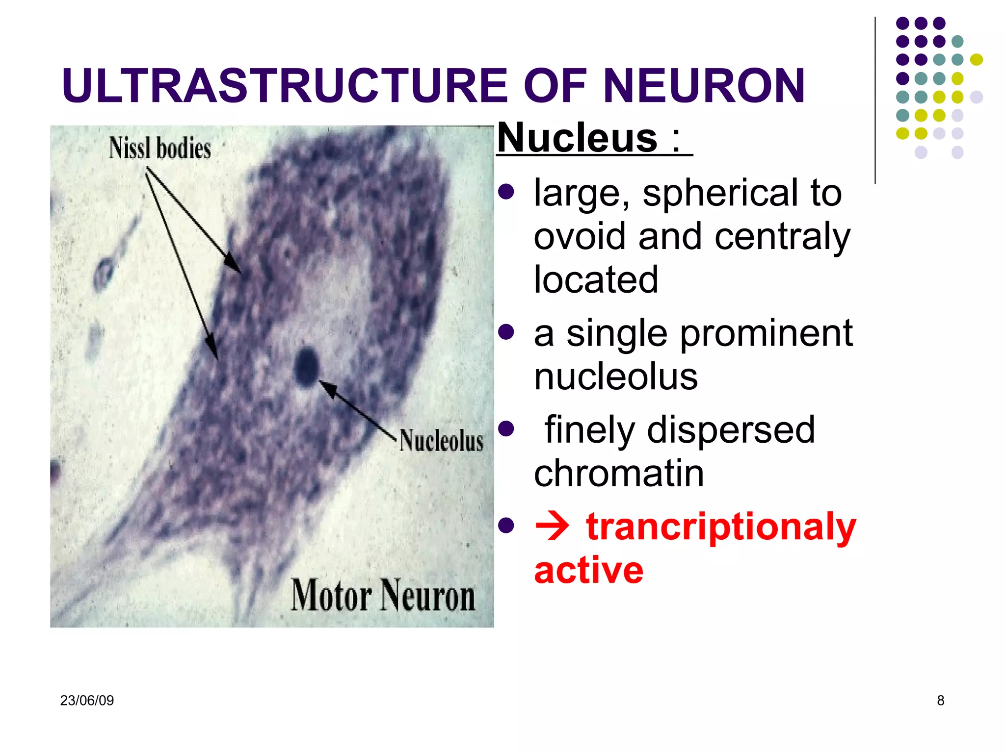
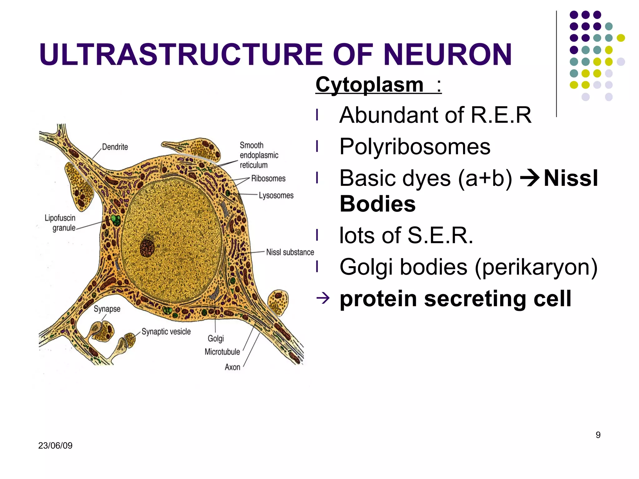
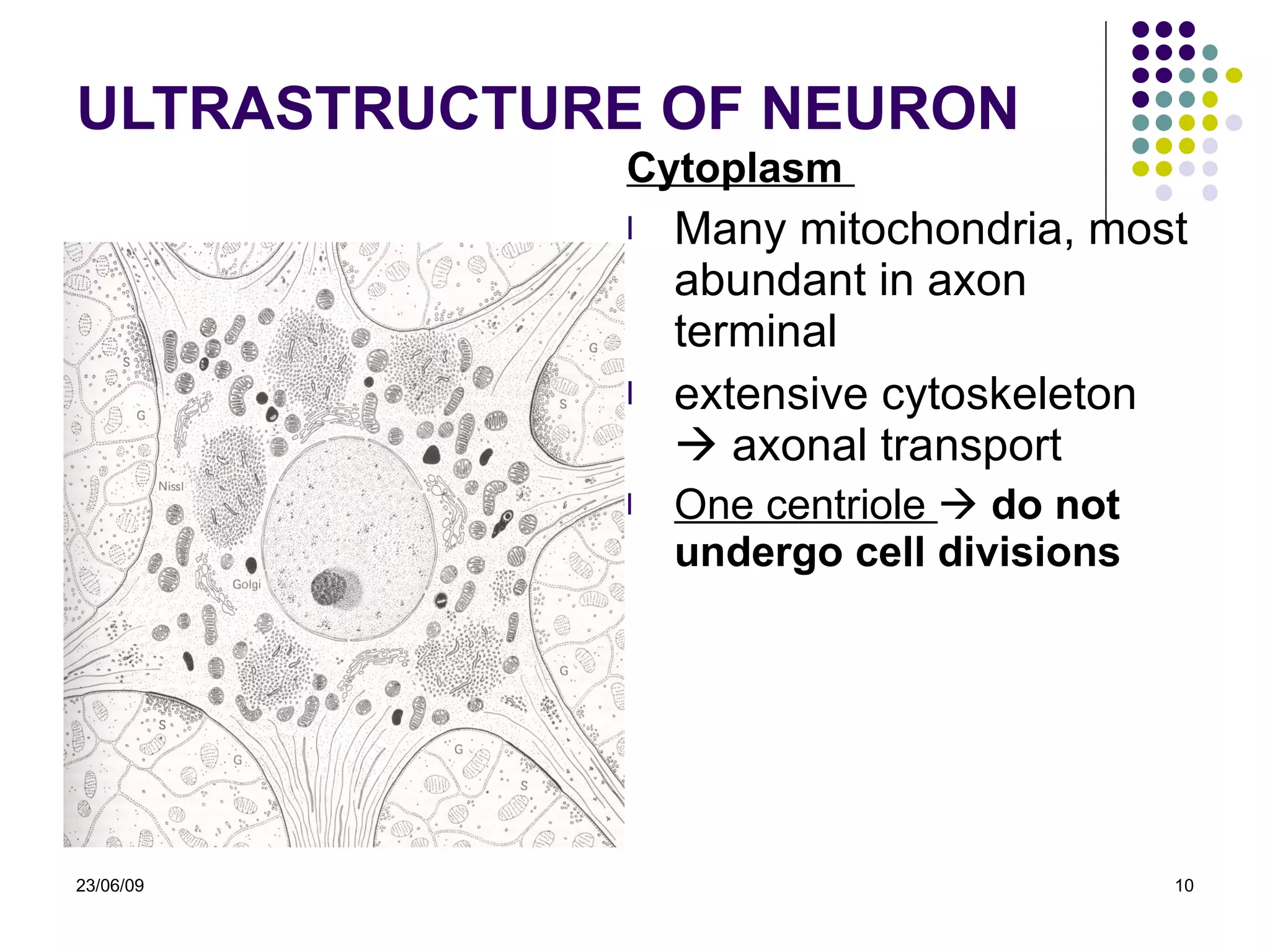
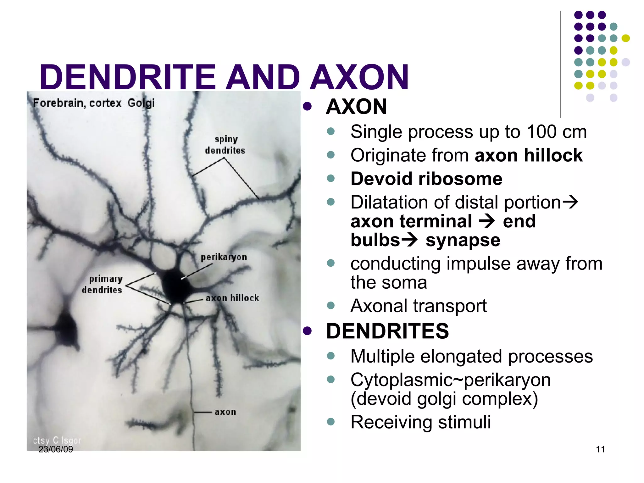
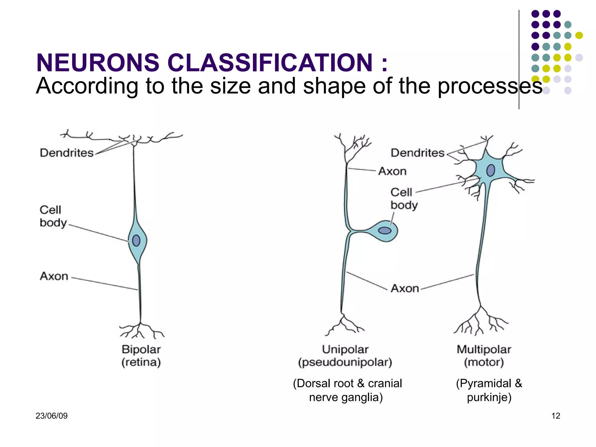
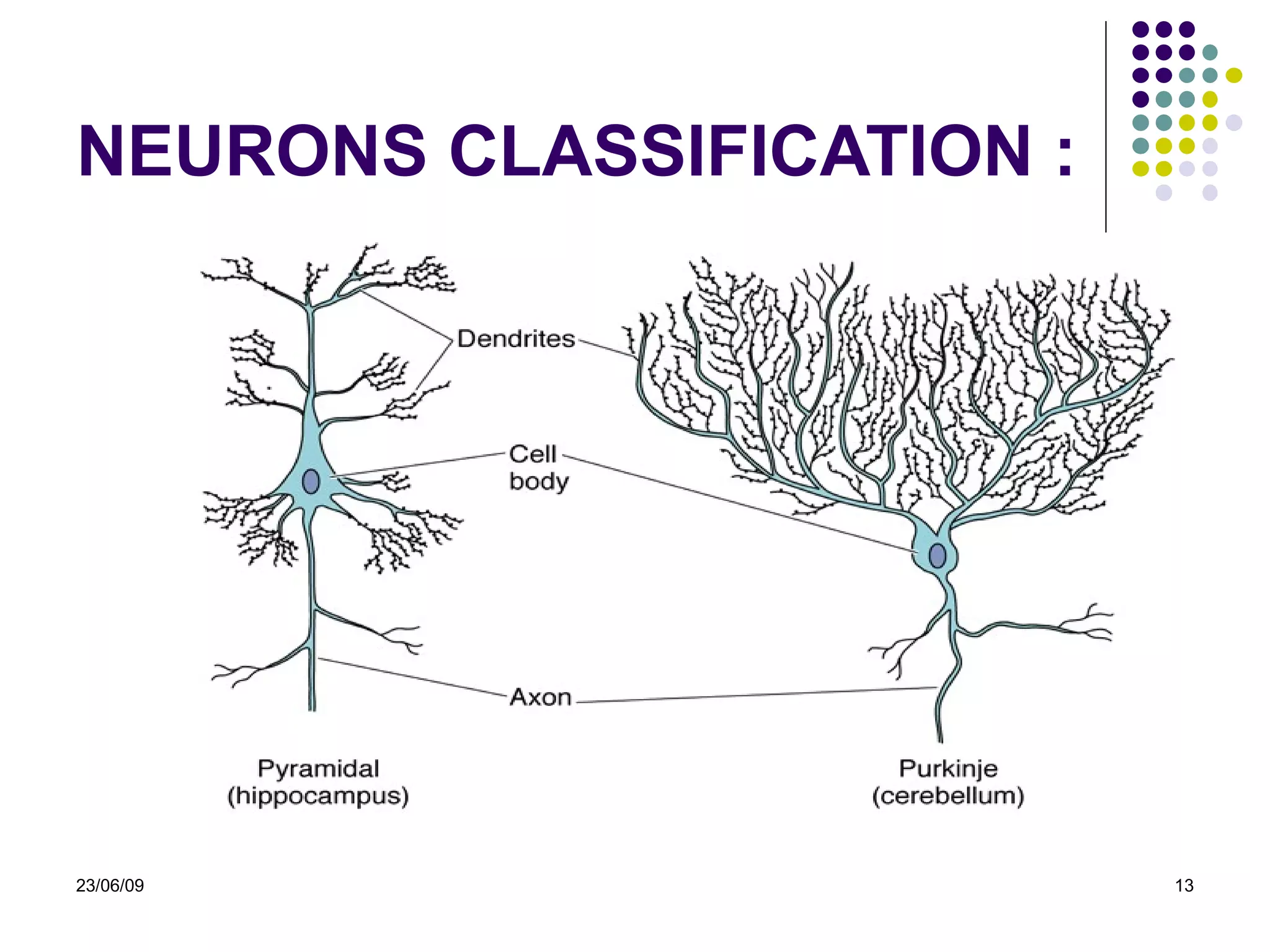
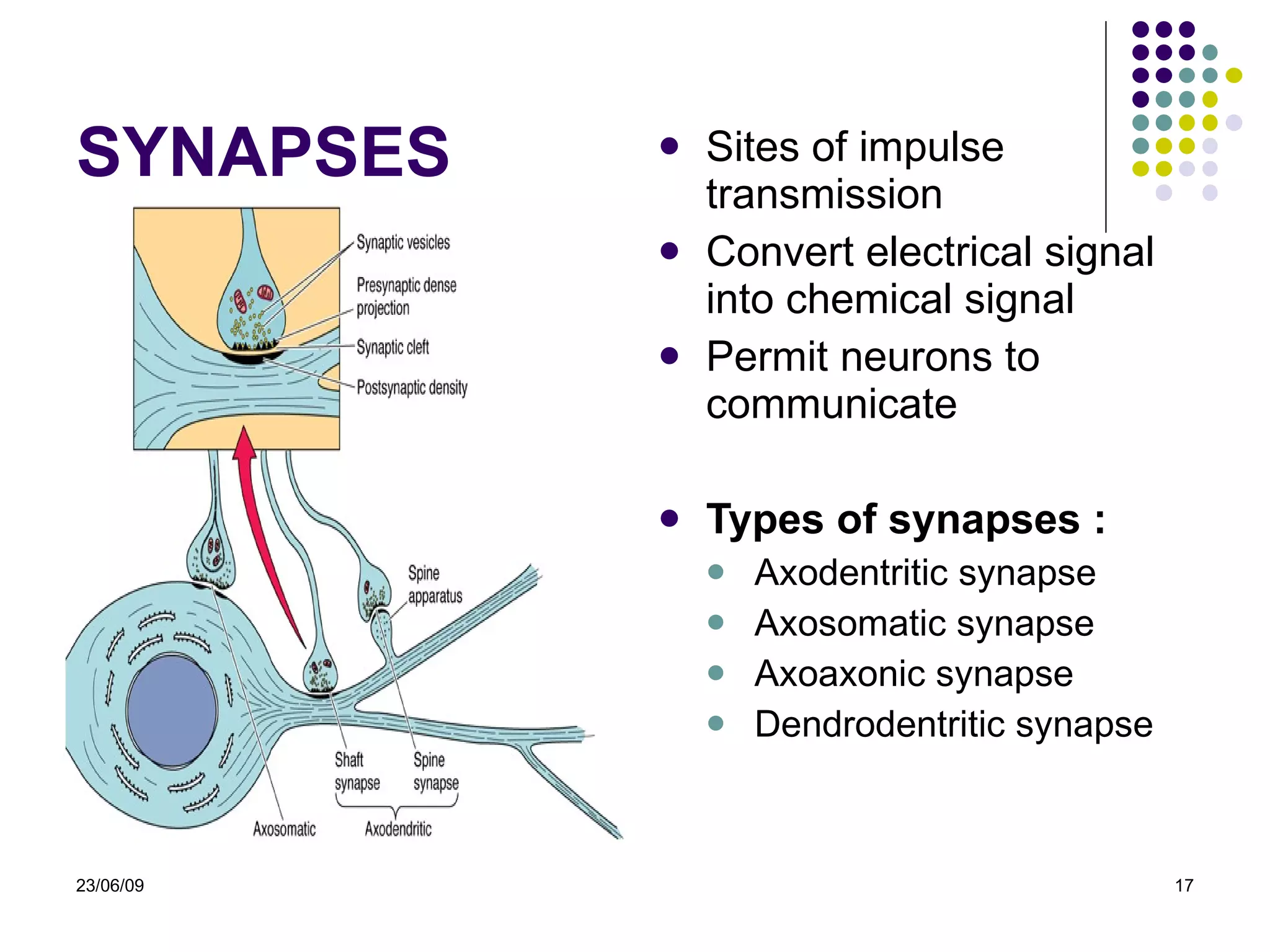
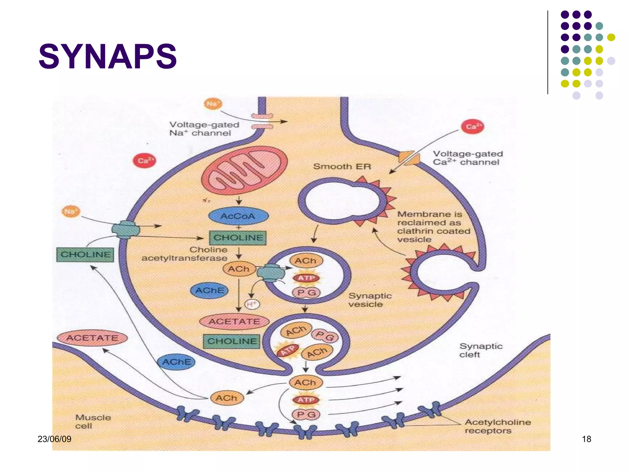
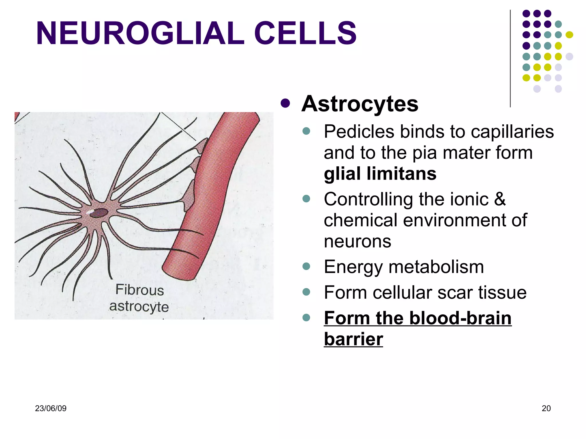
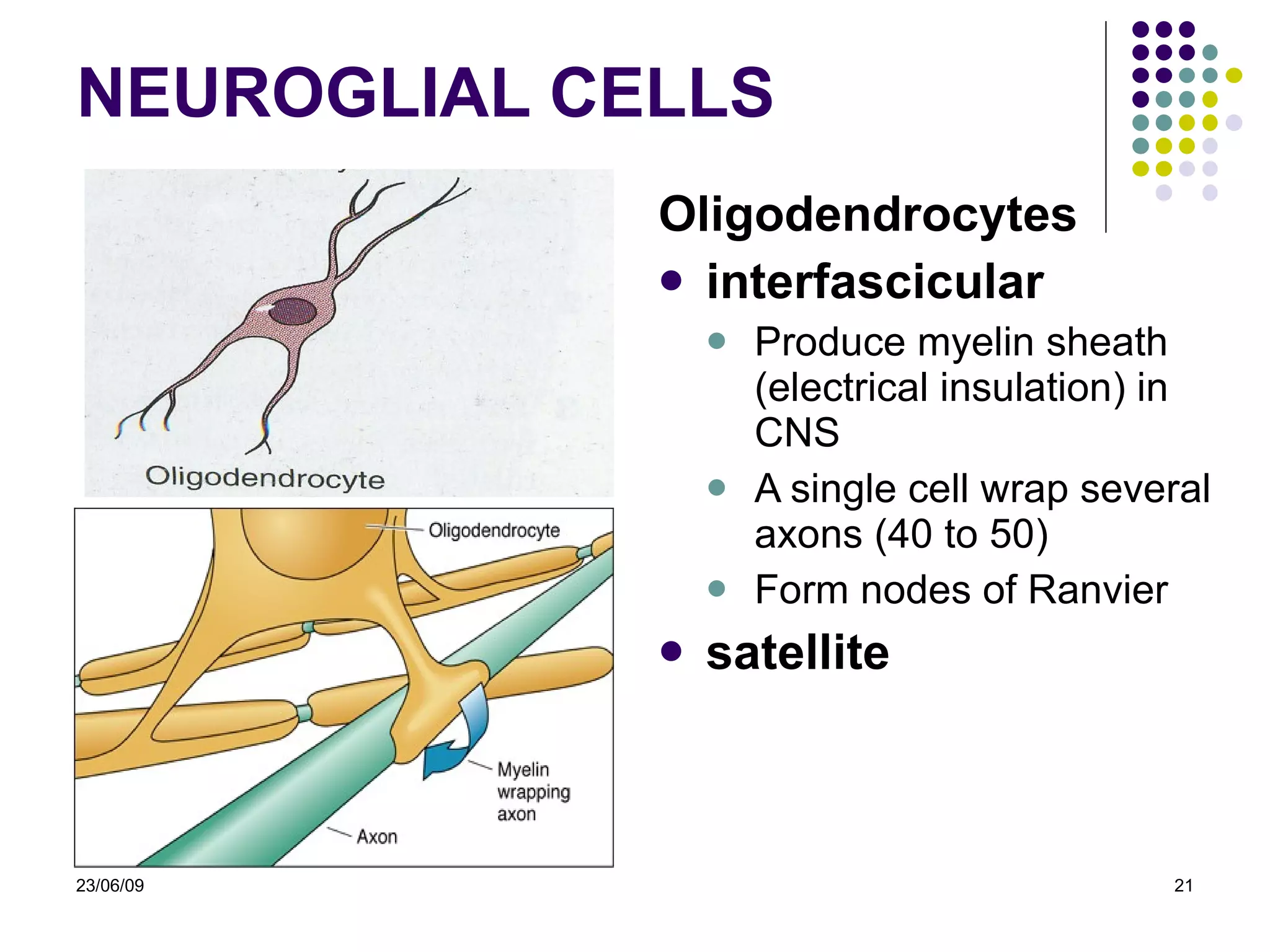
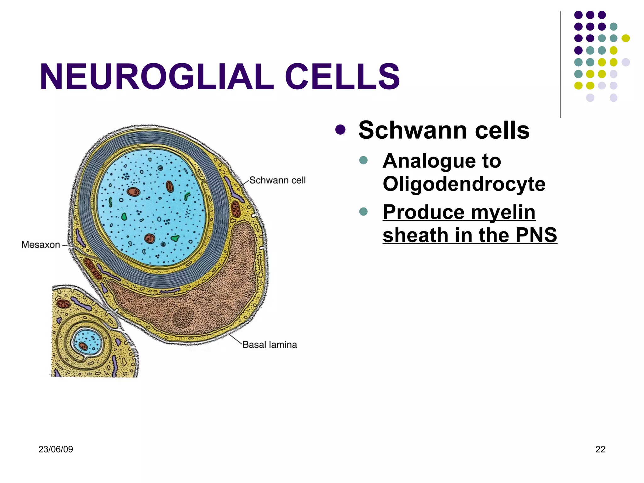
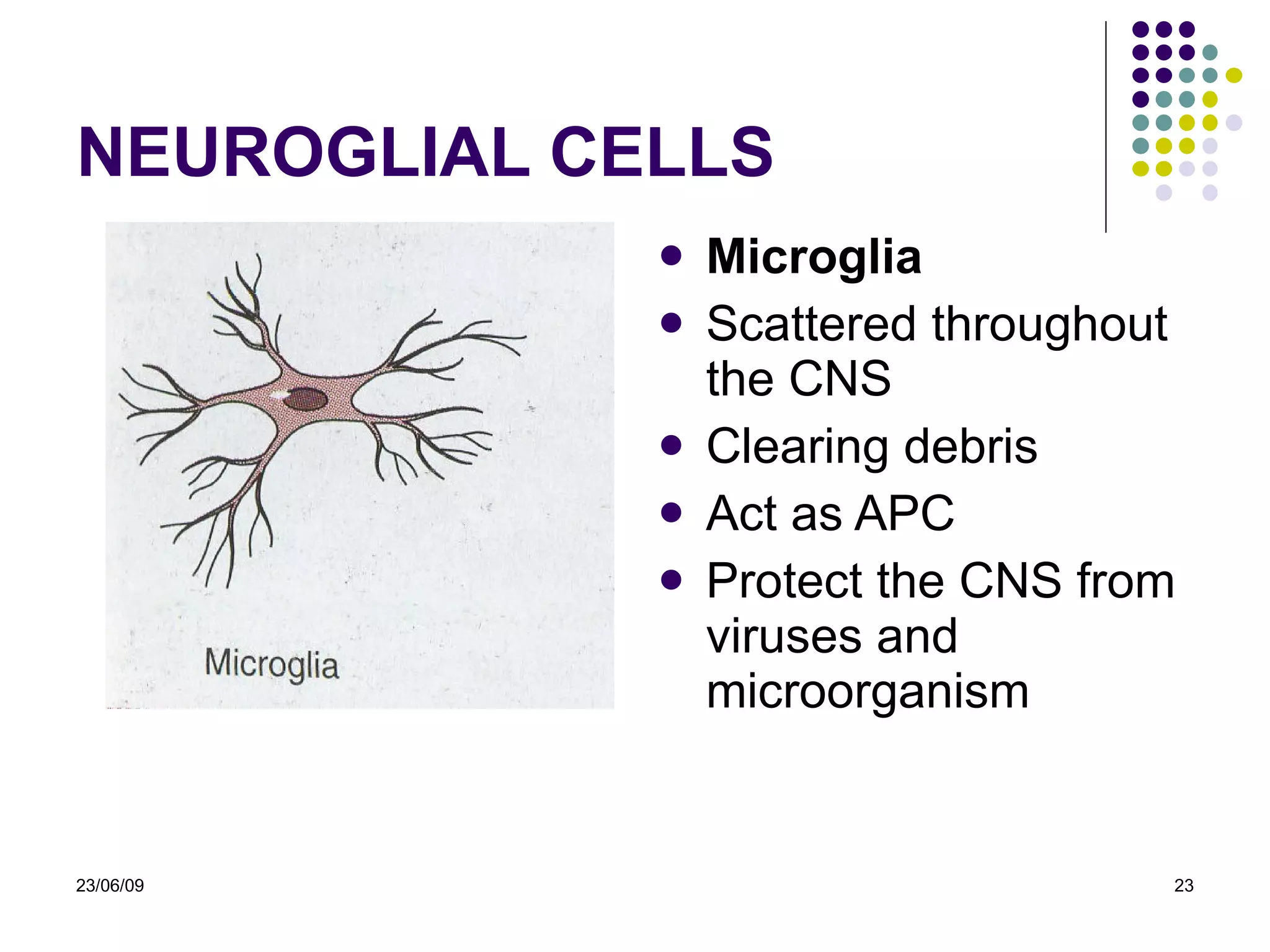
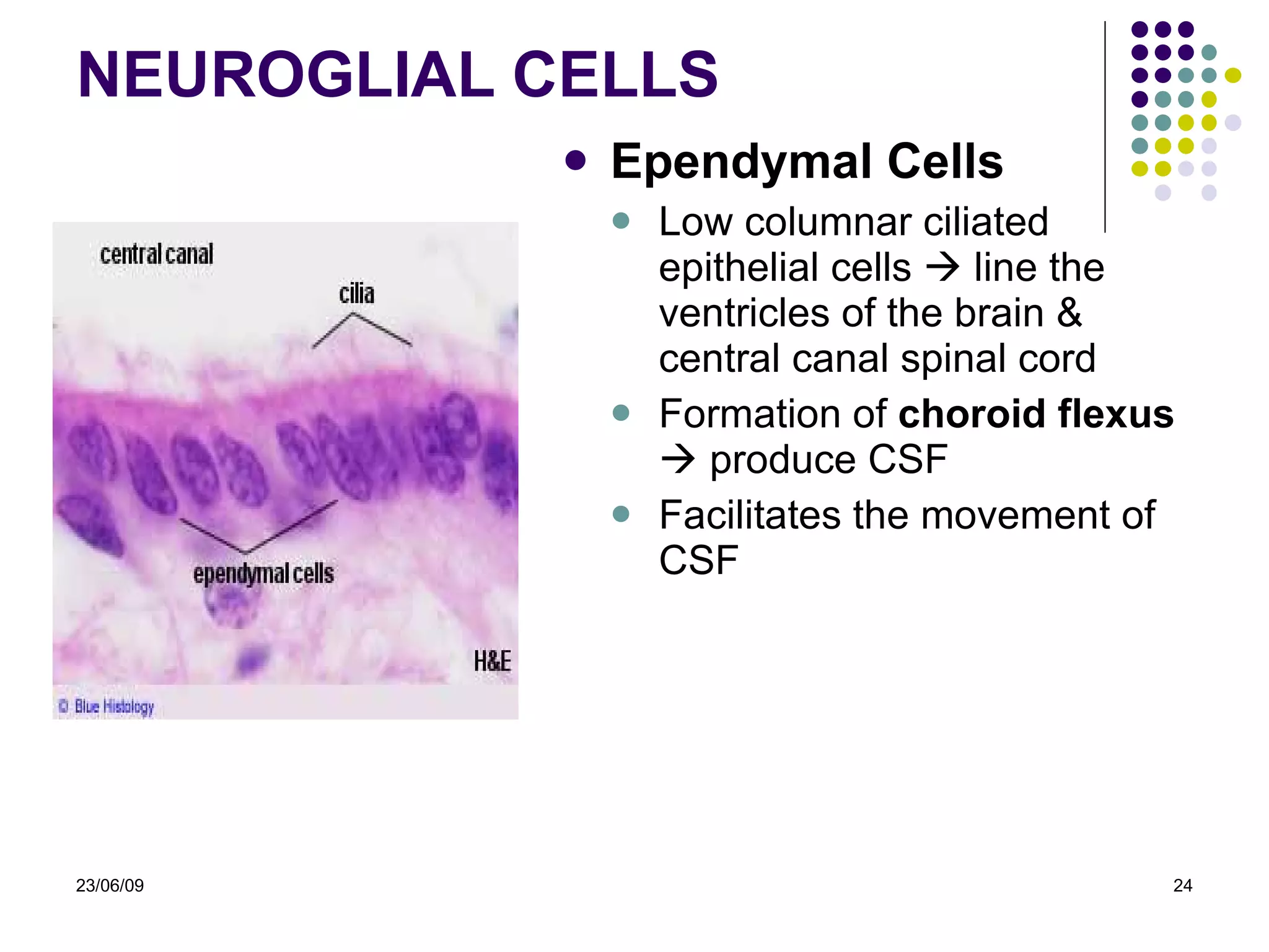
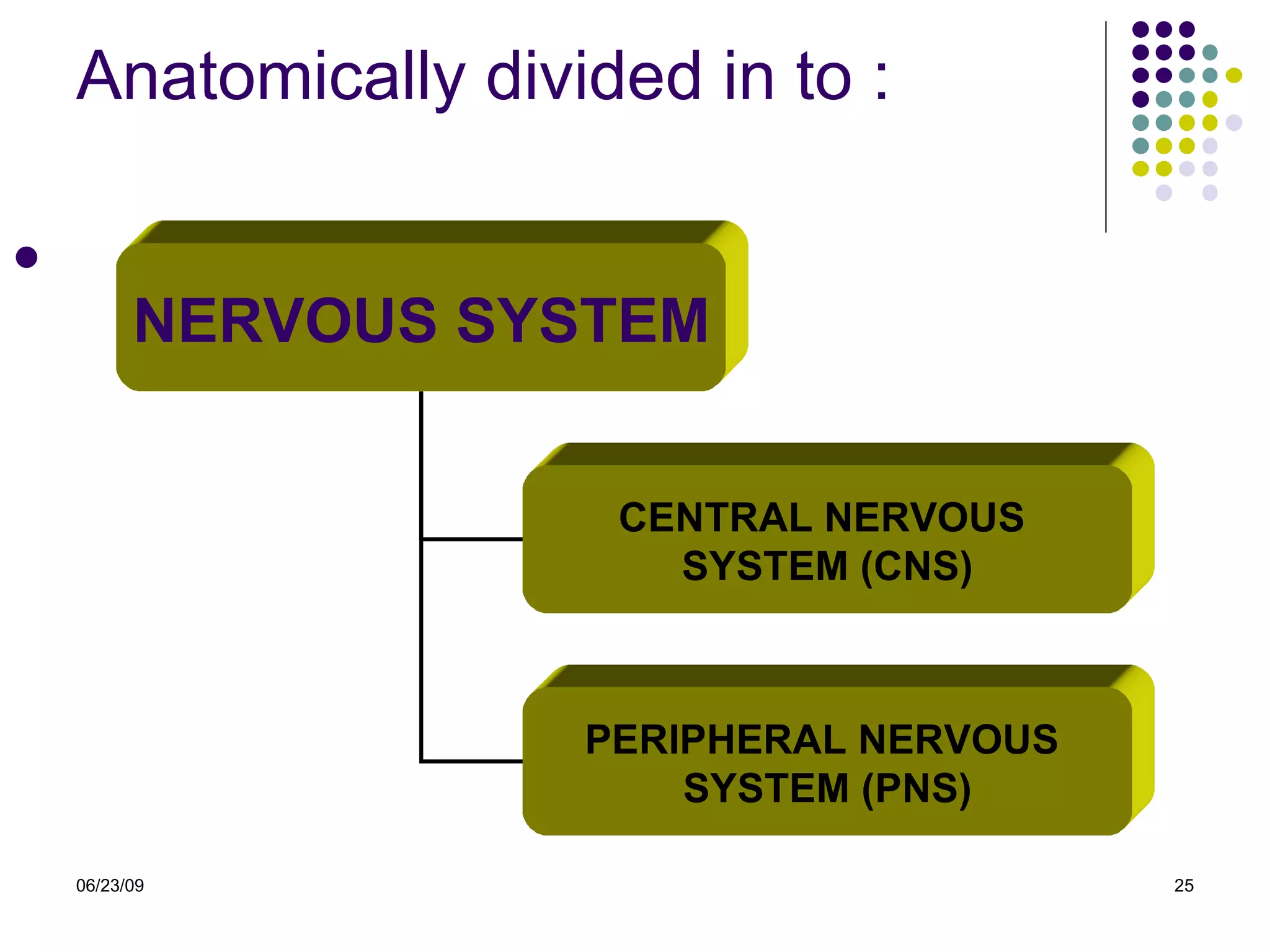
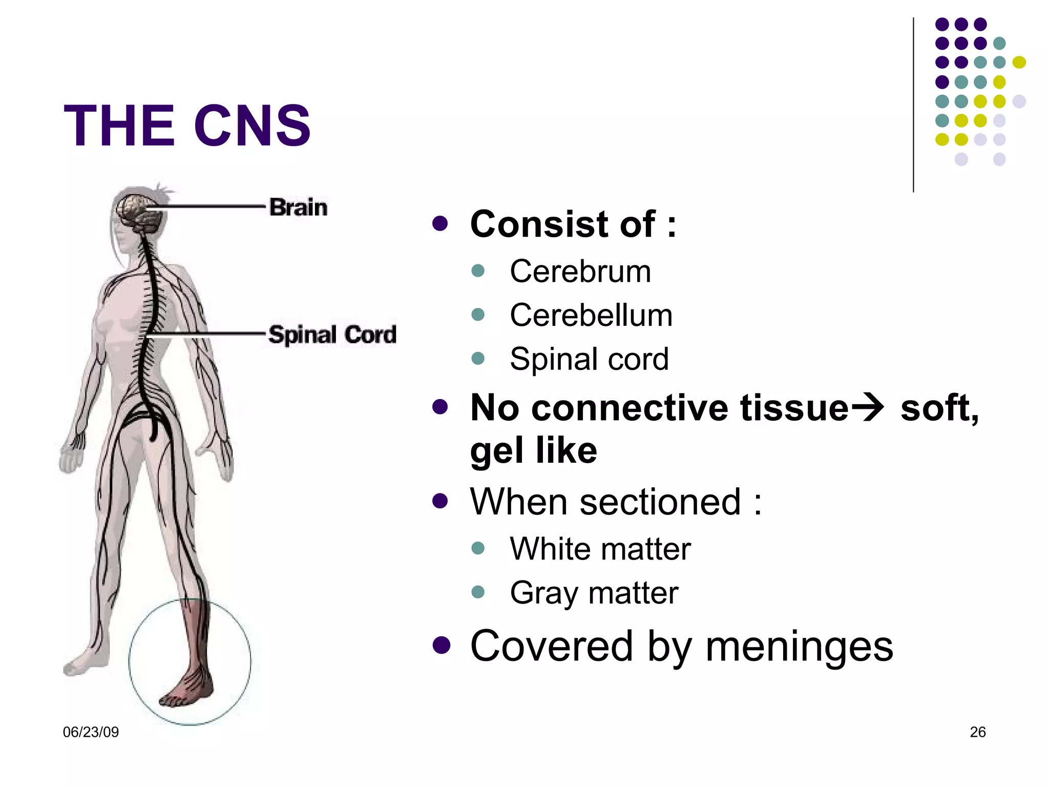
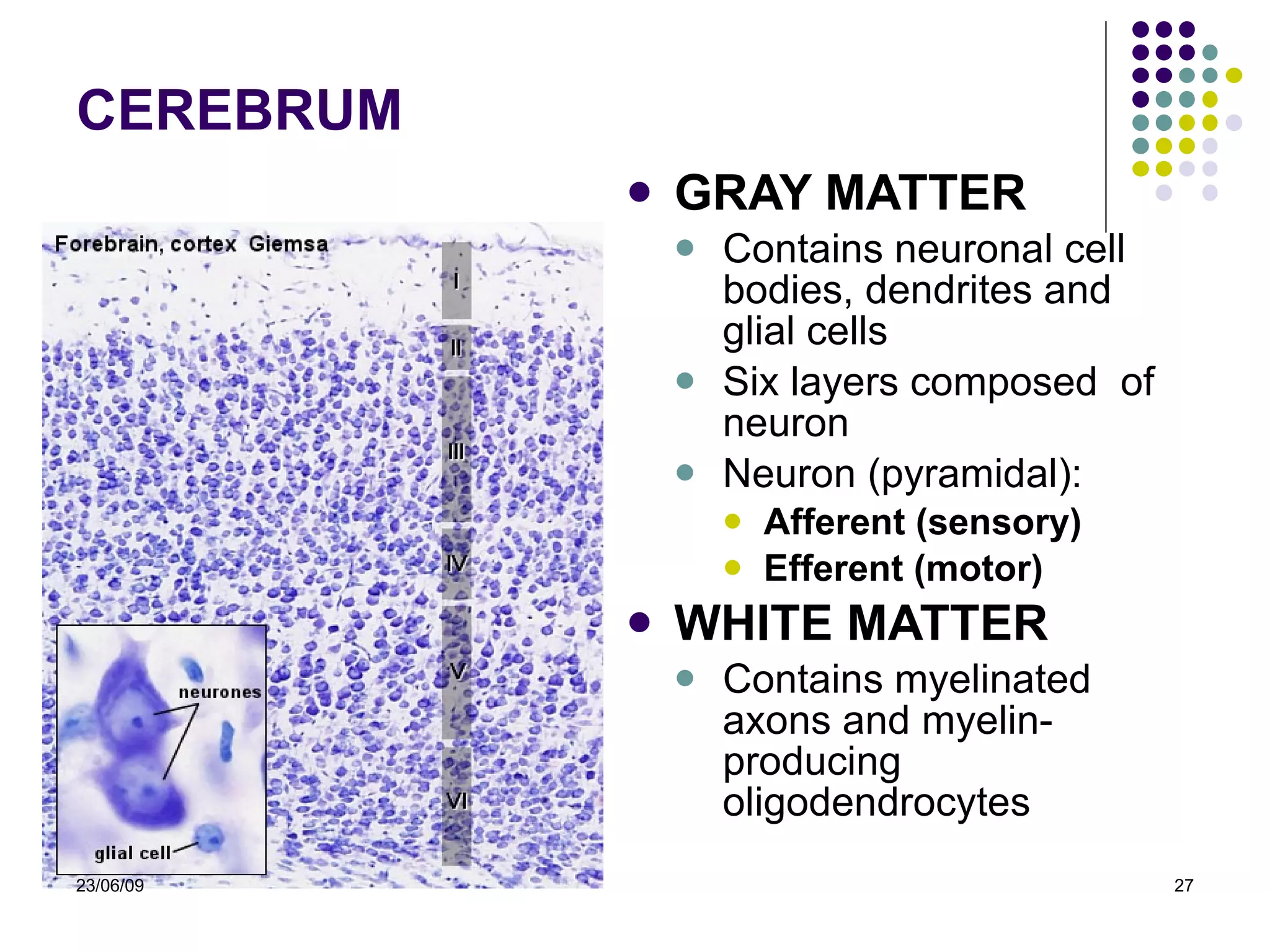
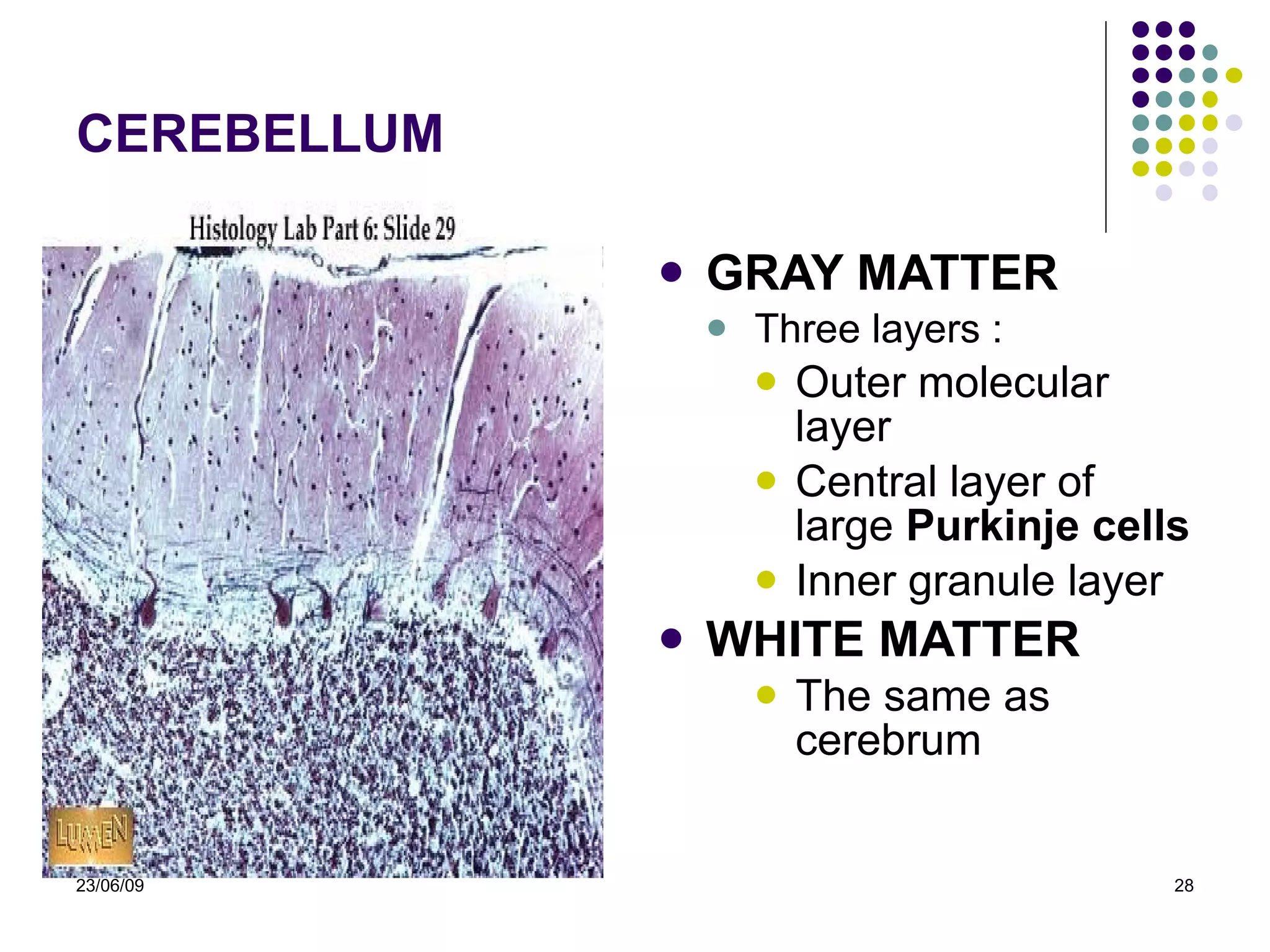
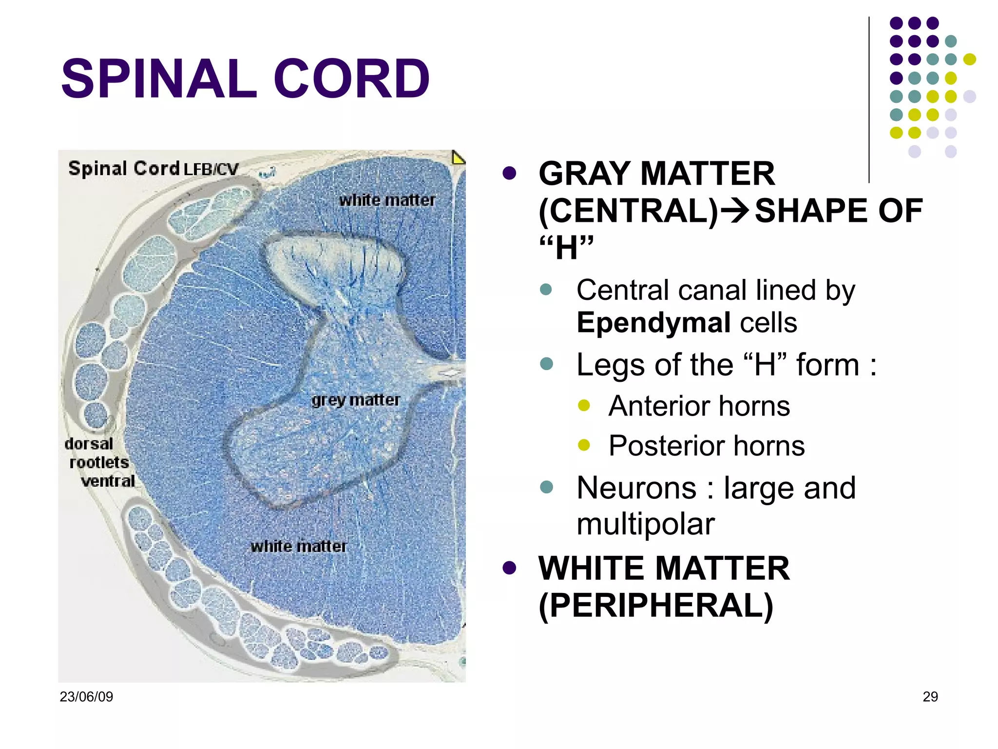
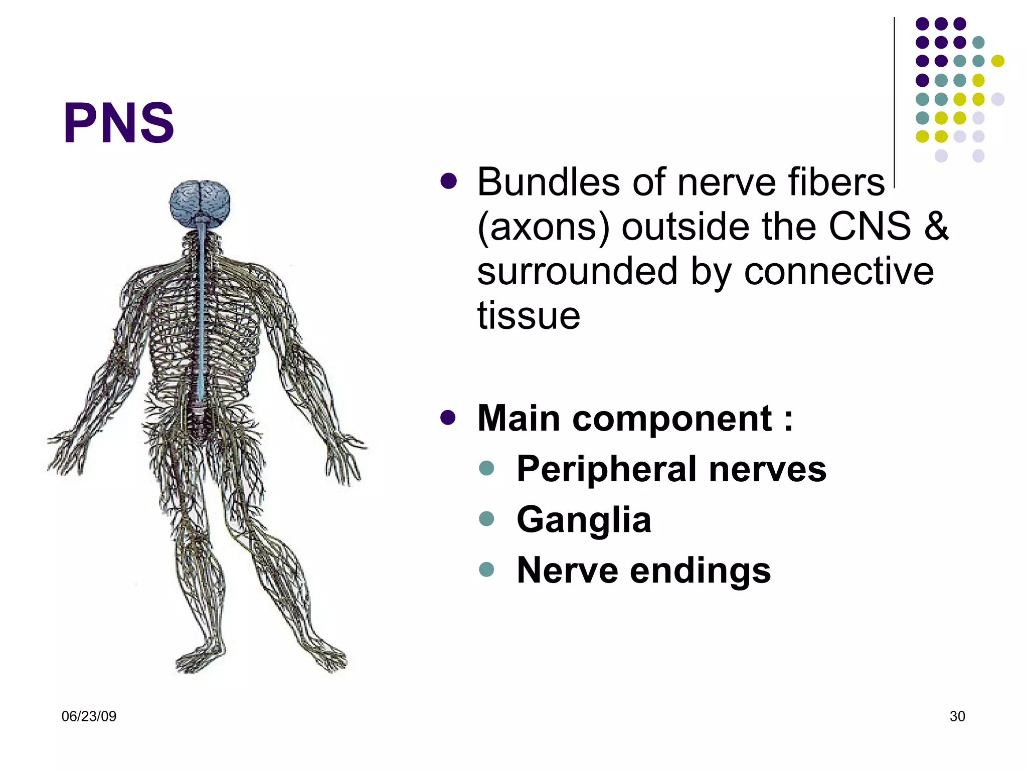
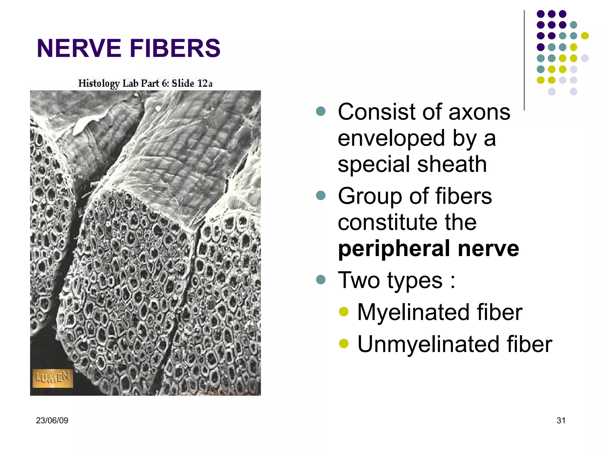
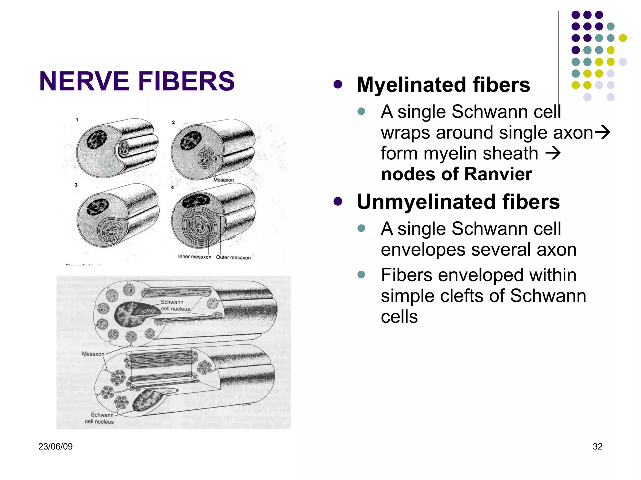
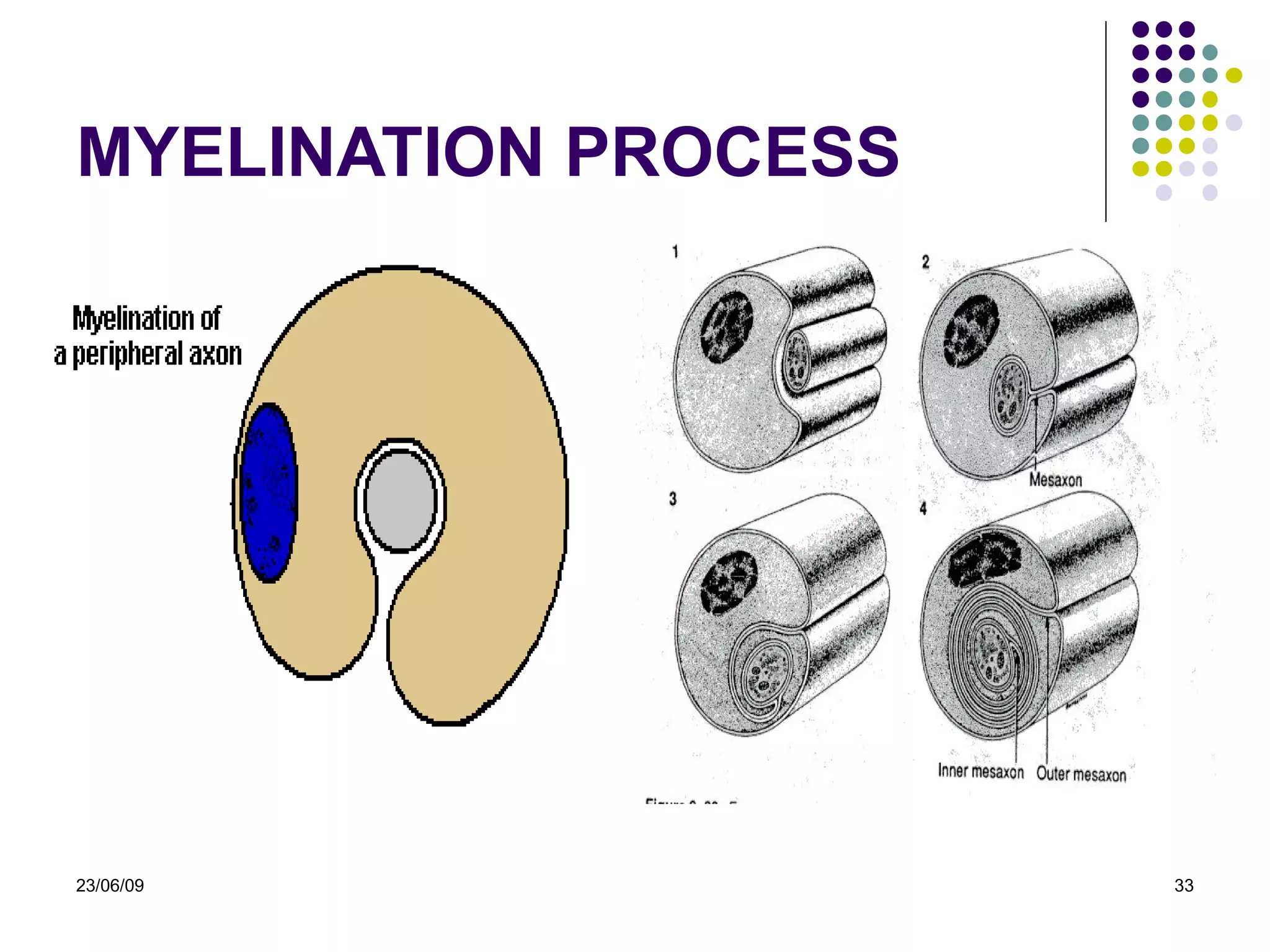
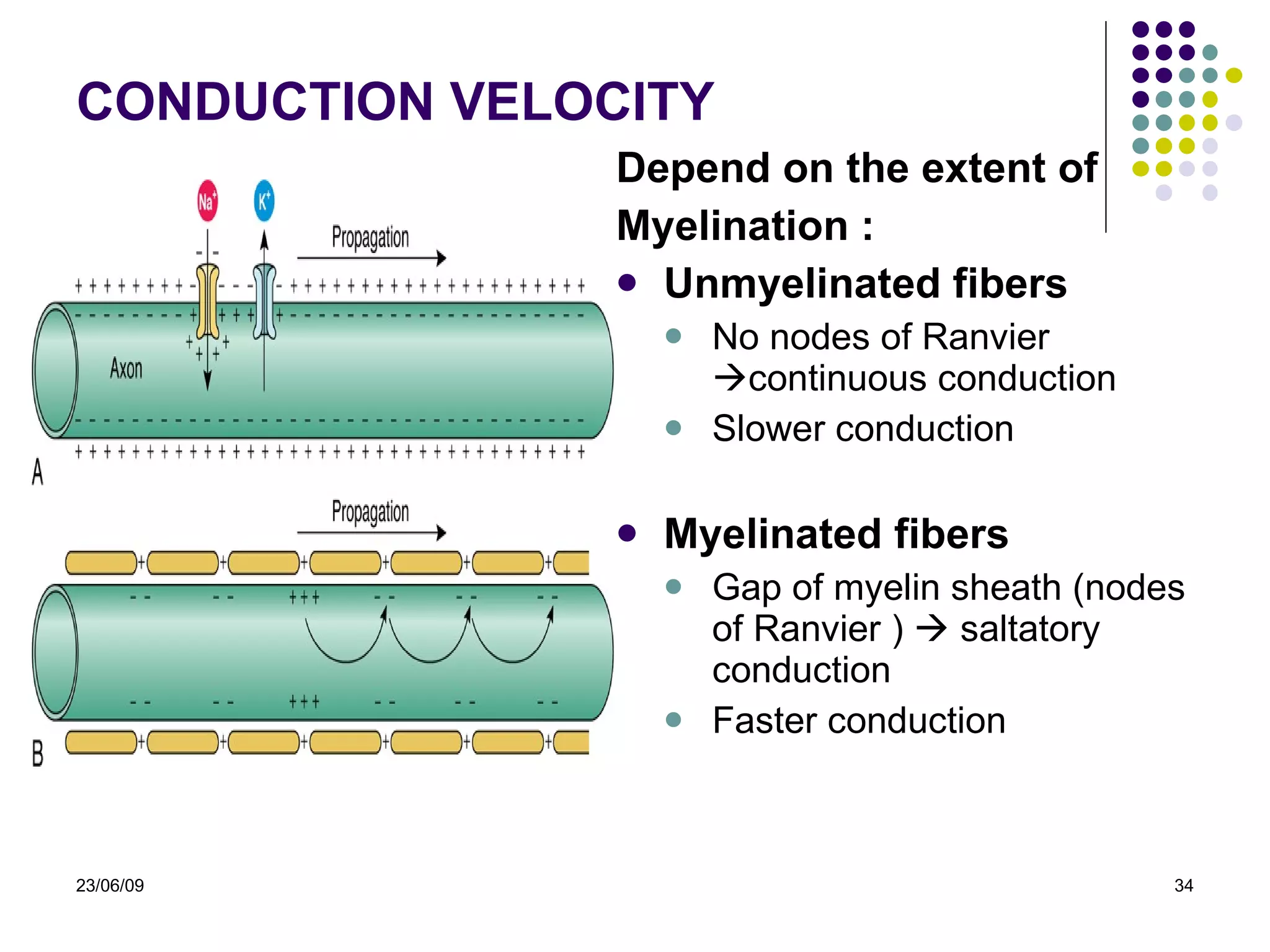
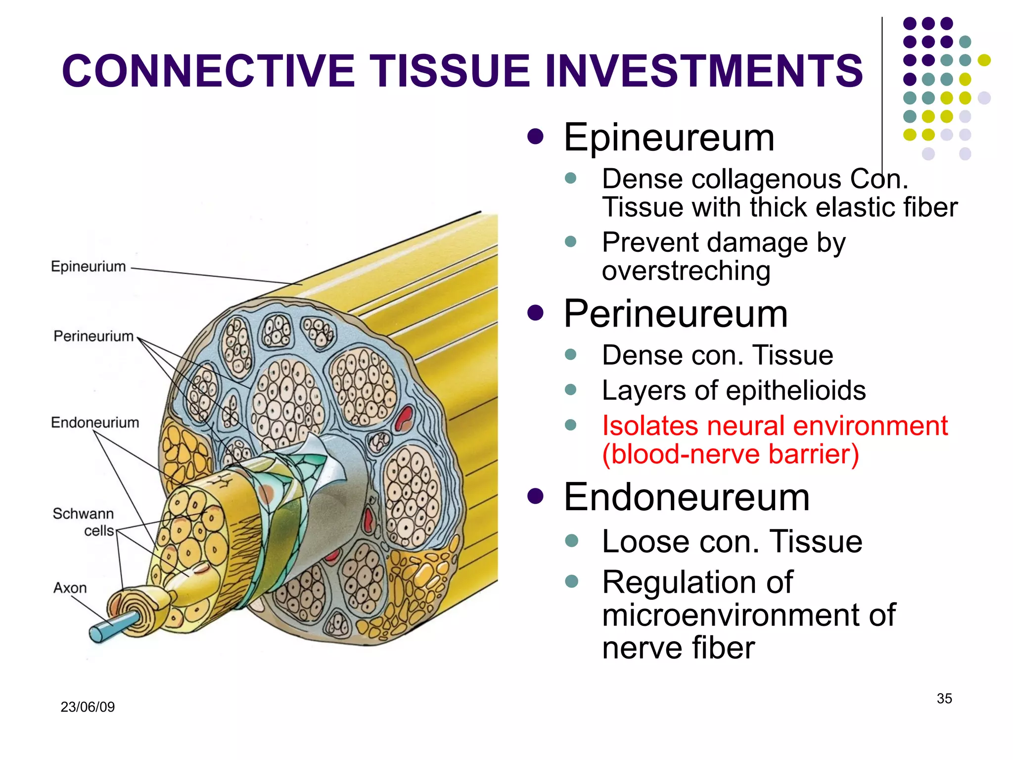
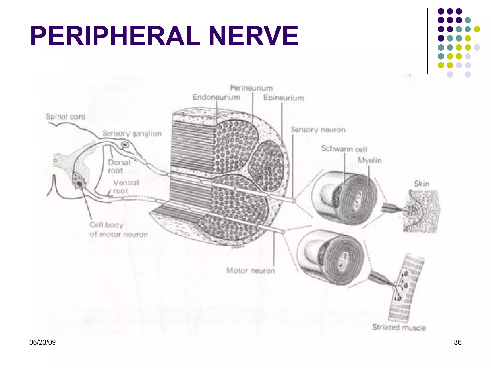
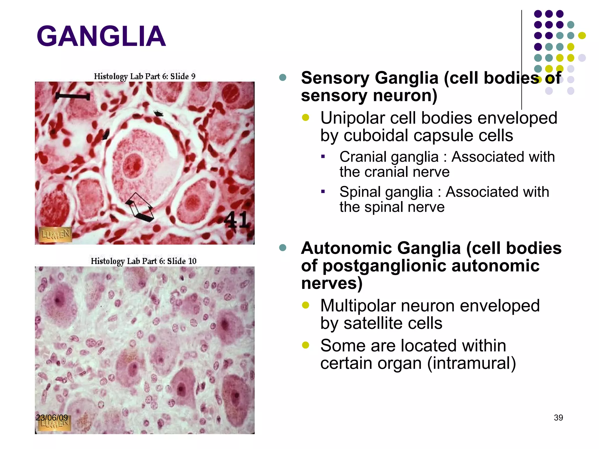
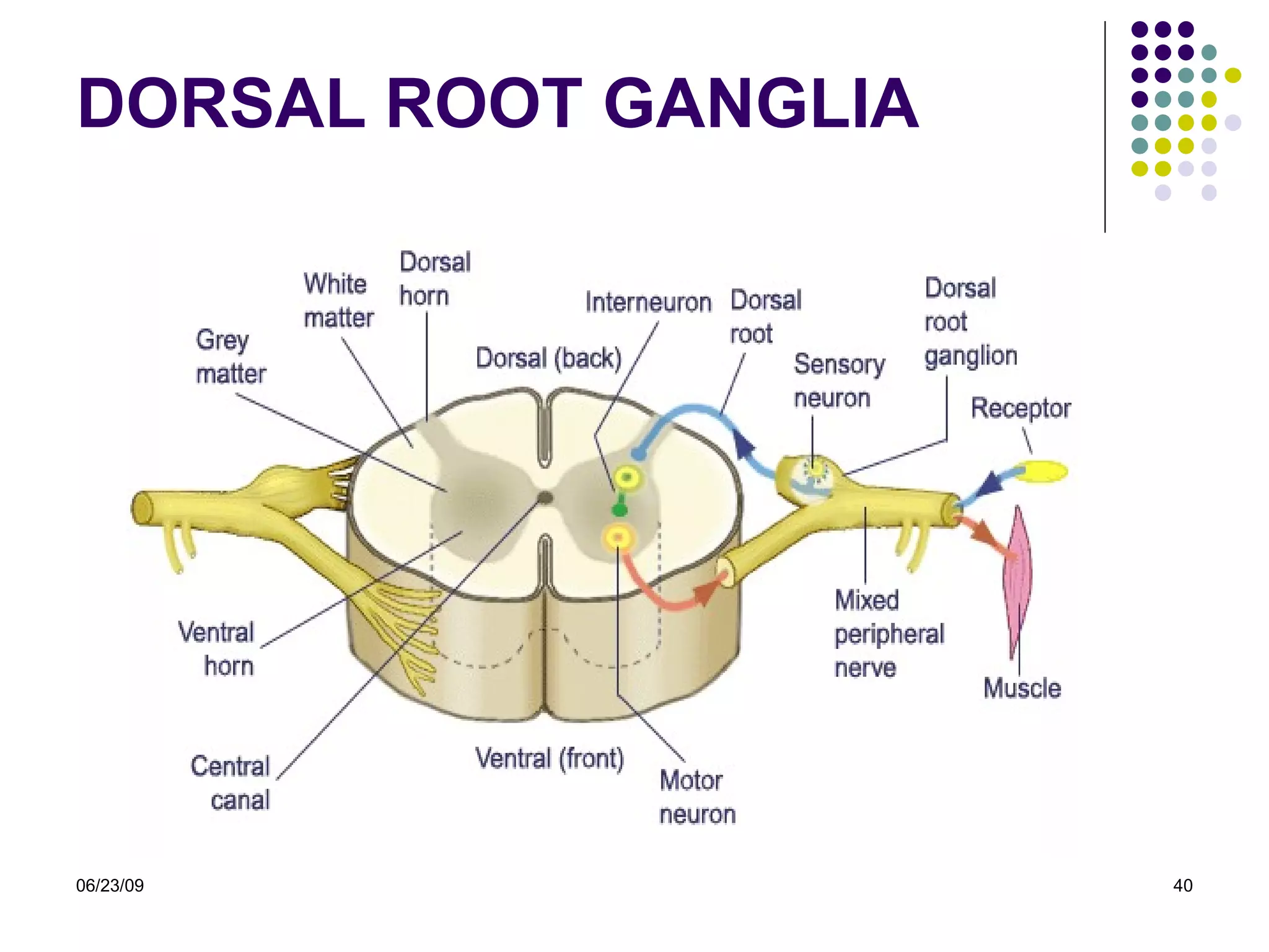
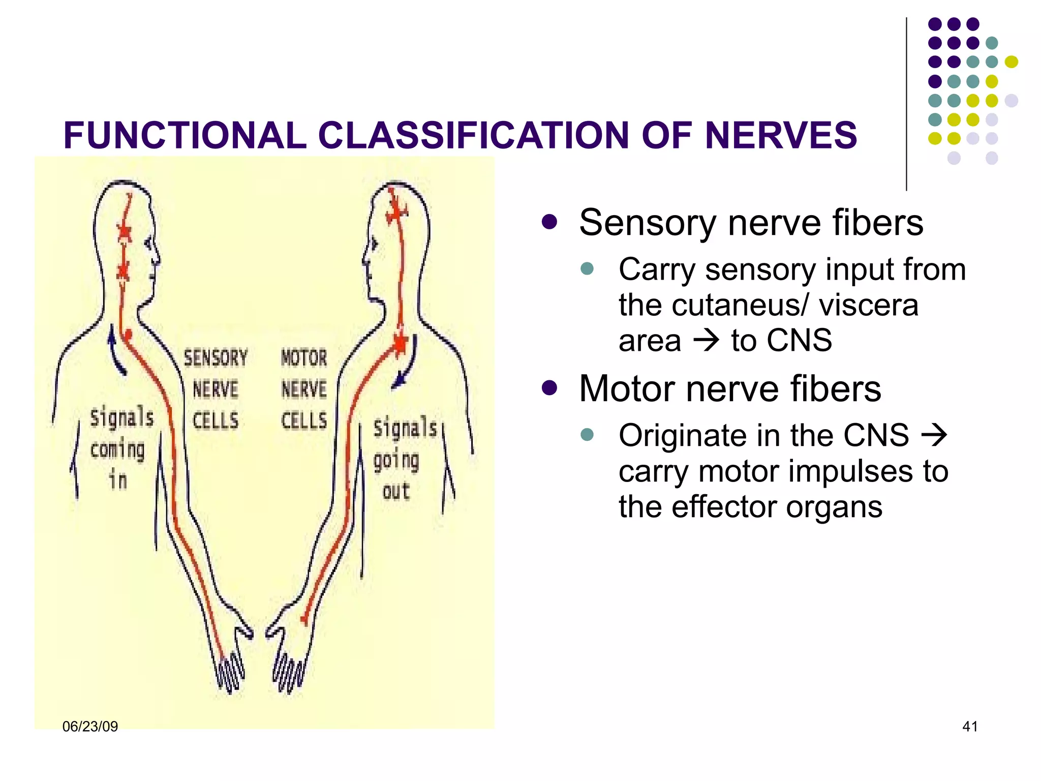
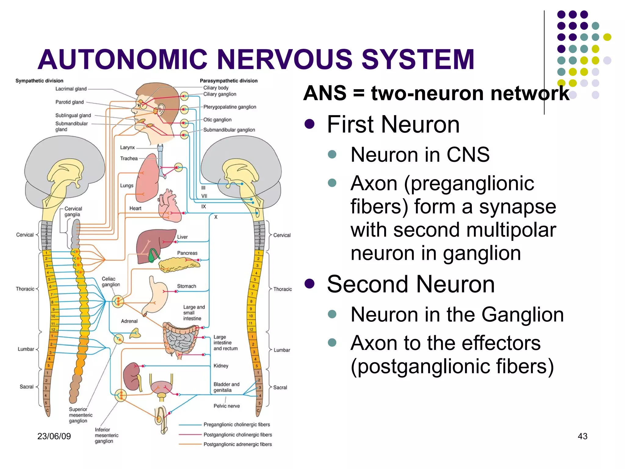
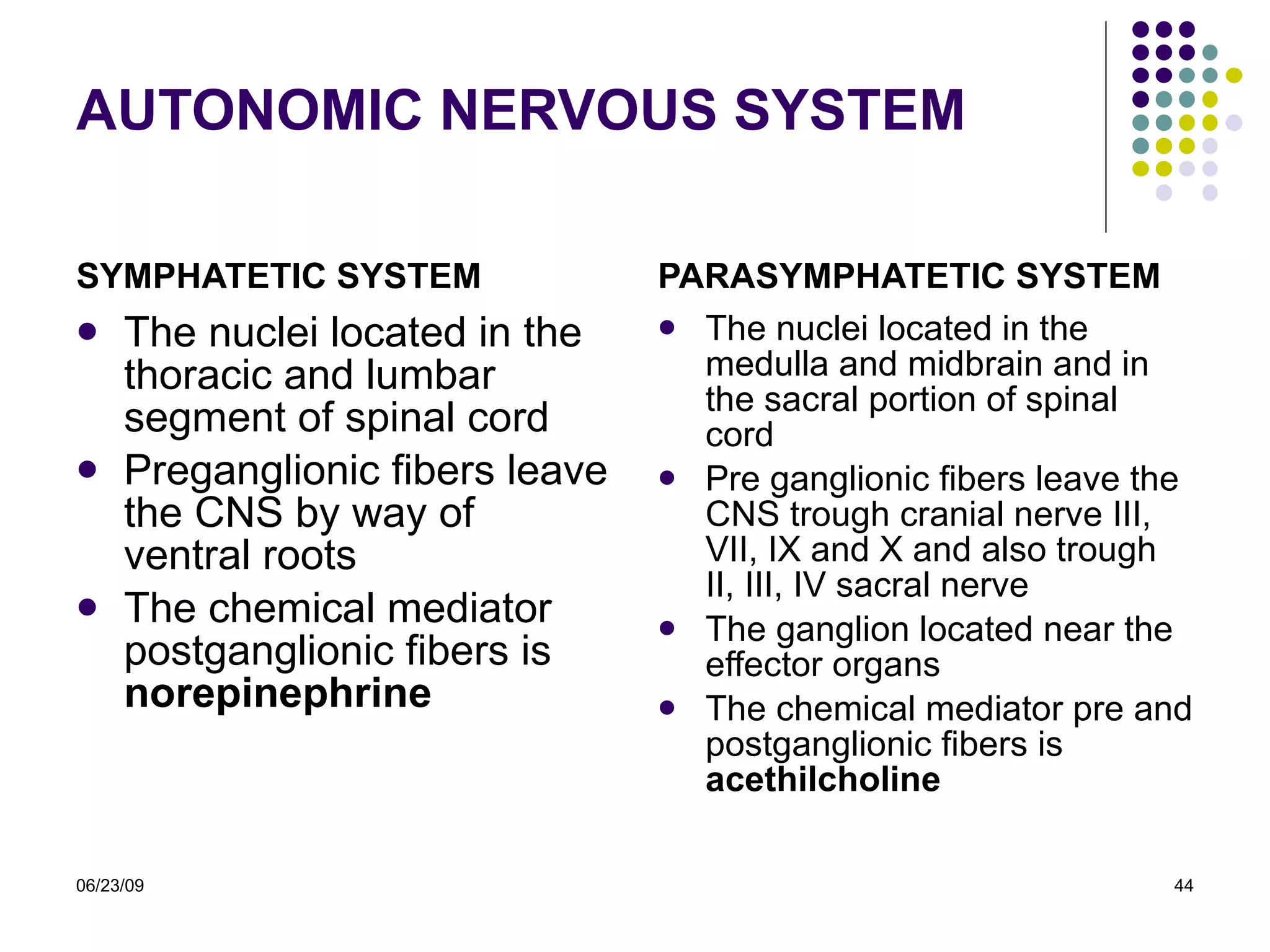
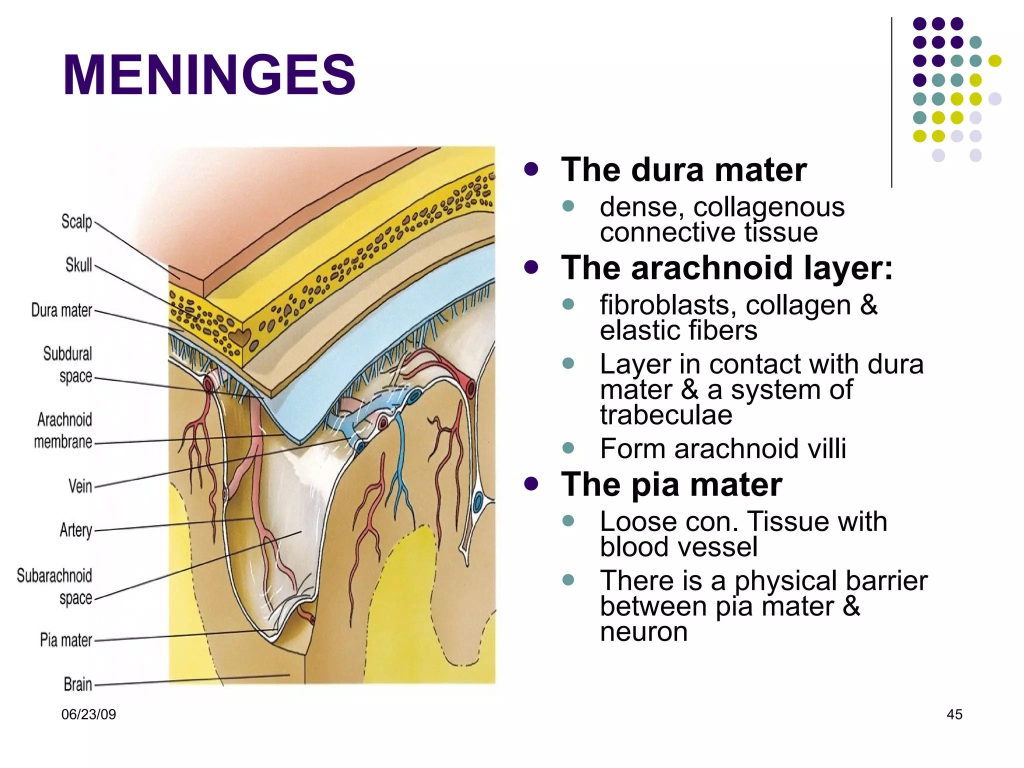
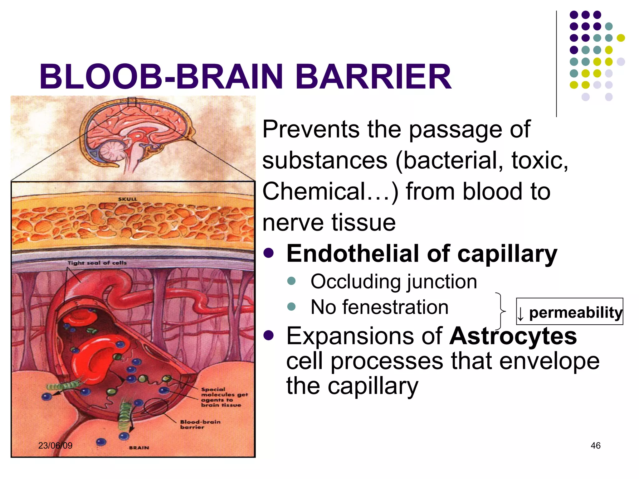
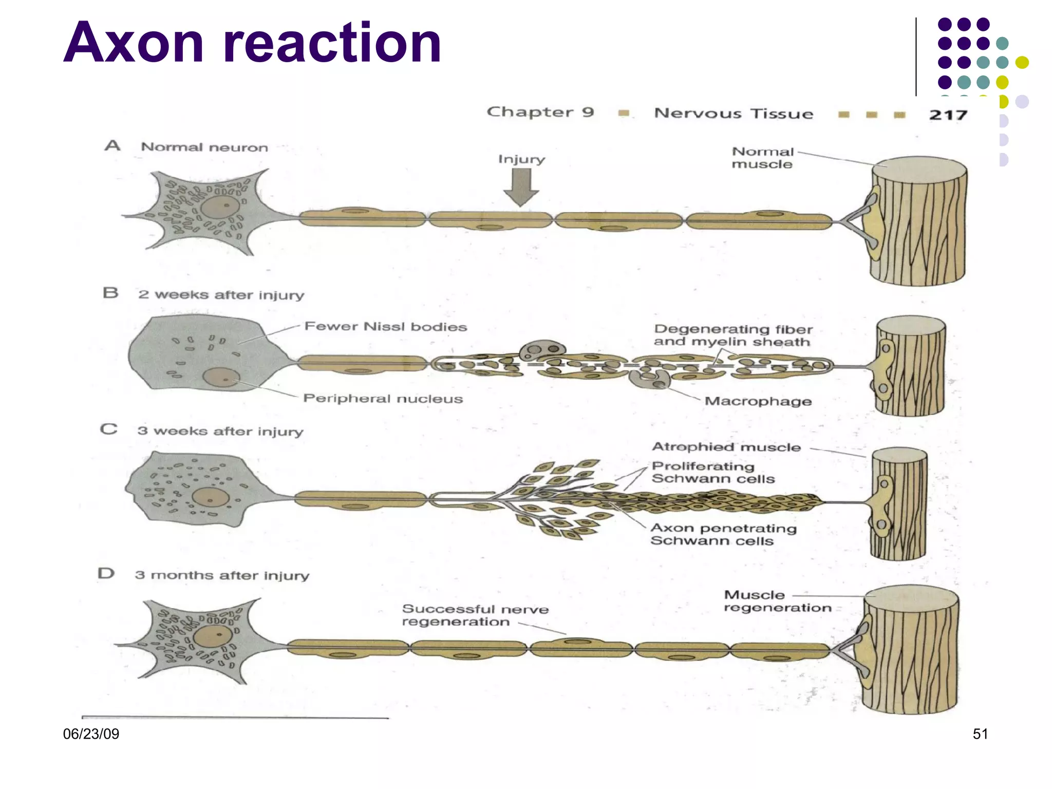
HISTOLOGY OF NERVOUS SYSTEM DR. I WAYAN SUGIRITAMA,M.Kes HISTOLOGY DEPARTMENT MEDICAL FACULTY OF UDAYANA UNIVERSITY
NERVOUS SYSTEM The most complex system in the human body Formed by network more than 100 million neuron Each neuron has a thousand  interconnection     a very complex system for communication Nerve tissue is distribute throughout the body, anatomically divide into :  CNS  &  PNS Structurally consist :  nerve cells  &  glial cells 23/06/09
CONTENTS Cells of nervous system Synaptic communication Central nervous system & Peripheral nervous system & associated structure Regeneration of nervous system 23/06/09
REFFERENCES Color Textbook Histology, third edition, leslie P. Gartner Basic Histology, tenth edition, L. Carlos Junqueira,2003 Histology and Cell Biology, second edition,2007 Elsevier’s Integrated Histology,2007 23/06/09
CELLS OF NERVOUS SYSTEM 23/06/09 NEURON NEUROGLIA
STRUCTURE OF  NEUR ON Principle cells of Nervous Tissue Consist of  3  parts  : CELL BODY  (perikaryon/soma) A single  AXON Multiple  DENDRITES ø  5-150  µm 23/06/09
CELL BODY (PERIKARYON) Central portion of the cell Generally are polygonal Different shape and size    characteristic regions of nervous system Contain : Nucleus Perinuclear cytoplasm 23/06/09
ULTRASTRUCTURE OF NEURON Nucle us  :  large,  spherical to ovoid and centraly located a single prominent nucleolus finely dispersed chromatin    trancriptionaly active    23/06/09
ULTRASTRUCTURE OF NEURON Cytoplasm   : A bundant  of  R.E.R Polyribosomes  Basic dyes (a+b)   Nissl Bodies  lots of  S.E.R. Golgi bodies  (perikaryon) protein secreting cell 23/06/09
ULTRASTRUCTURE OF NEURON Cytoplasm   M any mitochondria,  most abundant in axon terminal extensive cytoskeleton     axonal transport One centriole     do not undergo cell divisions 23/06/09
DENDRITE AND AXON AXON Single process  u p to 100 cm  Originate from  axon hillock Devoid ribosome Dilatation of  distal portion    axon terminal    end bulbs   synapse conducting impulse  away from the soma Axonal transport DENDRITES Multiple elongated processes Cytoplasmic~perikaryon (devoid golgi complex) Receiving stimuli  23/06/09
NEURONS CLASSIFICATION : According to the size and shape of the processes  (Dorsal root & cranial nerve ganglia)  (Pyramidal & purkinje) 23/06/09
NEURONS CLASSIFICATION : 23/06/09
NEURONS CLASSIFICATION : According to their function : Sensory Neuron (afferent ) Receive sensory input    conduct impulses to CNS Motor Neuron (Efferent) CNS    conduct impulses to muscles, glands and other neurons Interneuron In the CNS as interconnectors, establish neuronal circuit between sensory and motor neuron  23/06/09
NEURON GROUPING CORTEX Neuron form six layers on the cerrebrum Form three layers on the cerrebellum NUCLEI In subcortical region (thalamus, midbrain, brainstem and spinal cord) neuron form irregular cluster    nuclei GANGLION Cluster of neuron outside the CNS 23/06/09
FUNCTION OF NEURON R eceptive Receptor receive stimuli and tranduce into nerve impulse and transferro to other neuron I ntegrative Processing impulse on the higher center M otor Initiating motor respons and tranduse impulse to the effector 23/06/09
SYNAPS ES Sites of impulse transmission Convert electrical signal into chemical signal Permit neurons to communicate Types of synapses : Axodentritic synapse Axosomatic synapse Axoaxonic synapse Dendrodentritic synapse 23/06/09
SYNAPS 23/06/09
NEURO GLIAL CELLS Metabolic  and  mechanical  support   for  neuron 10 times abundant than neurons Neuroglial cells undergo mitosis Classification : Oligodendrocytes Astrocytes Ependymal Cells Microglia Schwan cells   CNS PNS 23/06/09
NEURO GLIAL CELLS Astrocytes Pedicles binds  to capillaries and to the pia mater  form  glial limitans Controlling the ionic  &  chemical env ironment  of neurons Energy metabolism Form cellular scar tissue Form the blood-brain barrier   23/06/09
NEURO GLIAL CELLS Oligodendrocytes interfascicular Produce myelin sheath (electrical insulation) in CNS A single cell wrap several axons (40 to 50) Form nodes of Ranvier satellite 23/06/09
NEURO GLIAL CELLS Schwann cells Analogue  to Oligodendrocyte Produce myelin sheath in the PN S 23/06/09
NEURO GLIAL CELLS Microglia Scattered throughout the CNS Clearing debris Act as APC Protect the CNS from viruses and microorganism 23/06/09
NEURO GLIAL CELLS Ependymal Cells Low columnar ciliated epithelial cells    line the  ventricles of the brain & central canal spinal cord Formation of  choroid flexus    produce CSF Facilitates the movement of CSF 23/06/09
Anatomically divided in to : 06/23/09 NERVOUS SYSTEM CENTRAL NERVOUS  SYSTEM (CNS) PERIPHERAL NERVOUS  SYSTEM (PNS)
THE CNS Consist of : Cerebrum Cerebellum Spinal cord No connective tissue   soft, gel like When sectioned : White matter Gray matter Covered by meninges 06/23/09
CEREBRUM GRAY MATTER Contains neuronal cell bodies, dendrites and glial cells Six layers composed  of neuron Neuron  (pyramidal) : Afferent (sensory) Efferent (motor) WHITE MATTER Contains myelinated axons and myelin-producing oligodendrocytes  23/06/09
CEREBELLUM GRAY MATTER Three layers : Outer molecular layer Central layer of large  Purkinje cells Inner granule layer WHITE MATTER The same as cerebrum   23/06/09
SPINAL CORD GRAY MATTER (CENTRAL)  SHAPE OF “H” Central canal lined by  Ependymal  cells Legs of the “H” form : Anterior horns Posterior horns Neurons : large and multipolar WHITE MATTER (PERIPHERAL) 23/06/09
PNS Bundles of nerve fibers (axons) outside the CNS & surrounded by connective tissue Main component : Peripheral nerves Ganglia Nerve endings 06/23/09
NERVE FIBERS Consist of axons enveloped by a special sheath Group of fibers constitute the  peripheral nerve Two types : Myelinated fiber Unmyelinated fiber 23/06/09
NERVE FIBERS Myelinated fibers A single  Schwann cell wraps around  single  axon   form myelin  sheath     nodes of Ranvier Unmyelinated fibers A single Schwann cell envelopes several axon Fibers enveloped within simple clefts of Schwann cells 23/06/09
MYELINATION PROCESS 23/06/09
CONDUCTION VELOCITY Depend on the extent of  Myelination : Unmyelinated fibers No nodes of Ranvier   continuous conduction Slower conduction Myelinated fibers Gap of myelin sheath  ( nodes of Ranvier  )    saltatory conduction Faster conduction 23/06/09
CONNECTIVE TISSUE INVESTMENTS Epineureum   Dense collagenous Con. Tissue with thick elastic fiber Prevent damage by overstreching Perineureum   Dense con. Tissue Layers of epithelioids Isolates neural environment (blood-nerve barrier) Endoneureum   Loose con. Tissue Regulation of microenvironment of nerve fiber 23/06/09
PERIPHERAL NERVE 06/23/09
PERIPHERAL NERVE 23/06/09
GANGLIA Ovoid structure containing neuronal cell bodies, glial cells supported by connective tissue Function : Relay stations to transmit impulses Types : Sensory ganglia Autonomic ganglia  23/06/09
GANGLI A Sensory Ganglia (cell bodies of sensory neuron) Unipolar cell bodies enveloped by cuboidal capsule cells Cranial ganglia :  Associated with the cranial nerve Spinal ganglia :  Associated with the spinal nerve Autonomic Ganglia (cell bodies of postganglionic autonomic nerves) Multipolar neuron enveloped by satellite cells Some are located within certain organ (intramural)  23/06/09
DORSAL ROOT GANGLIA 06/23/09
FUNCTIONAL CLASSIFICATION OF  NERVES Sensory nerve fibers Carry sensory input from the cutaneus/ viscera area    to CNS Motor nerve fibers Originate in the CNS    carry motor impulses to the effector organs 06/23/09
FUNCTIONAL CLASSIFICATION OF MOTOR COMPONENT Somatic nervous system Provide motor impulses to skeletal muscles Cells bodies (multipolar) originate in the motor nuclei of the brain or ventral horn of spinal cord The axon travel to the skeletal muscle via cranial or spinal nerve Autonomic nervous system  Provide motor impulses to the smooth muscle of viscera, cardiac muscle, secretory cells  HOMEOSTASIS 06/23/09
AUTONOMIC NERVOUS SYSTEM ANS = two-neuron network First Neuron  Neuron in CNS  Axon (preganglionic fibers) form a synapse with second multipolar neuron in ganglion Second Neuron Neuron in the Ganglion Axon to the effectors (postganglionic fibers) 23/06/09
AUTONOMIC NERVOUS SYSTEM SYMPHATETIC SYSTEM The nuclei located in the thoracic and lumbar segment of spinal cord Preganglionic fibers leave the CNS by way of ventral roots The chemical mediator postganglionic fibers is  norepinephrine PARASYMPHATETIC SYSTEM The nuclei located in the medulla and midbrain and in the sacral portion of spinal cord Pre ganglionic fibers leave the CNS trough cranial nerve III, VII, IX and X and also trough II, III, IV sacral nerve The ganglion located near the effector organs  The chemical mediator pre and postganglionic fibers is  acethilcholine 06/23/09
MENINGES The dura mater   dense, collagenous connective tissue The arachnoid layer : fibroblasts, collagen & elastic fibers Layer in contact with dura mater & a system of trabeculae Form arachnoid villi The pia mater Loose con. Tissue with blood vessel  There is a physical barrier between pia mater & neuron 06/23/09
BLOOB-BRAIN BARRIER Prevents the passage of  substances (bacterial, toxic,  Chemical…) from blood to  nerve tissue Endothelial of capillary Occluding junction No fenestration Expansions of  Astrocytes  cell processes that envelope the capillary ↓  permeability 23/06/09
CHOROID FLEXUS  Choroid flexus co nsist  of loose conn .  tissue of the pia matter, covered by a  simple/ low columnar epith .    ion transporting cells Elaborate CSF    fill the ventricles   subarachnoid space (arachnoid villi)  venous CSF function    metabolism and protective of CNS 23/06/09
CEREBROSPINAL FLUID Composition  clear,  low density & protein content High Na, K, Cl Function Metabolic Liquid cushion Blood-CSF barrier Zonulae occludents 23/06/09
NERVE REGENERATION Diseases affecting Schwann cells  loss of myelin (segmental demylination) Nerve injury    induced axonal degeneration (wallerian degeneration)   followed by regeneration Axon reaction : Local changes Anterograte changes Retrograte changes 06/23/09
Axon reaction Local reaction Severe ends of axon retract & cuts membrane fuses Macrophage & fibroblasts infiltration Anterograde reaction Axon terminal degeneration Schwann cells proliferation form a column of Schwann  (endoneureum is essential for proliferation of Schwann cells) Retrograde reaction Chromatolysis in perikaryon Several sprouts of axons emerge from proximal stump Axon sprouts enter the schwan tubes and guided to reach the target organ 22/04/2009
Axon reaction 06/23/09
REGENERATION OF CNS ?? Regeneration nerve fibers in CNS is not possible, because : An endoneureum is not present Oligodendrocytes do not proliferate Astrocytes deposit scar tissue (plaque) 23/06/09
THANK YOU 06/23/09

histologic structure of nervous system

  • 1.
    HISTOLOGY OF NERVOUSSYSTEM DR. I WAYAN SUGIRITAMA,M.Kes HISTOLOGY DEPARTMENT MEDICAL FACULTY OF UDAYANA UNIVERSITY
  • 2.
    NERVOUS SYSTEM Themost complex system in the human body Formed by network more than 100 million neuron Each neuron has a thousand interconnection  a very complex system for communication Nerve tissue is distribute throughout the body, anatomically divide into : CNS & PNS Structurally consist : nerve cells & glial cells 23/06/09
  • 3.
    CONTENTS Cells ofnervous system Synaptic communication Central nervous system & Peripheral nervous system & associated structure Regeneration of nervous system 23/06/09
  • 4.
    REFFERENCES Color TextbookHistology, third edition, leslie P. Gartner Basic Histology, tenth edition, L. Carlos Junqueira,2003 Histology and Cell Biology, second edition,2007 Elsevier’s Integrated Histology,2007 23/06/09
  • 5.
    CELLS OF NERVOUSSYSTEM 23/06/09 NEURON NEUROGLIA
  • 6.
    STRUCTURE OF NEUR ON Principle cells of Nervous Tissue Consist of 3 parts : CELL BODY (perikaryon/soma) A single AXON Multiple DENDRITES ø 5-150 µm 23/06/09
  • 7.
    CELL BODY (PERIKARYON)Central portion of the cell Generally are polygonal Different shape and size  characteristic regions of nervous system Contain : Nucleus Perinuclear cytoplasm 23/06/09
  • 8.
    ULTRASTRUCTURE OF NEURONNucle us : large, spherical to ovoid and centraly located a single prominent nucleolus finely dispersed chromatin  trancriptionaly active   23/06/09
  • 9.
    ULTRASTRUCTURE OF NEURONCytoplasm : A bundant of R.E.R Polyribosomes Basic dyes (a+b)  Nissl Bodies lots of S.E.R. Golgi bodies (perikaryon) protein secreting cell 23/06/09
  • 10.
    ULTRASTRUCTURE OF NEURONCytoplasm M any mitochondria, most abundant in axon terminal extensive cytoskeleton  axonal transport One centriole  do not undergo cell divisions 23/06/09
  • 11.
    DENDRITE AND AXONAXON Single process u p to 100 cm Originate from axon hillock Devoid ribosome Dilatation of distal portion  axon terminal  end bulbs  synapse conducting impulse away from the soma Axonal transport DENDRITES Multiple elongated processes Cytoplasmic~perikaryon (devoid golgi complex) Receiving stimuli 23/06/09
  • 12.
    NEURONS CLASSIFICATION :According to the size and shape of the processes (Dorsal root & cranial nerve ganglia) (Pyramidal & purkinje) 23/06/09
  • 13.
  • 14.
    NEURONS CLASSIFICATION :According to their function : Sensory Neuron (afferent ) Receive sensory input  conduct impulses to CNS Motor Neuron (Efferent) CNS  conduct impulses to muscles, glands and other neurons Interneuron In the CNS as interconnectors, establish neuronal circuit between sensory and motor neuron 23/06/09
  • 15.
    NEURON GROUPING CORTEXNeuron form six layers on the cerrebrum Form three layers on the cerrebellum NUCLEI In subcortical region (thalamus, midbrain, brainstem and spinal cord) neuron form irregular cluster  nuclei GANGLION Cluster of neuron outside the CNS 23/06/09
  • 16.
    FUNCTION OF NEURONR eceptive Receptor receive stimuli and tranduce into nerve impulse and transferro to other neuron I ntegrative Processing impulse on the higher center M otor Initiating motor respons and tranduse impulse to the effector 23/06/09
  • 17.
    SYNAPS ES Sitesof impulse transmission Convert electrical signal into chemical signal Permit neurons to communicate Types of synapses : Axodentritic synapse Axosomatic synapse Axoaxonic synapse Dendrodentritic synapse 23/06/09
  • 18.
  • 19.
    NEURO GLIAL CELLSMetabolic and mechanical support for neuron 10 times abundant than neurons Neuroglial cells undergo mitosis Classification : Oligodendrocytes Astrocytes Ependymal Cells Microglia Schwan cells  CNS PNS 23/06/09
  • 20.
    NEURO GLIAL CELLSAstrocytes Pedicles binds to capillaries and to the pia mater form glial limitans Controlling the ionic & chemical env ironment of neurons Energy metabolism Form cellular scar tissue Form the blood-brain barrier 23/06/09
  • 21.
    NEURO GLIAL CELLSOligodendrocytes interfascicular Produce myelin sheath (electrical insulation) in CNS A single cell wrap several axons (40 to 50) Form nodes of Ranvier satellite 23/06/09
  • 22.
    NEURO GLIAL CELLSSchwann cells Analogue to Oligodendrocyte Produce myelin sheath in the PN S 23/06/09
  • 23.
    NEURO GLIAL CELLSMicroglia Scattered throughout the CNS Clearing debris Act as APC Protect the CNS from viruses and microorganism 23/06/09
  • 24.
    NEURO GLIAL CELLSEpendymal Cells Low columnar ciliated epithelial cells  line the ventricles of the brain & central canal spinal cord Formation of choroid flexus  produce CSF Facilitates the movement of CSF 23/06/09
  • 25.
    Anatomically divided into : 06/23/09 NERVOUS SYSTEM CENTRAL NERVOUS SYSTEM (CNS) PERIPHERAL NERVOUS SYSTEM (PNS)
  • 26.
    THE CNS Consistof : Cerebrum Cerebellum Spinal cord No connective tissue  soft, gel like When sectioned : White matter Gray matter Covered by meninges 06/23/09
  • 27.
    CEREBRUM GRAY MATTERContains neuronal cell bodies, dendrites and glial cells Six layers composed of neuron Neuron (pyramidal) : Afferent (sensory) Efferent (motor) WHITE MATTER Contains myelinated axons and myelin-producing oligodendrocytes 23/06/09
  • 28.
    CEREBELLUM GRAY MATTERThree layers : Outer molecular layer Central layer of large Purkinje cells Inner granule layer WHITE MATTER The same as cerebrum 23/06/09
  • 29.
    SPINAL CORD GRAYMATTER (CENTRAL)  SHAPE OF “H” Central canal lined by Ependymal cells Legs of the “H” form : Anterior horns Posterior horns Neurons : large and multipolar WHITE MATTER (PERIPHERAL) 23/06/09
  • 30.
    PNS Bundles ofnerve fibers (axons) outside the CNS & surrounded by connective tissue Main component : Peripheral nerves Ganglia Nerve endings 06/23/09
  • 31.
    NERVE FIBERS Consistof axons enveloped by a special sheath Group of fibers constitute the peripheral nerve Two types : Myelinated fiber Unmyelinated fiber 23/06/09
  • 32.
    NERVE FIBERS Myelinatedfibers A single Schwann cell wraps around single axon  form myelin sheath  nodes of Ranvier Unmyelinated fibers A single Schwann cell envelopes several axon Fibers enveloped within simple clefts of Schwann cells 23/06/09
  • 33.
  • 34.
    CONDUCTION VELOCITY Dependon the extent of Myelination : Unmyelinated fibers No nodes of Ranvier  continuous conduction Slower conduction Myelinated fibers Gap of myelin sheath ( nodes of Ranvier )  saltatory conduction Faster conduction 23/06/09
  • 35.
    CONNECTIVE TISSUE INVESTMENTSEpineureum Dense collagenous Con. Tissue with thick elastic fiber Prevent damage by overstreching Perineureum Dense con. Tissue Layers of epithelioids Isolates neural environment (blood-nerve barrier) Endoneureum Loose con. Tissue Regulation of microenvironment of nerve fiber 23/06/09
  • 36.
  • 37.
  • 38.
    GANGLIA Ovoid structurecontaining neuronal cell bodies, glial cells supported by connective tissue Function : Relay stations to transmit impulses Types : Sensory ganglia Autonomic ganglia 23/06/09
  • 39.
    GANGLI A SensoryGanglia (cell bodies of sensory neuron) Unipolar cell bodies enveloped by cuboidal capsule cells Cranial ganglia : Associated with the cranial nerve Spinal ganglia : Associated with the spinal nerve Autonomic Ganglia (cell bodies of postganglionic autonomic nerves) Multipolar neuron enveloped by satellite cells Some are located within certain organ (intramural) 23/06/09
  • 40.
  • 41.
    FUNCTIONAL CLASSIFICATION OF NERVES Sensory nerve fibers Carry sensory input from the cutaneus/ viscera area  to CNS Motor nerve fibers Originate in the CNS  carry motor impulses to the effector organs 06/23/09
  • 42.
    FUNCTIONAL CLASSIFICATION OFMOTOR COMPONENT Somatic nervous system Provide motor impulses to skeletal muscles Cells bodies (multipolar) originate in the motor nuclei of the brain or ventral horn of spinal cord The axon travel to the skeletal muscle via cranial or spinal nerve Autonomic nervous system Provide motor impulses to the smooth muscle of viscera, cardiac muscle, secretory cells HOMEOSTASIS 06/23/09
  • 43.
    AUTONOMIC NERVOUS SYSTEMANS = two-neuron network First Neuron Neuron in CNS Axon (preganglionic fibers) form a synapse with second multipolar neuron in ganglion Second Neuron Neuron in the Ganglion Axon to the effectors (postganglionic fibers) 23/06/09
  • 44.
    AUTONOMIC NERVOUS SYSTEMSYMPHATETIC SYSTEM The nuclei located in the thoracic and lumbar segment of spinal cord Preganglionic fibers leave the CNS by way of ventral roots The chemical mediator postganglionic fibers is norepinephrine PARASYMPHATETIC SYSTEM The nuclei located in the medulla and midbrain and in the sacral portion of spinal cord Pre ganglionic fibers leave the CNS trough cranial nerve III, VII, IX and X and also trough II, III, IV sacral nerve The ganglion located near the effector organs The chemical mediator pre and postganglionic fibers is acethilcholine 06/23/09
  • 45.
    MENINGES The duramater dense, collagenous connective tissue The arachnoid layer : fibroblasts, collagen & elastic fibers Layer in contact with dura mater & a system of trabeculae Form arachnoid villi The pia mater Loose con. Tissue with blood vessel There is a physical barrier between pia mater & neuron 06/23/09
  • 46.
    BLOOB-BRAIN BARRIER Preventsthe passage of substances (bacterial, toxic, Chemical…) from blood to nerve tissue Endothelial of capillary Occluding junction No fenestration Expansions of Astrocytes cell processes that envelope the capillary ↓ permeability 23/06/09
  • 47.
    CHOROID FLEXUS Choroid flexus co nsist of loose conn . tissue of the pia matter, covered by a simple/ low columnar epith .  ion transporting cells Elaborate CSF  fill the ventricles  subarachnoid space (arachnoid villi)  venous CSF function  metabolism and protective of CNS 23/06/09
  • 48.
    CEREBROSPINAL FLUID Composition clear, low density & protein content High Na, K, Cl Function Metabolic Liquid cushion Blood-CSF barrier Zonulae occludents 23/06/09
  • 49.
    NERVE REGENERATION Diseasesaffecting Schwann cells  loss of myelin (segmental demylination) Nerve injury  induced axonal degeneration (wallerian degeneration)  followed by regeneration Axon reaction : Local changes Anterograte changes Retrograte changes 06/23/09
  • 50.
    Axon reaction Localreaction Severe ends of axon retract & cuts membrane fuses Macrophage & fibroblasts infiltration Anterograde reaction Axon terminal degeneration Schwann cells proliferation form a column of Schwann (endoneureum is essential for proliferation of Schwann cells) Retrograde reaction Chromatolysis in perikaryon Several sprouts of axons emerge from proximal stump Axon sprouts enter the schwan tubes and guided to reach the target organ 22/04/2009
  • 51.
  • 52.
    REGENERATION OF CNS?? Regeneration nerve fibers in CNS is not possible, because : An endoneureum is not present Oligodendrocytes do not proliferate Astrocytes deposit scar tissue (plaque) 23/06/09
  • 53.