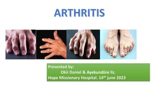
2. ARTHRITIS
Swelling and tenderness of one or more
joints causing pain and inflammation.
It is majorly categorized into;
• Rheumatoid Arthritis
• Osteoarthritis
3. RHEUMATOID ATHRITIS
Defns;
A systemic autoimmune disease of unknown cause with its primary manifestation in synovial tissues.
Alternatively: RA can be defined as Chronic and usually progressive inflammatory disorder of
unknown etiology x-terised by polyarticular symmetric joint involvement and systemic
manifestations.
-*THE Synovial tissues proliferate in an uncontrolled fashion resulting in stretching of tendon and
ligaments and erosions of bone.
RA is theorized to develop when a genetically susceptible individual experiences an external
trigger (e.g, cigarette smoking, infection, or trauma) that triggers an autoimmune reaction
5 Months of Disease 5 Years of Disease Rheumatoid Arthritis: 10 Years Later
4. Aetiology and pathophysiology
The cause of rheumatoid arthritis remains unclear with hormonal, genetic
and environmental factors playing a key role.
Genetic factors contribute 53–65% of the risk of developing this disease.
The HLA-DR4 allele is associated with both the development and severity of
rheumatoid arthritis.
Cigarette smoking is a strong risk factor for developing rheumatoid arthritis.
Pathologically, rheumatoid arthritis is characterized by the infiltration of a
variety of inflammatory cells into the joint.
The synovial membrane, which is normally acellular, becomes highly
vascularised and hypertrophied, creating a so-called pannus formation.
There is proliferation of synovial fibroblasts and an increase in the number
of inflammatory cells present within the joint.
5. Con’t
The inflammatory cells involved in rheumatoid arthritis include T-cells
(predominantly CD4 helper cells), B-cells, macrophages and plasma cells.
Cytokines are released by these cells which cause the synovium to release
proteolytic enzymes, resulting in the destruction of bone and cartilage.
Key cytokines involved in rheumatoid arthritis include tumour necrosis factor
(TNF)-a, interleukin-1, interleukin-6 and granulocyte macrophage colony-
stimulating factor (GM-CSF).
These play a crucial role in the pro inflammatory reaction.
6. CLINICAL PRESENTATION
The disease may present as a polyarticular arthritis with gradual onset,
extra-articular manifestations may be present.
The disease is usually insidious with predominant Signs and symptoms
of:
pain,
Tenderness,
warmth at the joints,
swollen joints, Joint stiffness that is usually worse in the mornings and
after inactivity. Fatigue, fever and loss of appetite. etc
7. Investigations
Inflammatory markers, including C-reactive protein (CRP) and erythrocyte
sedimentation rate (ESR), are usually but not always elevated in active disease
and are useful for monitoring response to treatment.
Rheumatoid factor (RF), CBC, Cyclic citrullinated peptide (CCP), antinuclear
antibody (ANA) are among the other investigations.
NB: -Those patients who do not have a detectable RF are said to be
‘seronegative’. RF is not specific to rheumatoid arthritis, its is also present in
patients with chronic lung and liver disease, other connective tissue diseases,
neoplasia, infections
Radiographies such ultrasound, x-rays for aparticular suspected joint can be
also done.
8. American College of Rheumatology (formerly American Rheumatism
Association) revised classification criteria for rheumatoid arthritis
Criterion Description
1. Morning
stiffness
Morning stiffness in and around the joints, lasting at least
one hour before maximal improvement
2. Arthritis
of three or
more joint
areas
At least three joint areas (out of 14 possible areas; right or
left PIP, MCP, wrist, elbow, knee, ankle, MTP joints)
simultaneously have had soft tissue swelling or fluid (not
bony overgrowth alone)
3. Arthritis of
hand joints
At least one area swollen (as defined above) in a wrist, MCP,
or PIP joint
Diagnosis is based on the ACR criterion as shown in the table
below:
9. 4. Symmetric
arthritis
Simultaneous involvement of the same joint areas (as defined
above) on both sides of the body (bilateral involvement of
PIPs, MCPs, or MTPs, without absolute symmetry is
acceptable).
5. Rheumatoid
nodules
Subcutaneous nodules over bony prominences or extensor
surfaces, or in juxta-articular regions as observed by a physician
6. Serum
rheumatoid
factor
Demonstration of abnormal amounts of serum rheumatoid
factor by any method for which the result has been positive in
less than 5 percent of normal control subjects.
7.
Radiographic
changes
Radiographic changes typical of rheumatoid arthritis on
posteroanterior hand or wrist radiographs, which must include
erosions or unequivocal bony decalcification localised in, or
most marked adjacent to, the involved joints (osteoarthritis
changes alone do not qualify).
10. Con’t…
• NB:
• For classification purposes, a patient is said to have rheumatoid
arthritis if he or she has satisfied at least four of the above seven
criteria. Criteria 1 through 4 must be present for at least 6 weeks.
Patients with two clinical diagnoses are not excluded. Designation as
classic, definite, or probable rheumatoid arthritis is not to be made.
11. GOAL OF THERAPY
The ultimate goal of RA treatment is to:-
Induce a complete remission, although this may be difficult to
achieve.
Reduce joint swelling, stiffness, and pain;
Preserve range of motion and joint function;
Improve quality of life;
Prevent systemic complications; and slow destructive joint changes.
12. Management strategies for RA
Non-pharmacological approach Pharmacological approach
-Education
Heat and cold therapies
-Psychological support and employment
counselling
-Occupational therapy
-Physiotherapy
-Surgical interventions, such as
synovectomy and arthroplasty, may be
useful to relieve pain and restore
function.
-NSAIDs, cox-2 selective ihibitors
-Corticosteroids
-DMARDS:-
Conventional
e.g, Methotrexate, Hydrochloroquine,
Sulfasalazine, Leflunomide, Gold, Azathioprine,
Minocycline, leflunomide.
Biologicals
e.g, >TNFα antagonists like Etanercept (Enbrel),
Infliximab (Remicade), Adalimumab (Humira),
>Interleukin-1 antagonist like Anakinra (Kineret),
>Suppress T-Cell activation like Abatacept
(Orencia)
>Anti B-Cell monoclonal antibody like Rituximab
(Rituxan)
13. OSTEOARTHRITIS
• Osteoarthritis (OA) is a degenerative disorder with inflammatory components arising from
the biochemical breakdown of articular (hyaline) cartilage in the synovial joints.
• OA primarily affects the weight-bearing diarthrodial joints of the peripheral and axial
skeleton, and it’s characterized by progressive deterioration and loss of articular cartilage,
resulting in osteophyte formation, pain, limitation of motion, deformity, and progressive
disability. The current view holds that osteoarthritis involves not only the articular cartilage
but the entire joint organ, including the subchondral bone and synovium.
• It is the most common type of joint disease and the leading cause of chronic disability in
older adults
• NB;Inflammation may or may not be present in the affected joints.
14. OSTEOATHRITIS
• It is a chronic degenerative
disorder of the synovial joints in
which there is progressive
softening and
erosion/disintegration of the
articular cartilage.
15. OSTEOARTHRITIS
• It occurs when the protective cartilage that
cushions the ends of the bones wears
down over time.
• Although osteoarthritis can damage any
joint, the disorder most commonly affects
joints in your hands, knees, hips and spine.
16.
17. RISK FACTORS
•Joint injury or overuse—Injury or overuse, such as knee bending and repetitive stress on a joint, can
damage a joint and increase the risk of OA in that joint.
•Age—The risk of developing OA increases with age.
•Gender—Women are more likely to develop OA than men, especially after age 50.
•Obesity—Extra weight puts more stress on joints, particularly weight-bearing joints like the hips and
knees. This stress increases the risk of OA in that joint. Obesity may also have metabolic effects that
increase the risk of OA.
•Genetics—People who have family members with OA are more likely to develop OA. People who have
hand OA are more likely to develop knee OA.
•Race— Some Asian populations have lower risk for OA.
•Life style-the more active people are likely to develop OA.
18. SIGNS AND SYMPTOMS
Signs and symptoms
• Deep, achy joint pain exacerbated by extensive use - The disease’s
primary symptom
• Reduced range of motion and crepitus - Frequently present
• Stiffness during rest (gelling) - May develop, with morning joint
stiffness usually lasting for less than 30 minutes
• Swelling
19. CONT
Osteoarthritis of the hand
• Distal interphalangeal (DIP) joints are most often affected
• Proximal interphalangeal (PIP) joints and the carpometacarpal (cmc) joints
at the base of the thumb are also typically involved
• Heberden nodes, which represent palpable osteophytes in the DIP joints,
are more characteristic in women than in men
• Inflammatory changes are typically absent, less pronounced, or go
unnoticed
21. NONPHARMACOLOGIC INTERVENTIONS
• The cornerstones of osteoarthritis therapy, nonpharmacologic interventions
include the following:
• Patient education, Heat and cold, Weight loss
• Exercise, Physical therapy
• Muscle training (eg, quadriceps strengthening for knee OA)
• Occupational therapy (use of assistive devices for independence in ADL)
• Unloading in certain joints (eg, knee and hip)
22. PHARMACOLOGIC THERAPY
Topical capsaicin
Topical nonsteroidal anti-inflammatory
drugs (NSAIDs)
Oral NSAIDs, Tramadol,
Glucosamine and chondroitins
Acetaminophen
Intra-articular corticosteroid injections.
(methylprednisolone acetate or
triamcinolone Hexacetonide)
Surgery
Arthroscopy
Osteotomy
Arthroplasty - Particularly with knee or
hip osteoarthritis
Fusion
23. CON’T….
NB. Glucosamine and chondroitin are dietary supplements that were shown
to stimulate proteoglycan synthesis from articular cartilage in vitro. SOME of
the examples of drugs in this classes include: Jointace capsules, osteomin
tabs, osteocare, osteoflex tabs etc
Hyaluronate Injections. High-molecular-weight hyaluronic acid is a
constituent of normal cartilage that provides lubrication with motion and
shock absorbency during rapid movements.