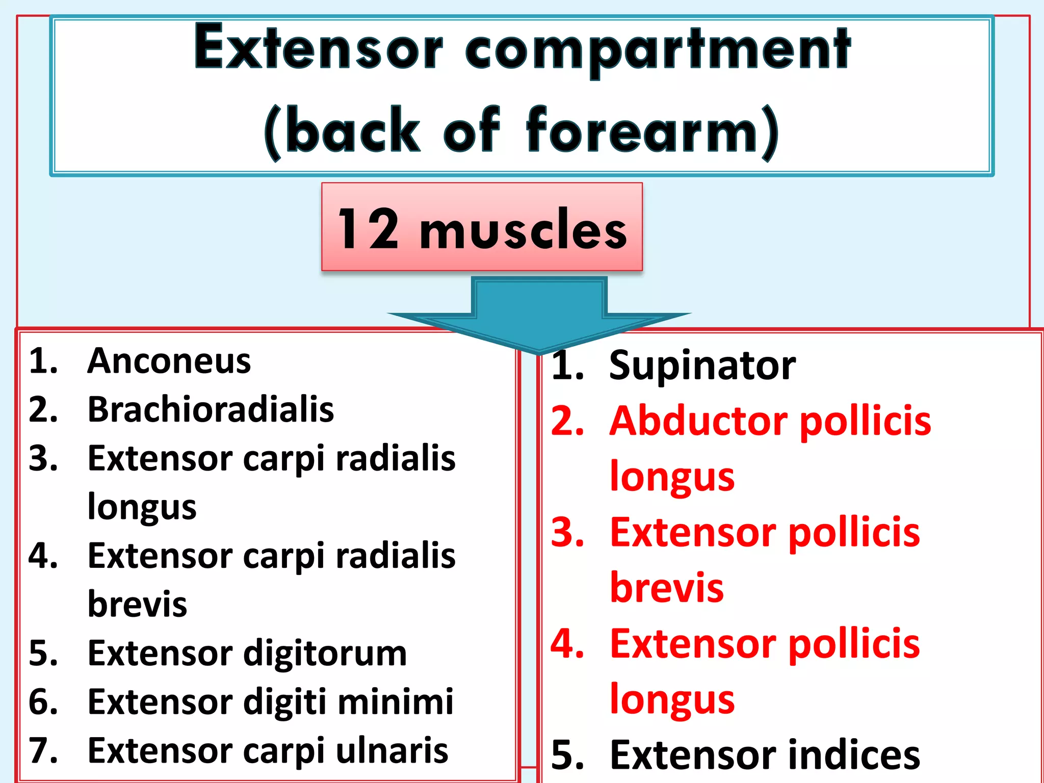
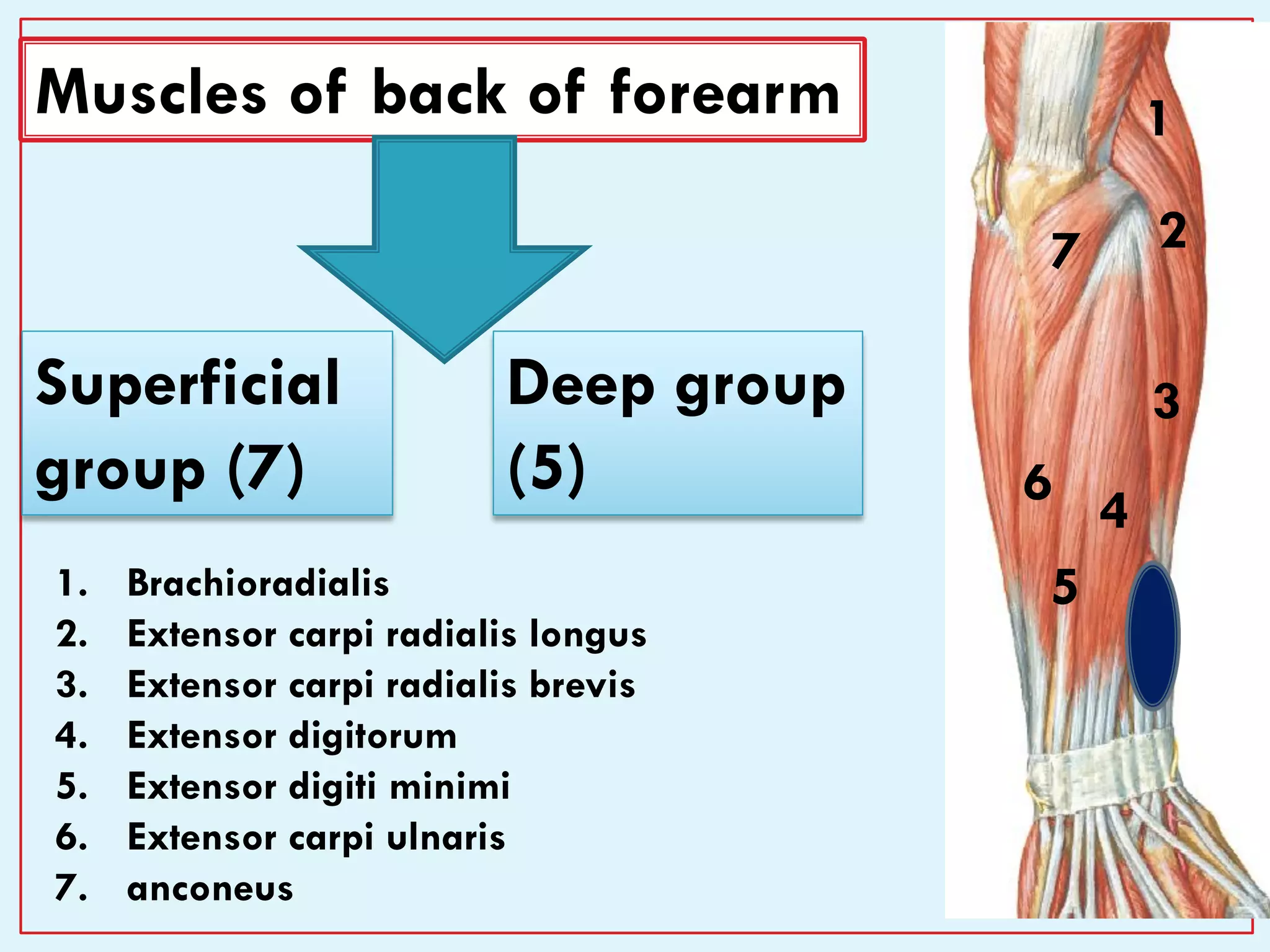
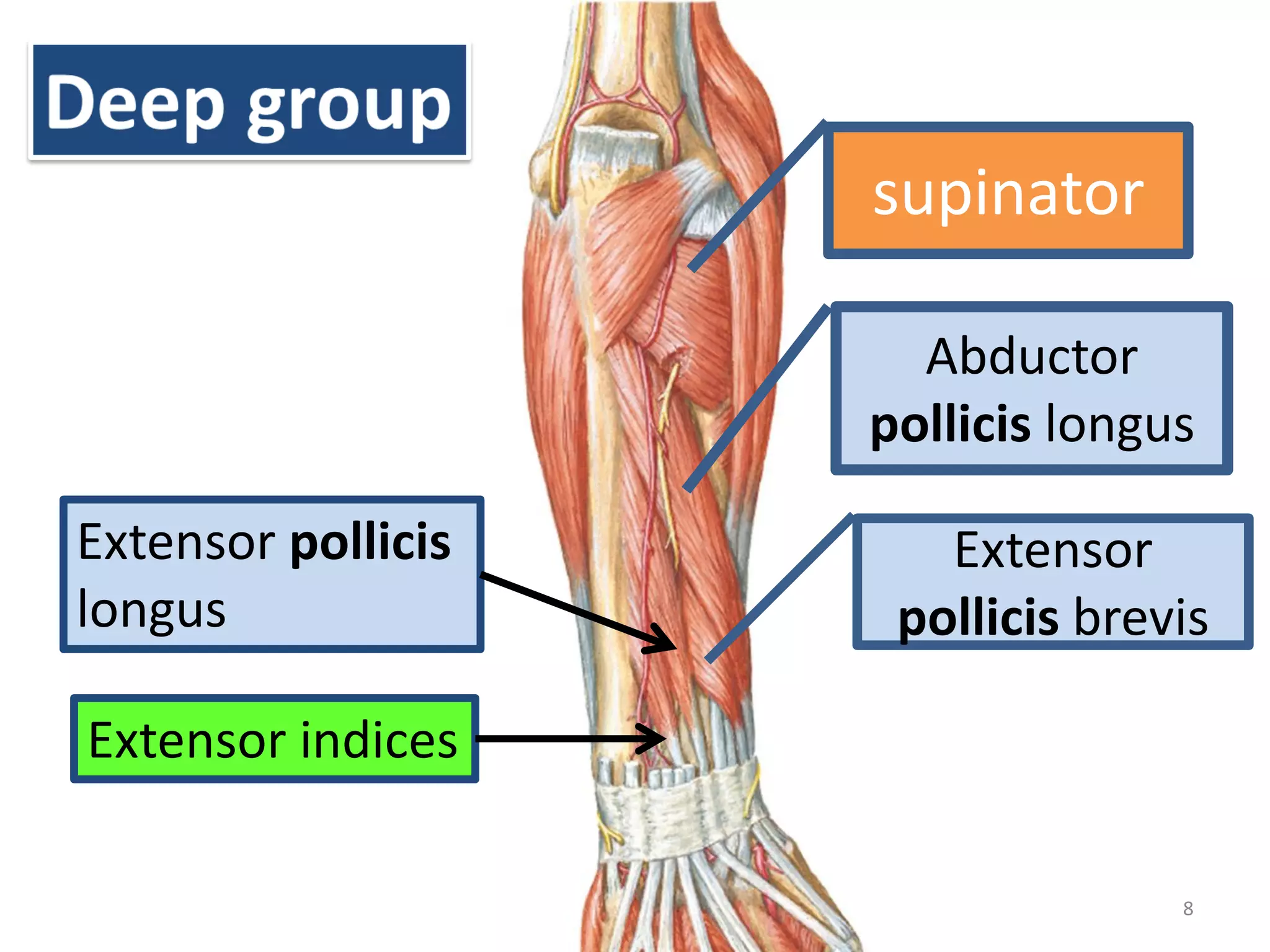
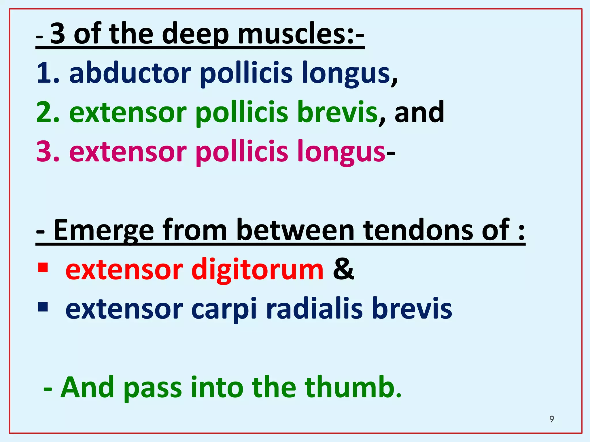
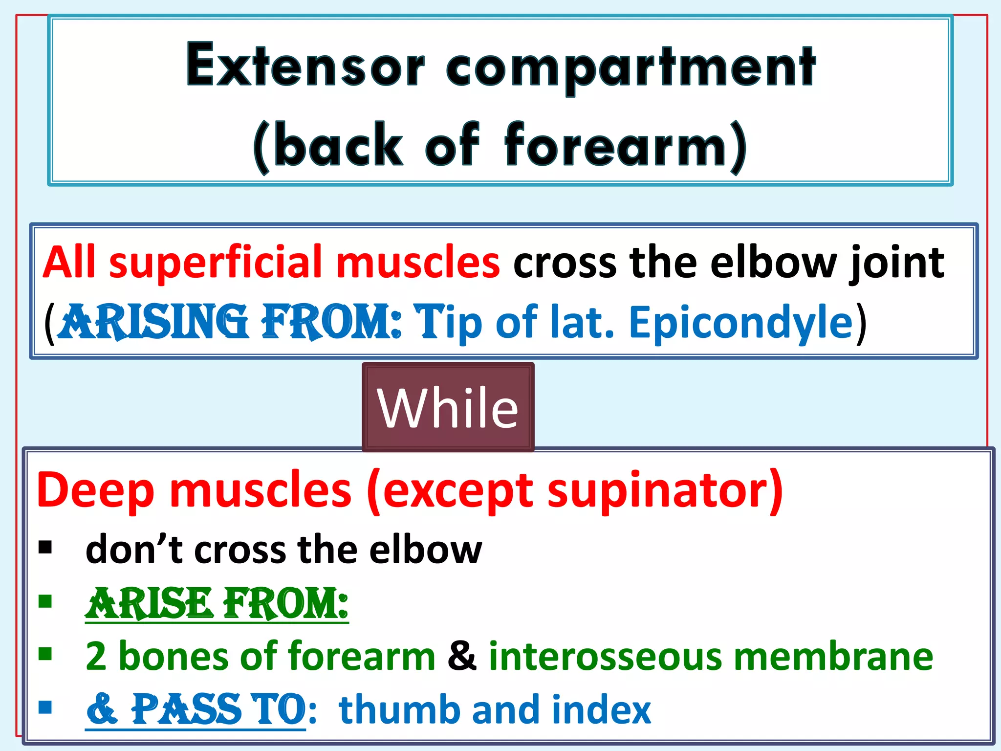
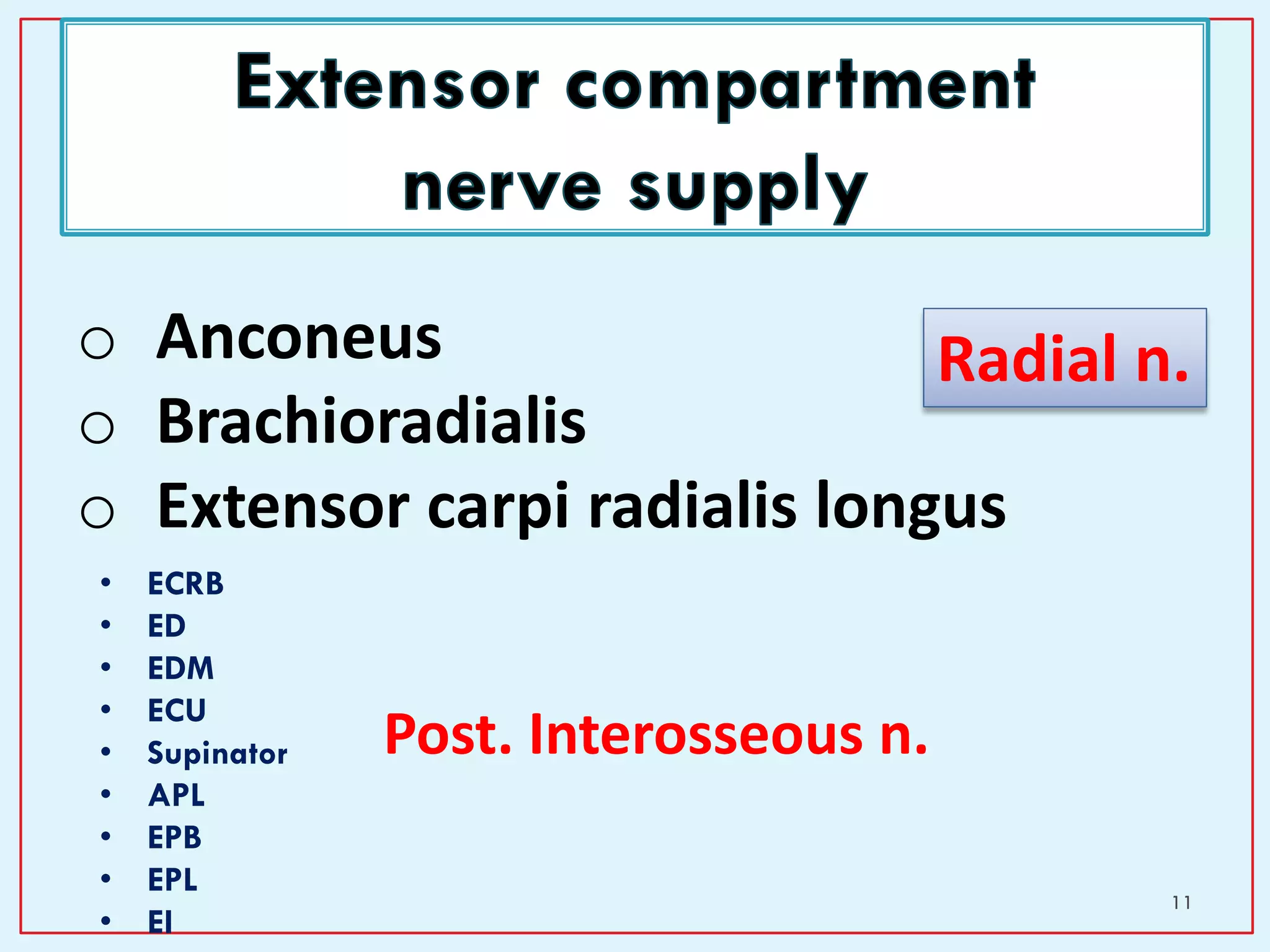
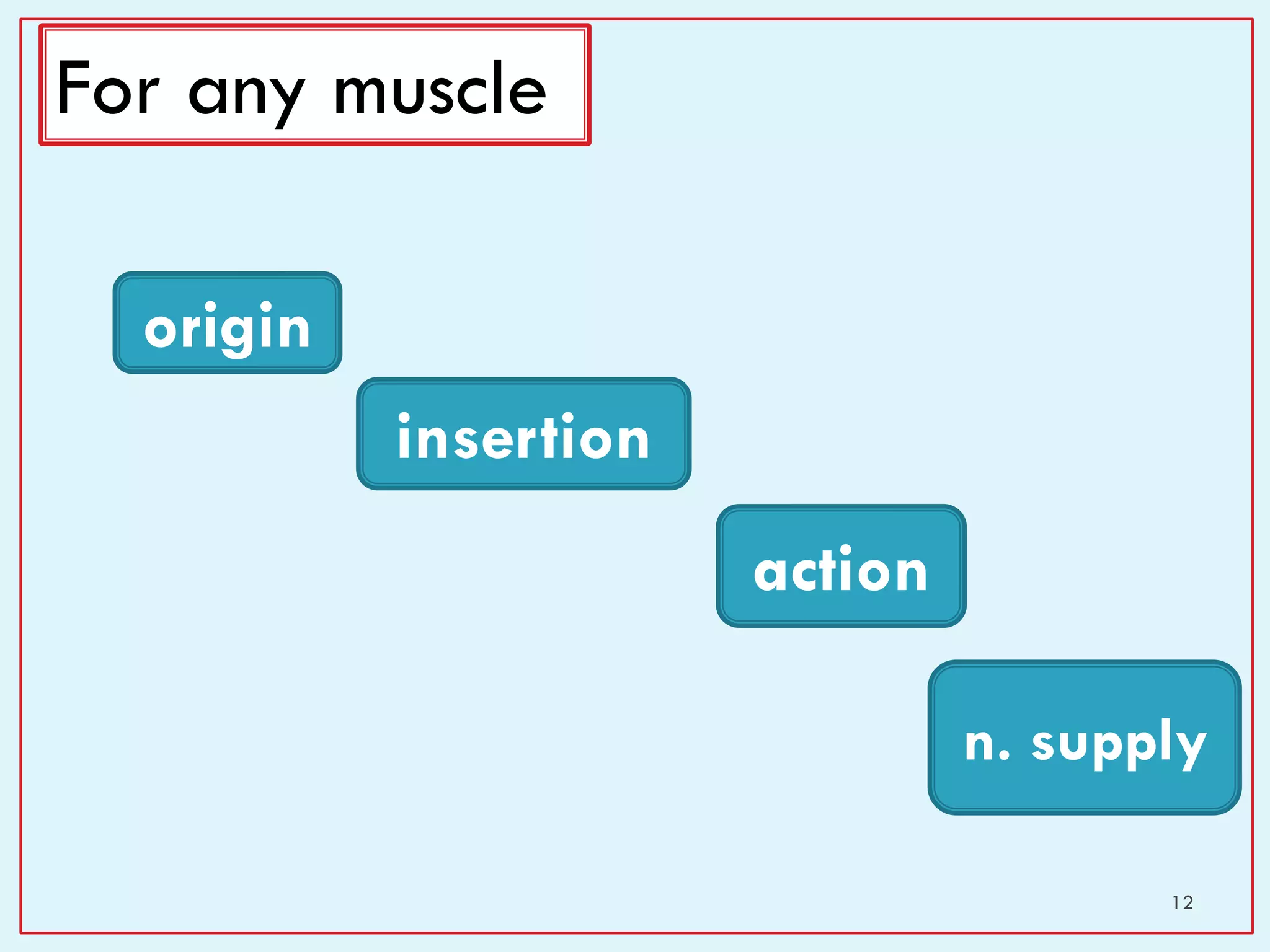
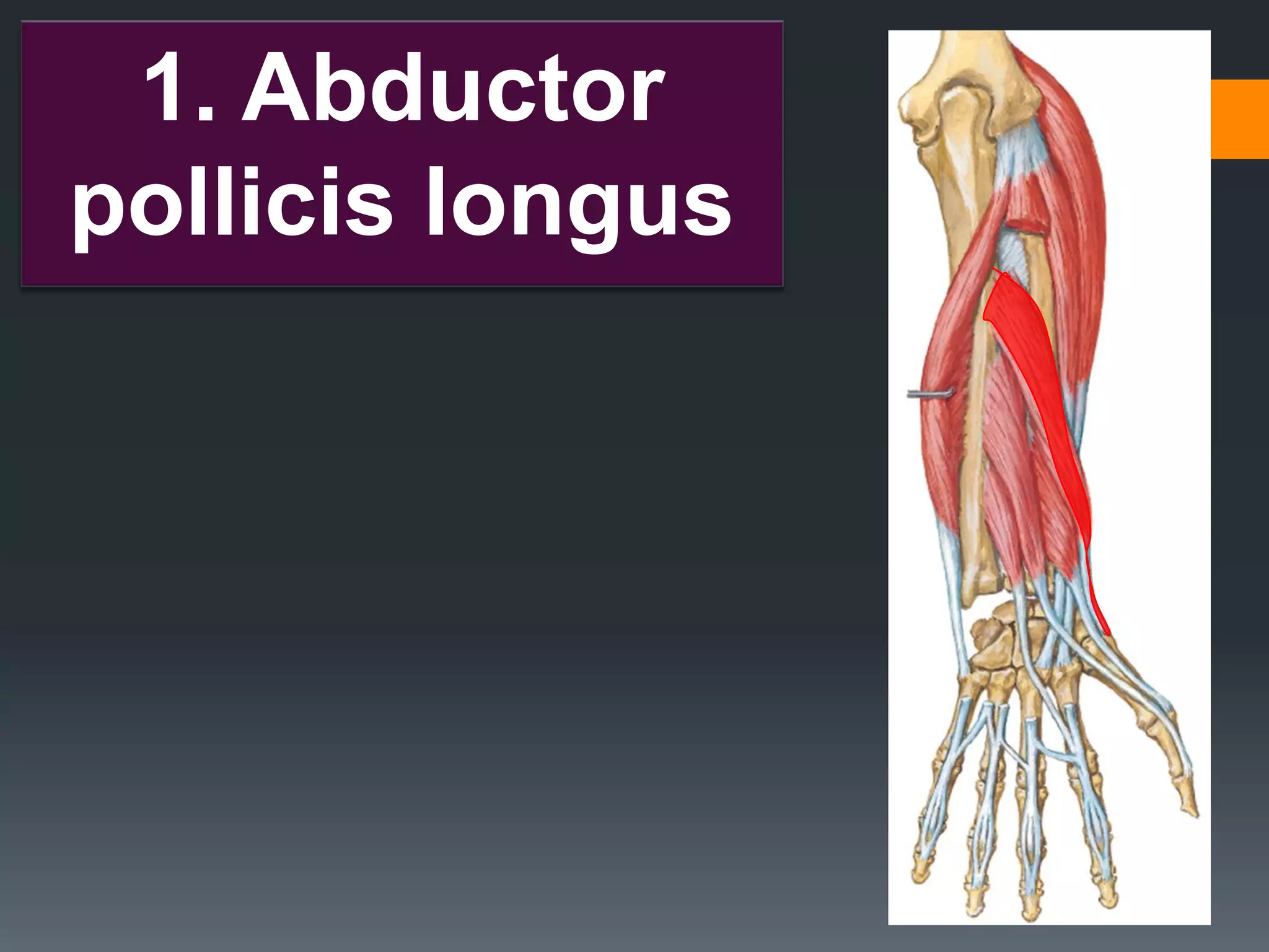
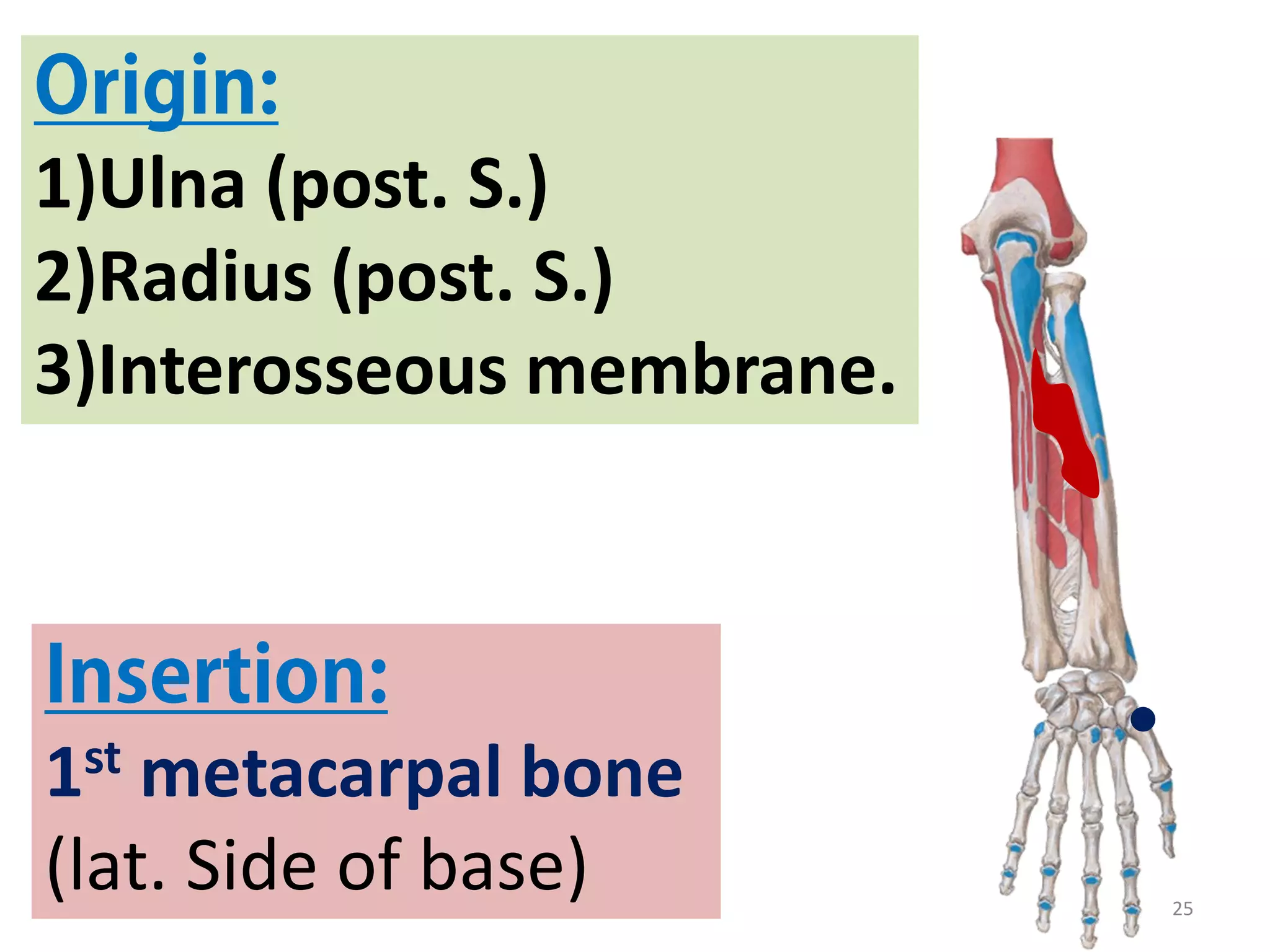
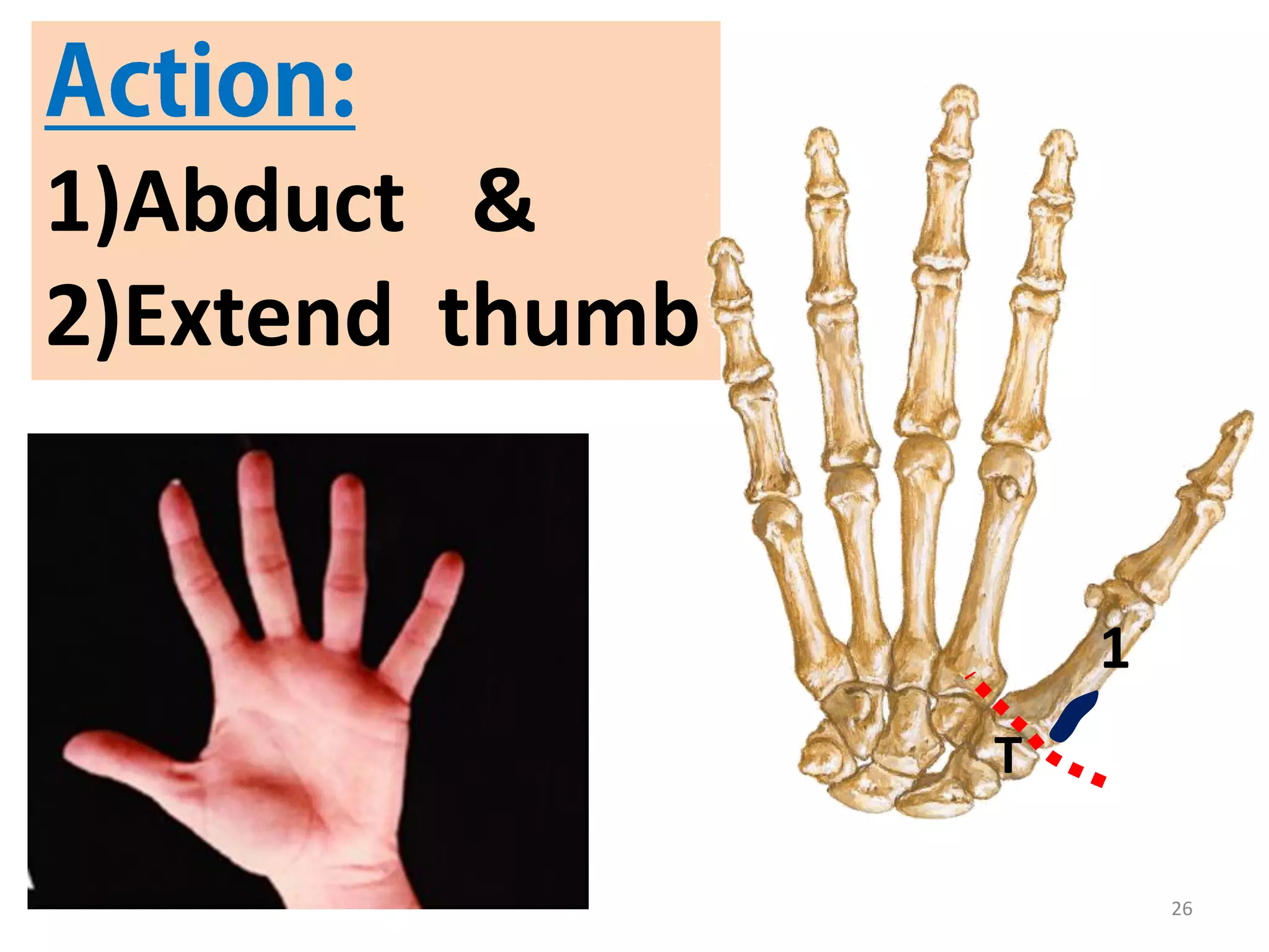
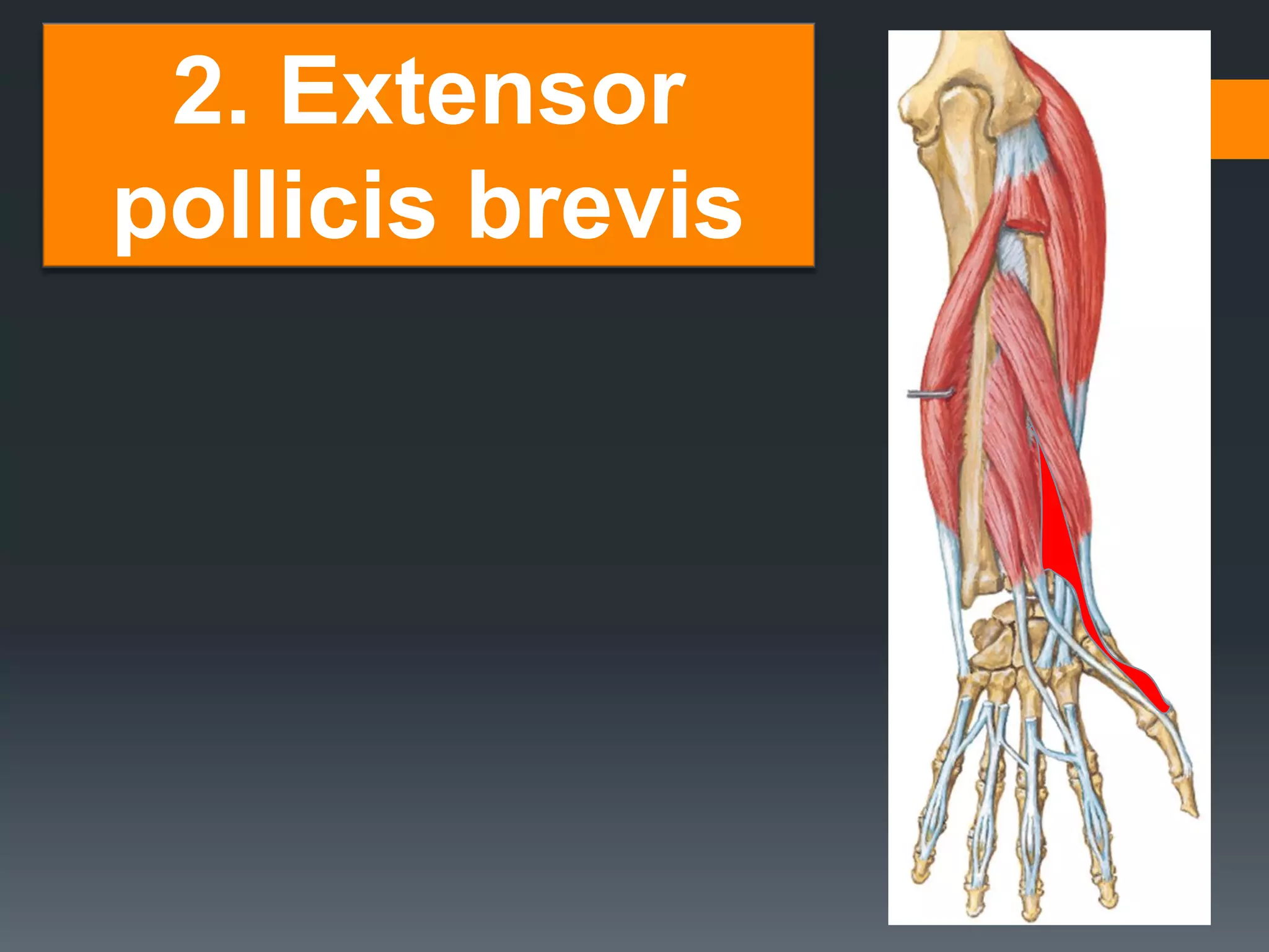
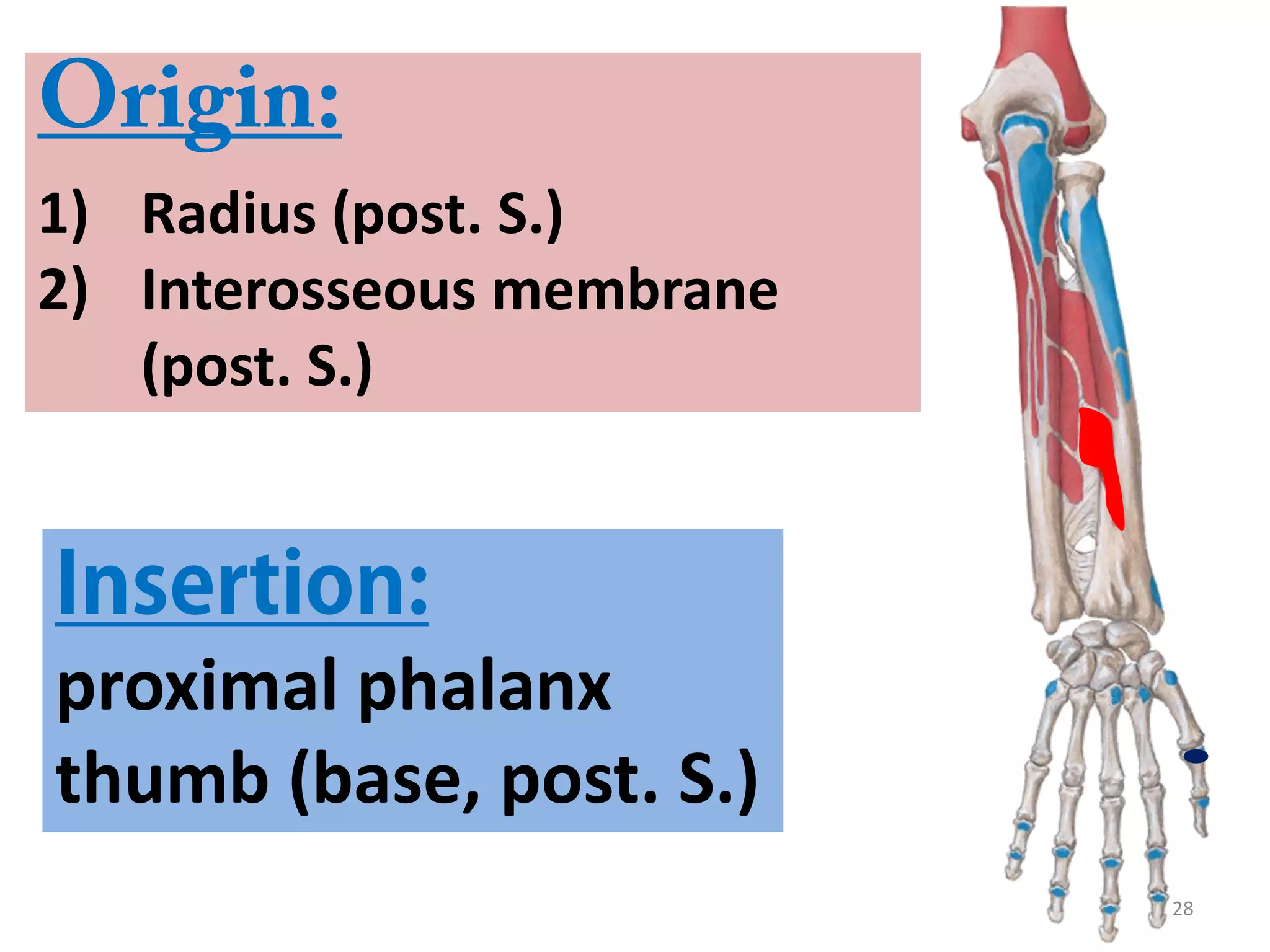
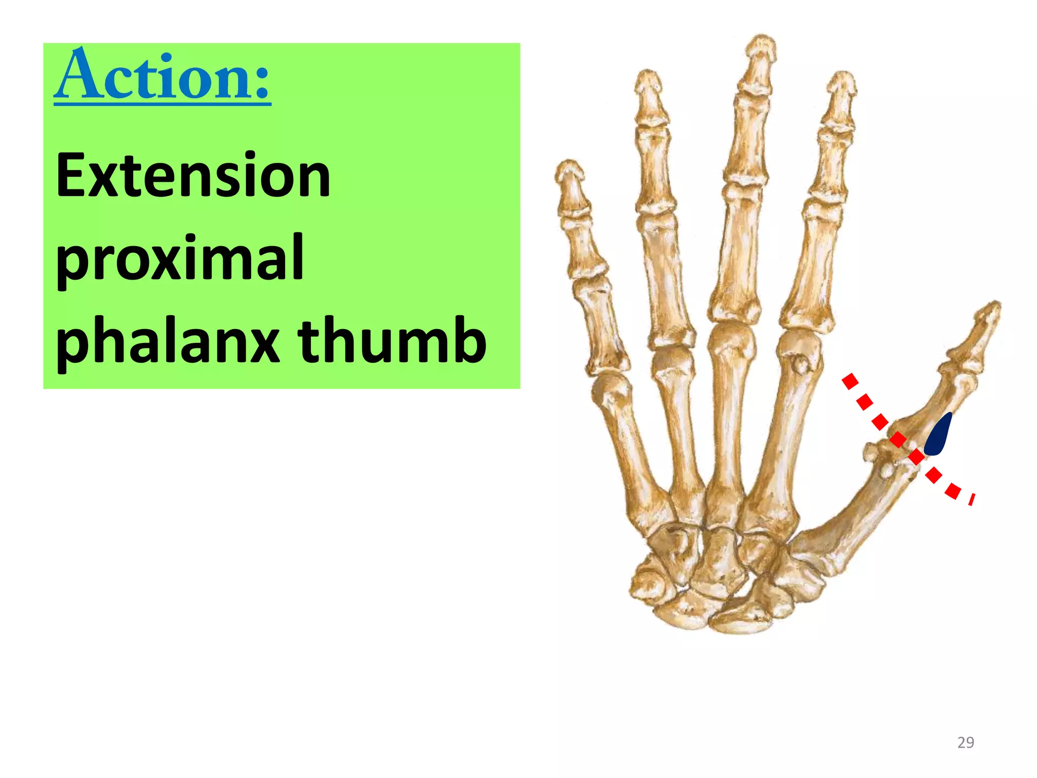
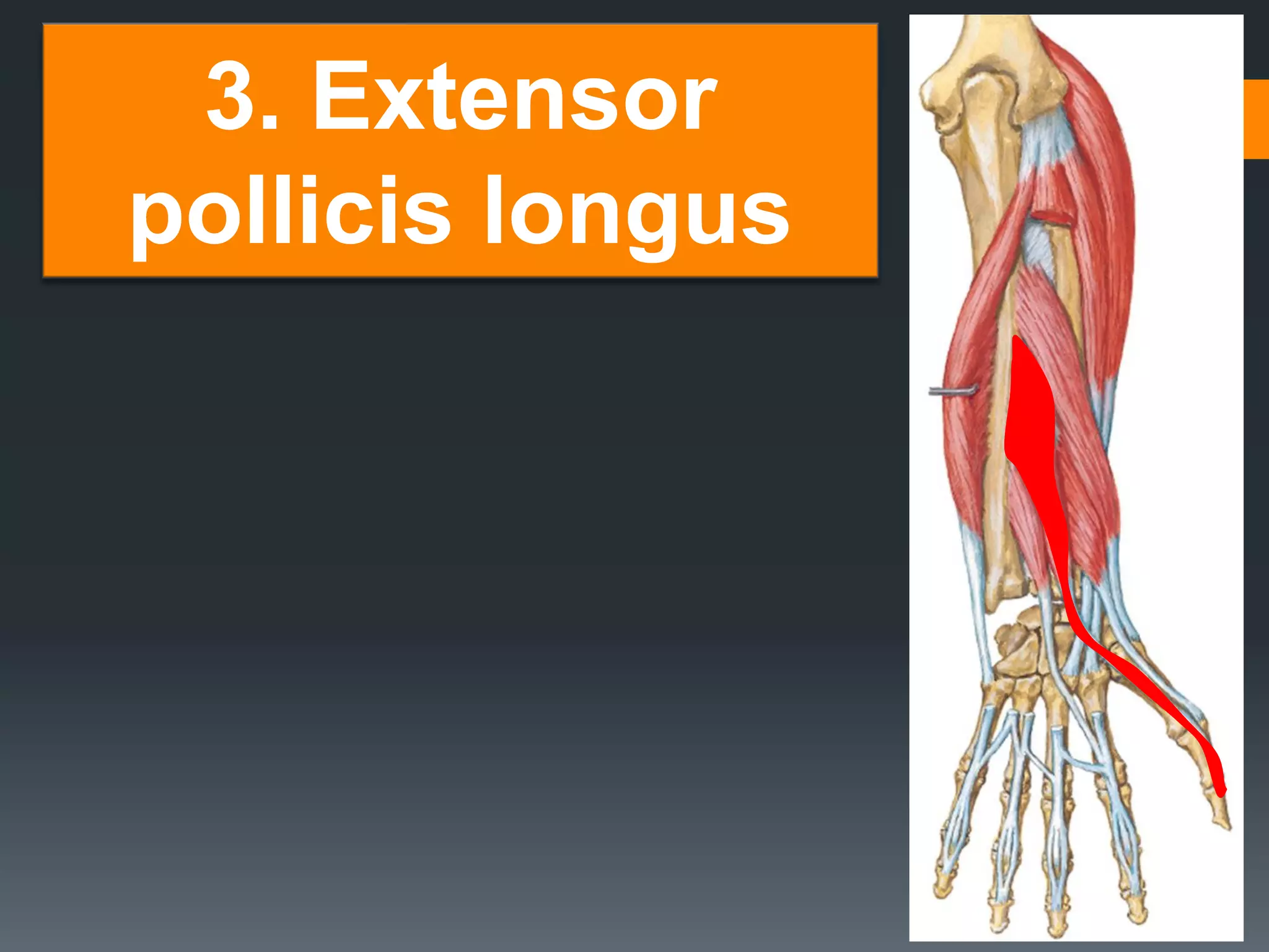
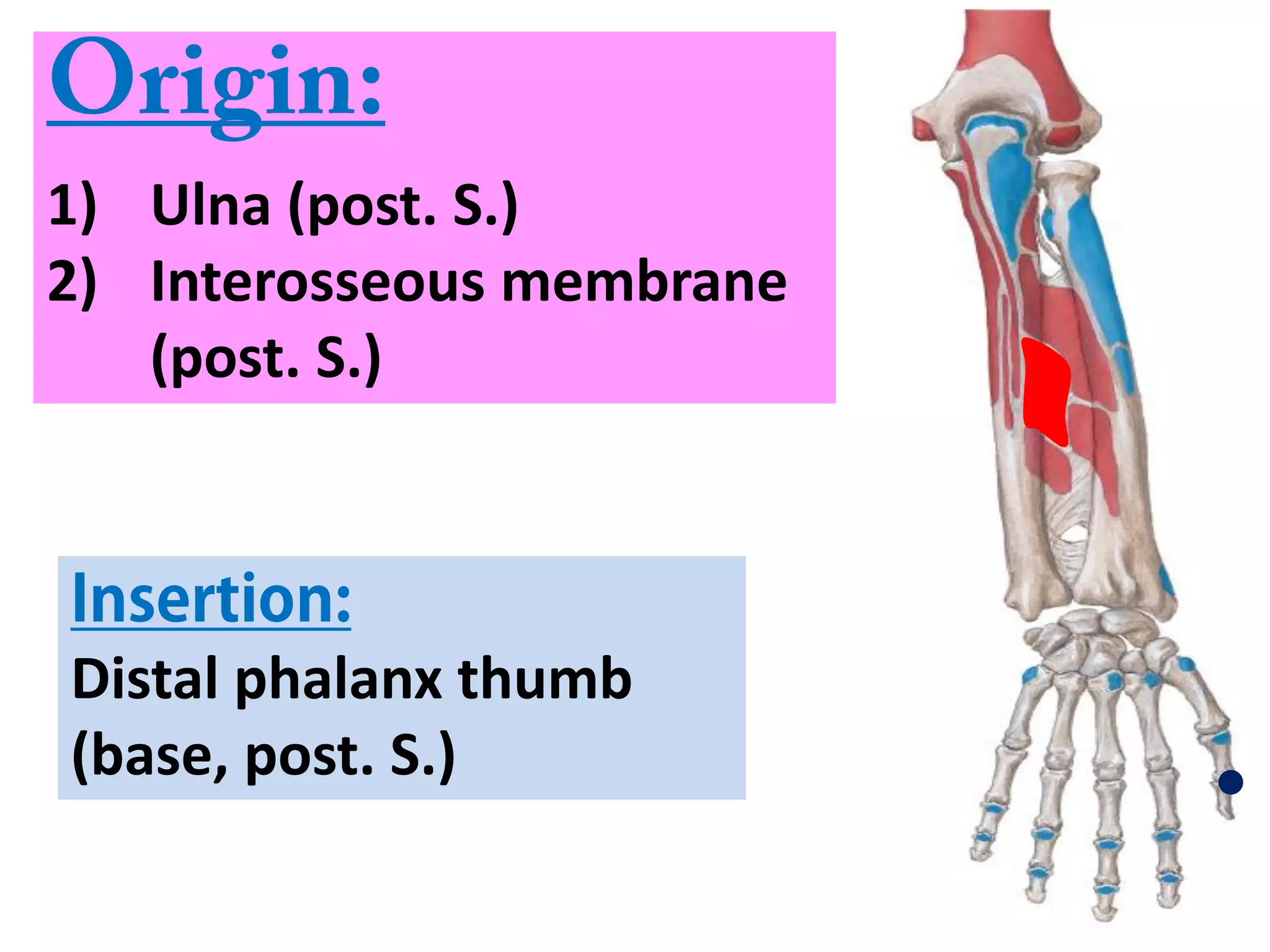
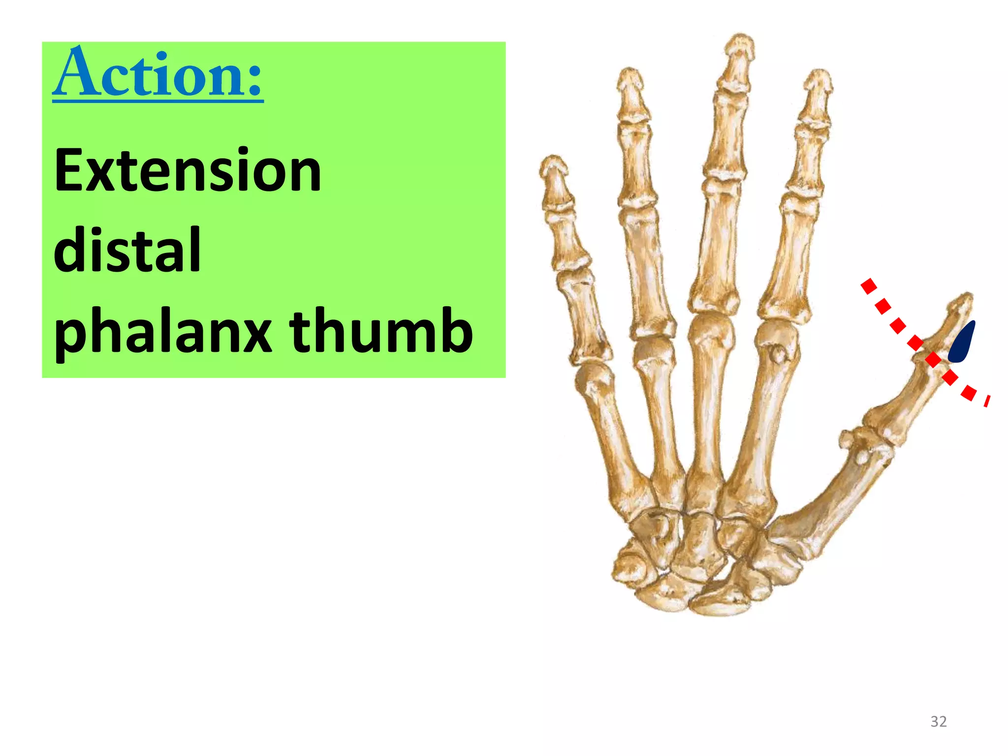
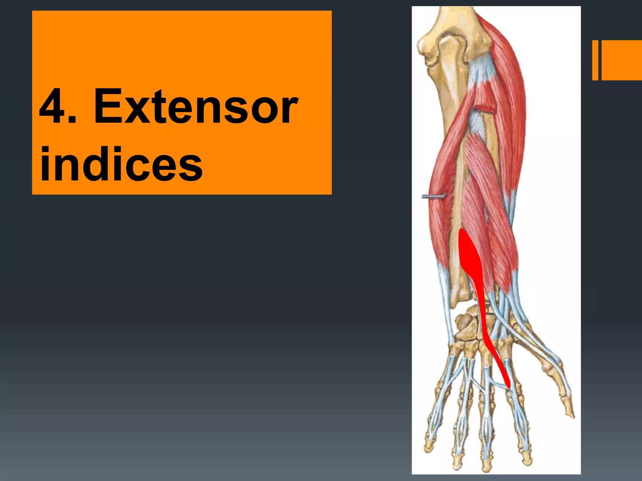
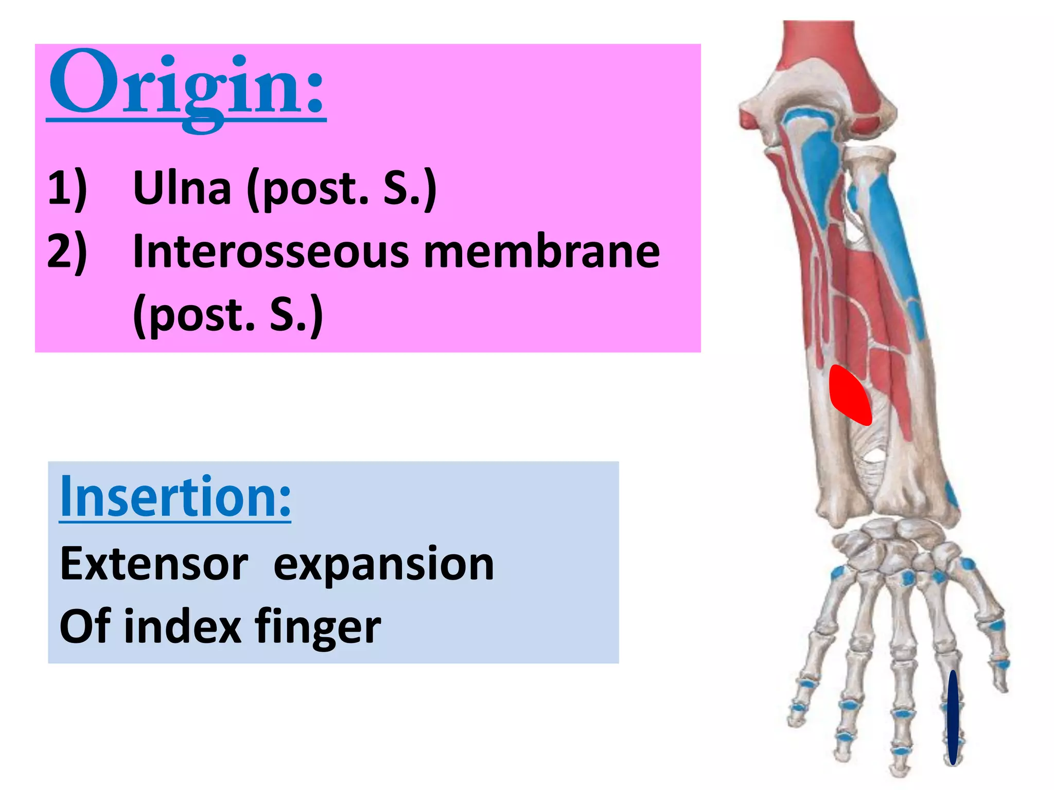
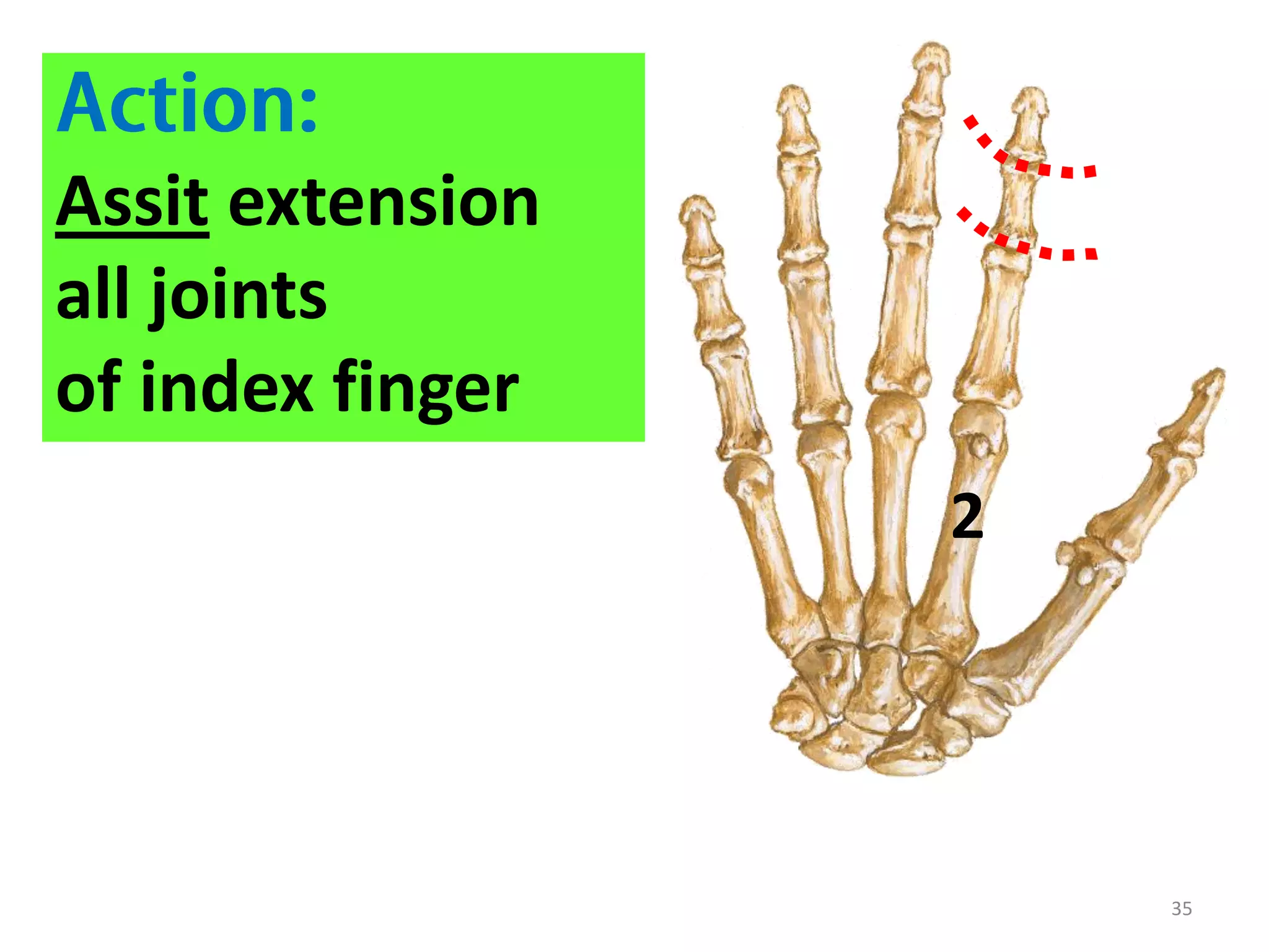
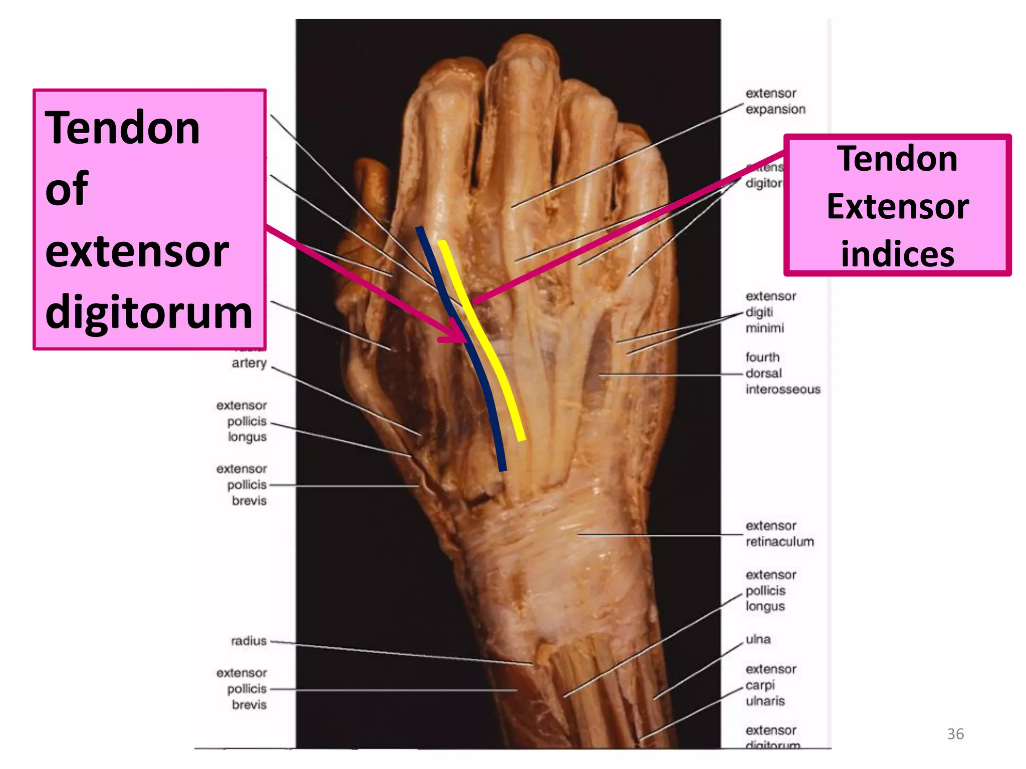
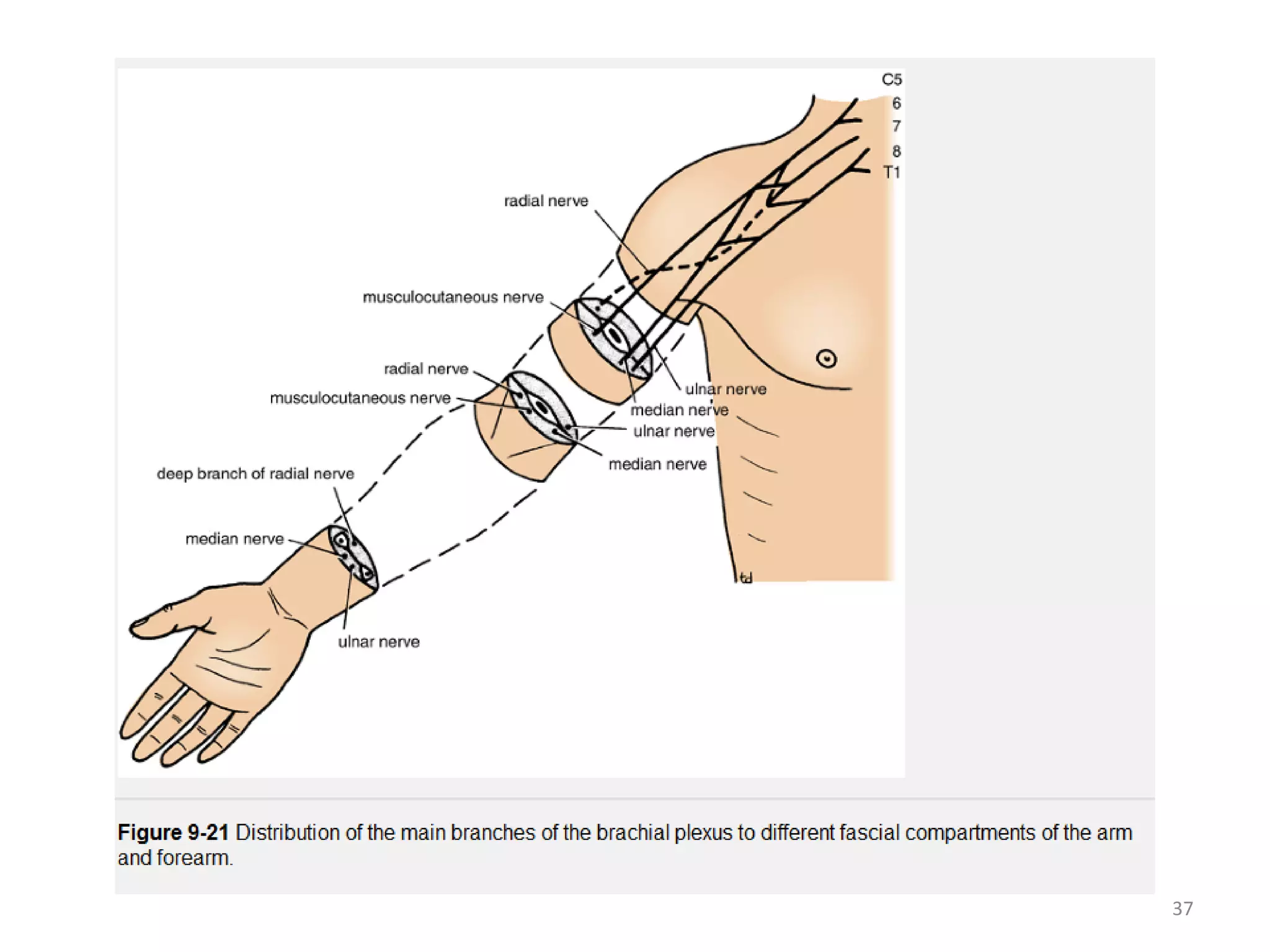
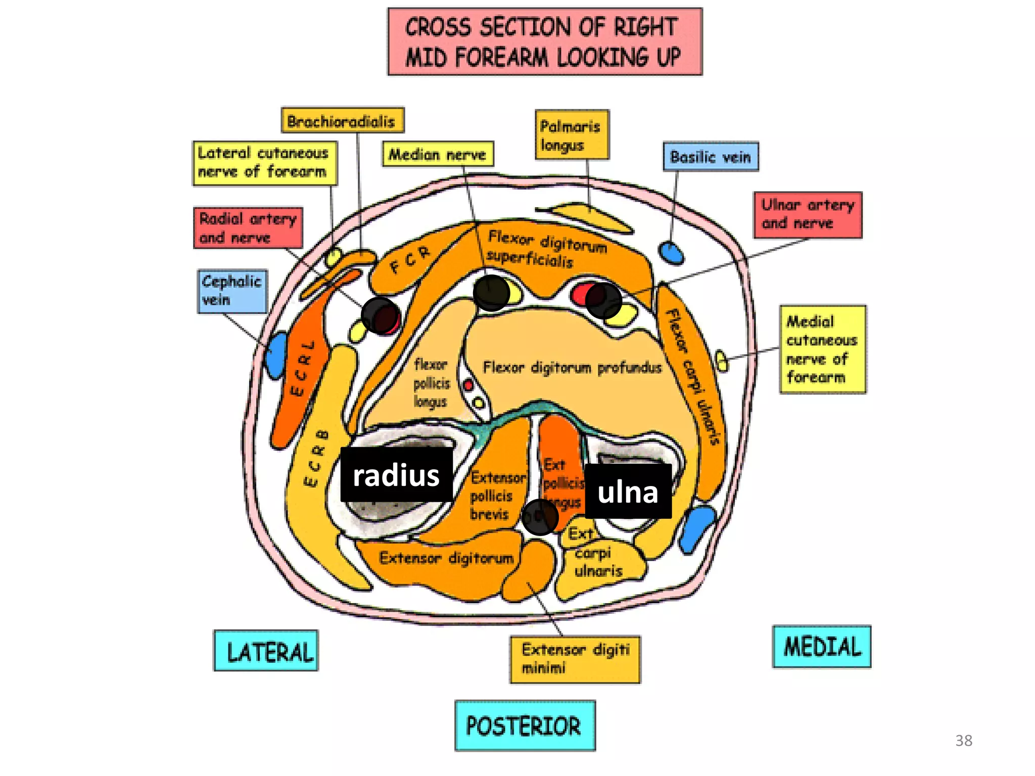
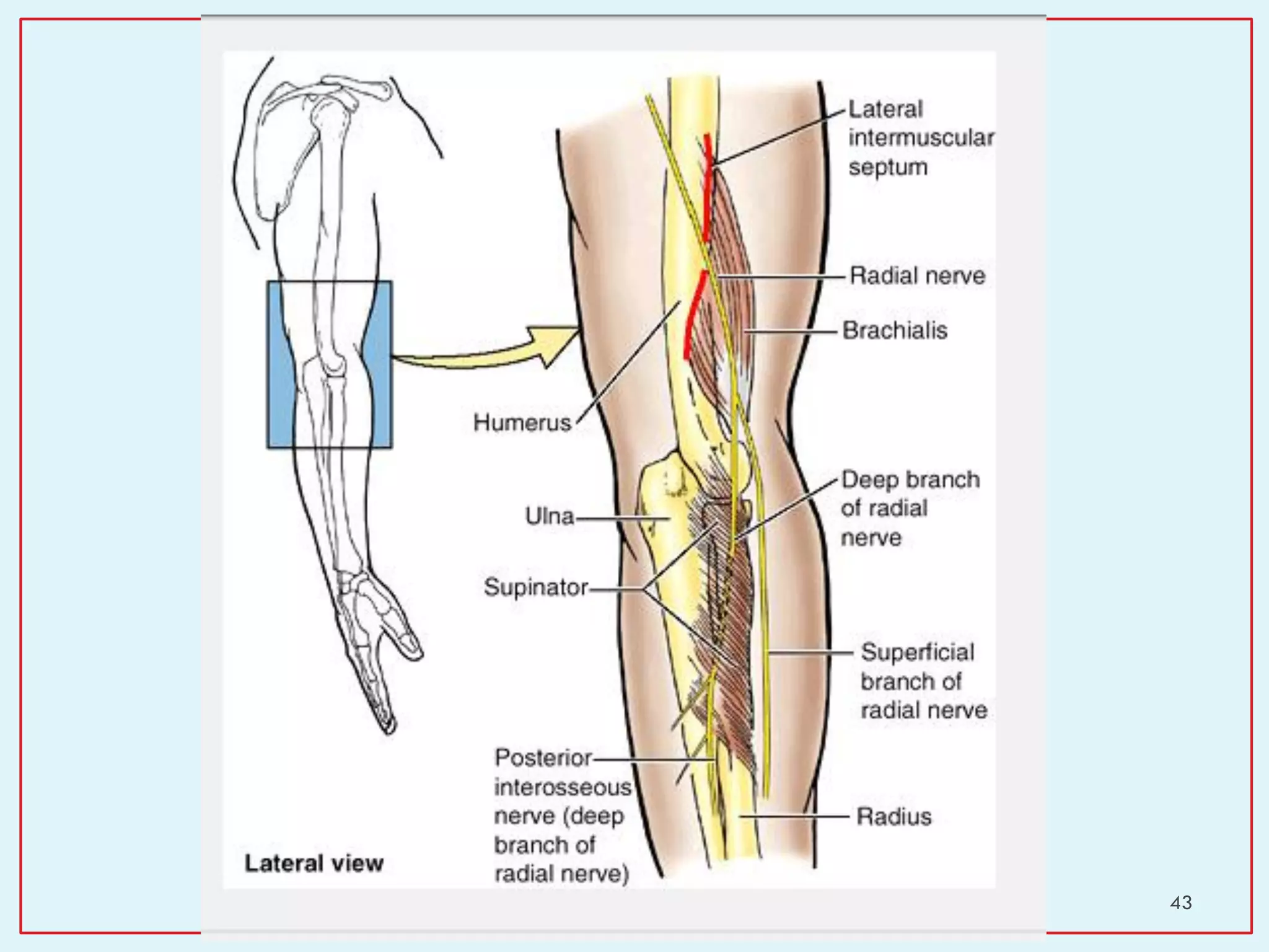
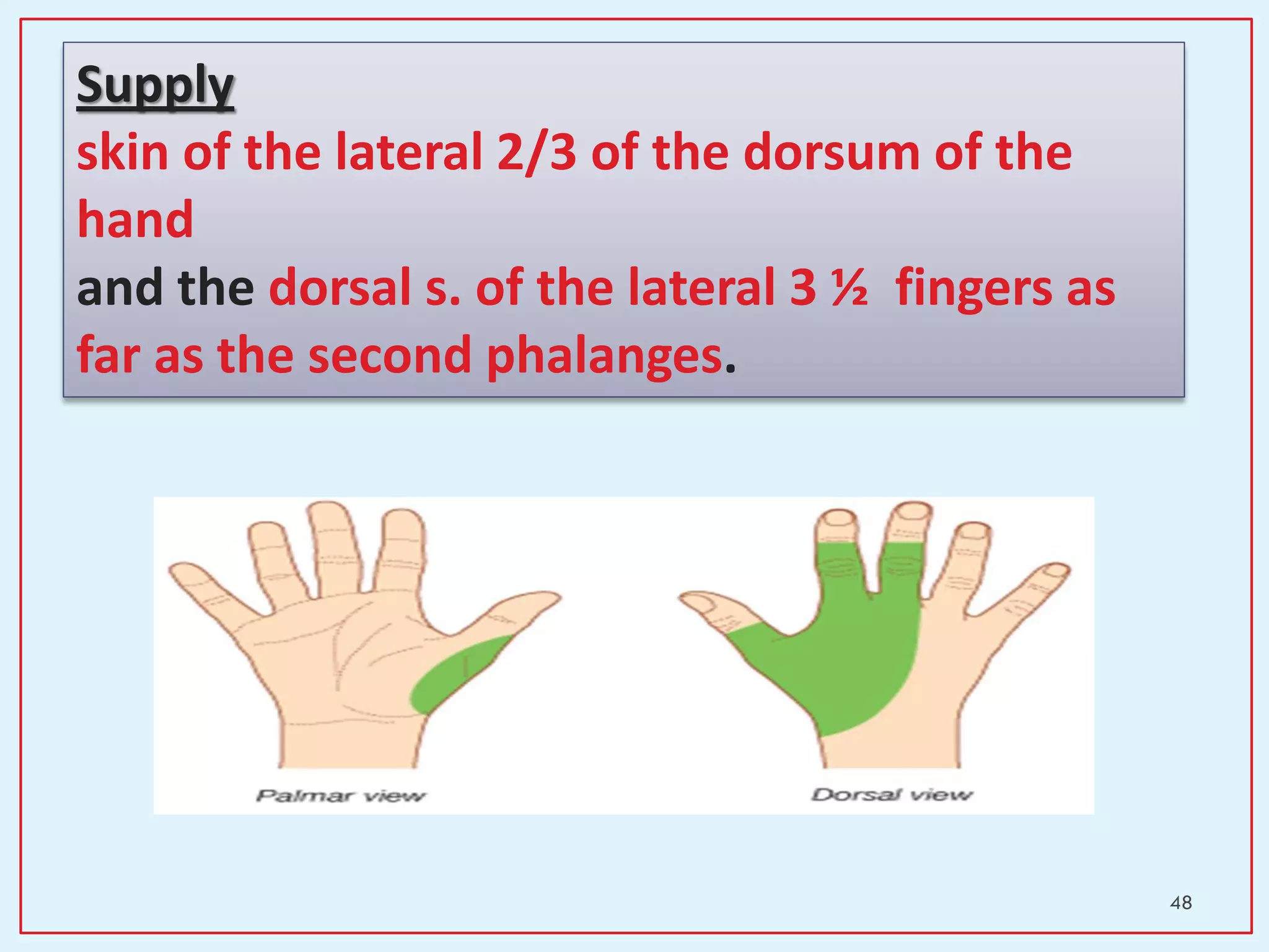
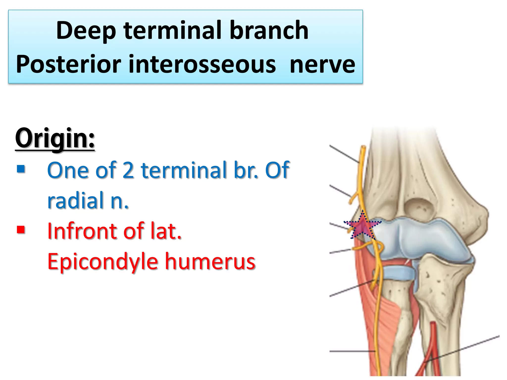
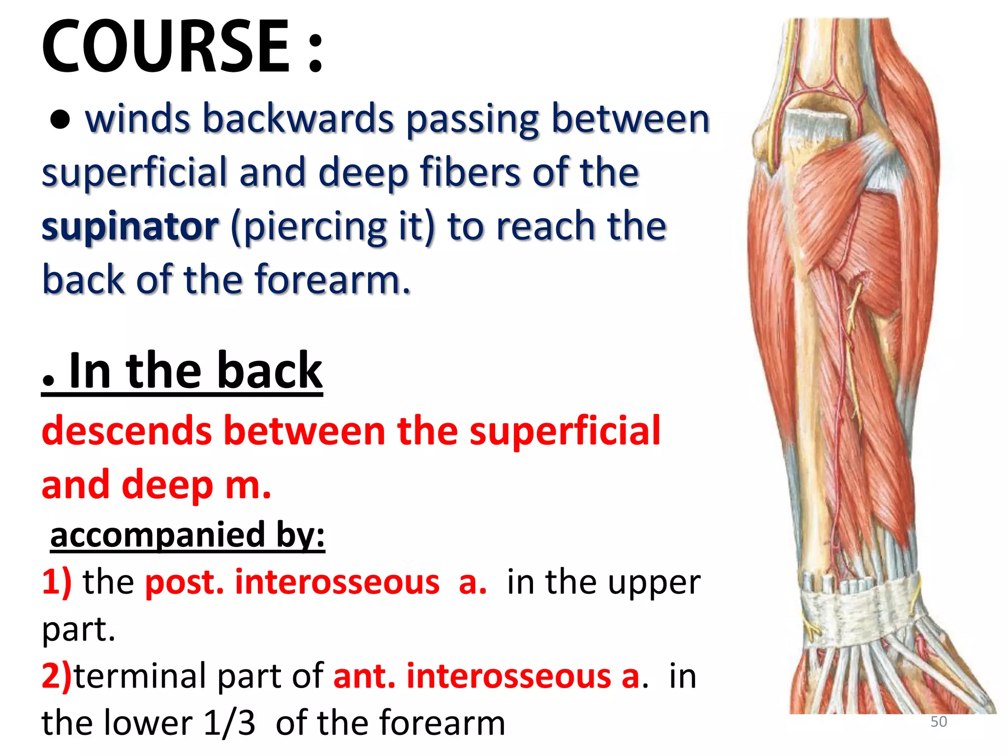
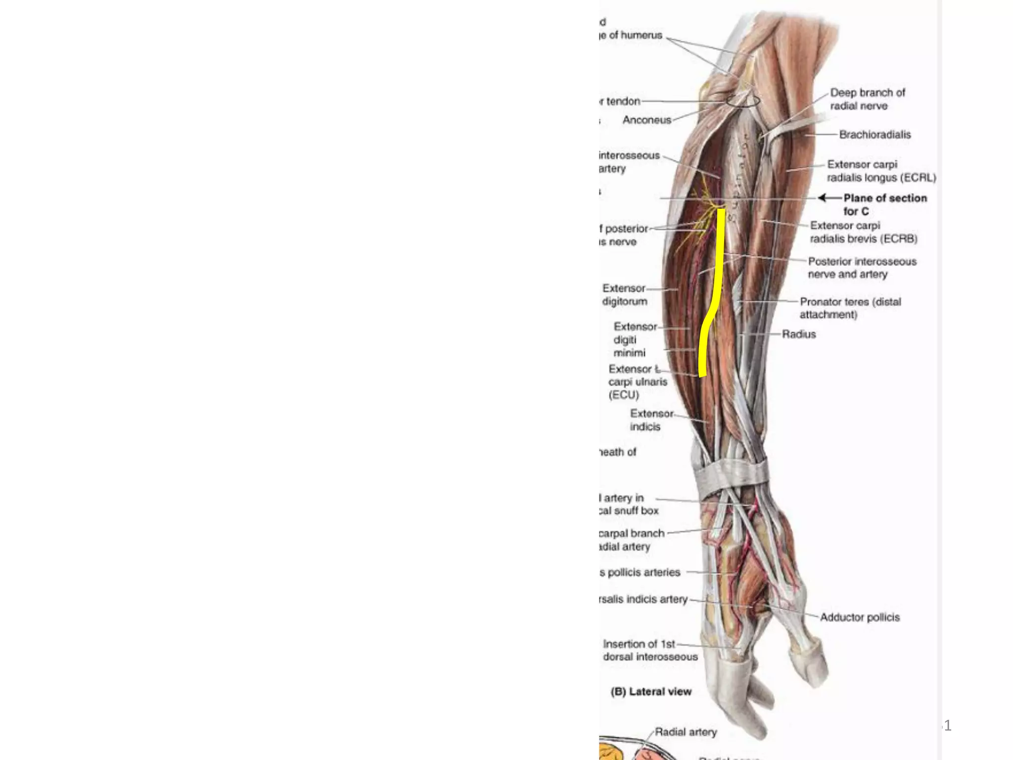
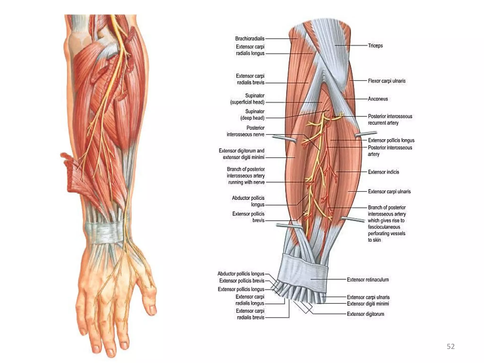
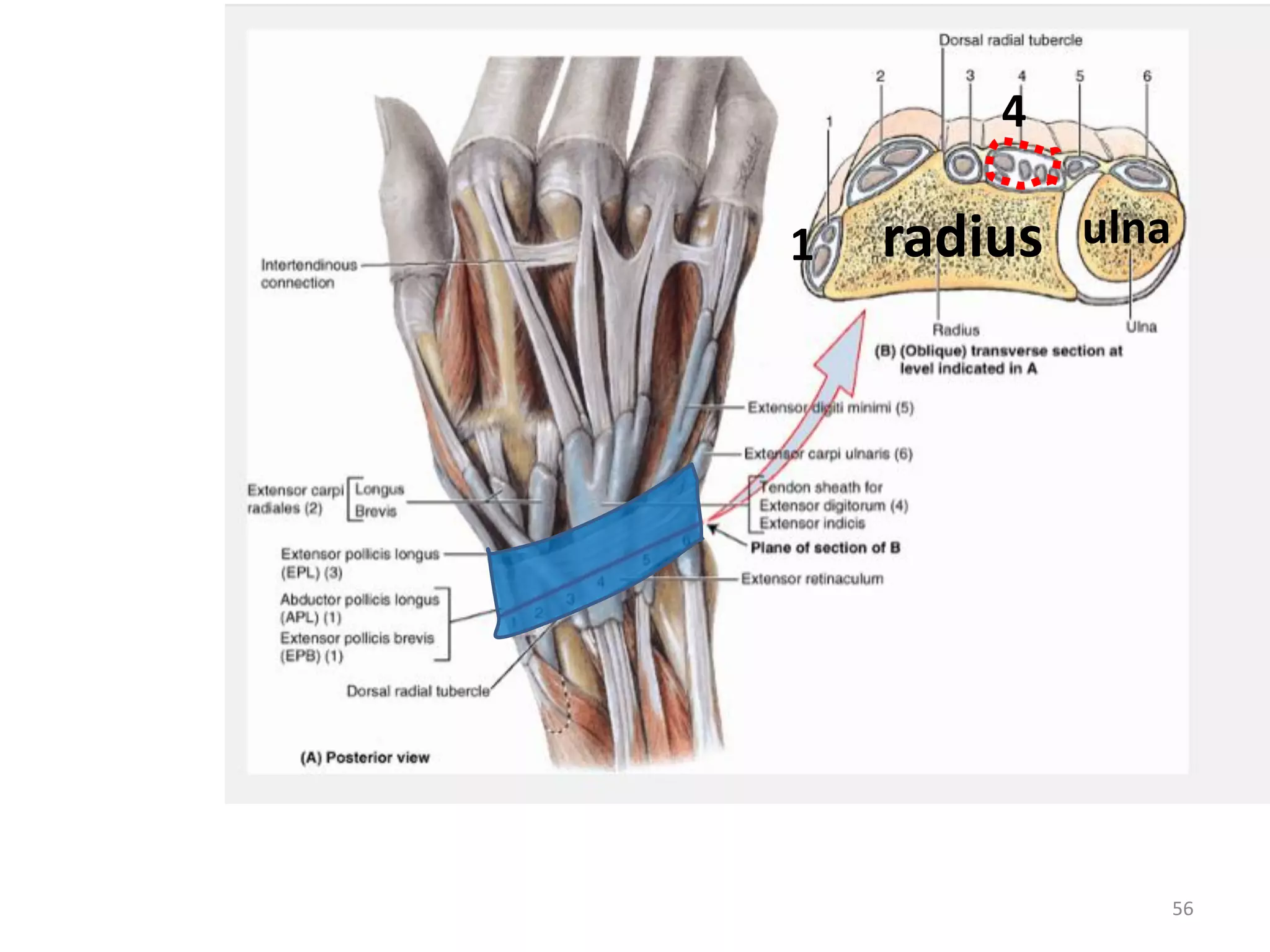
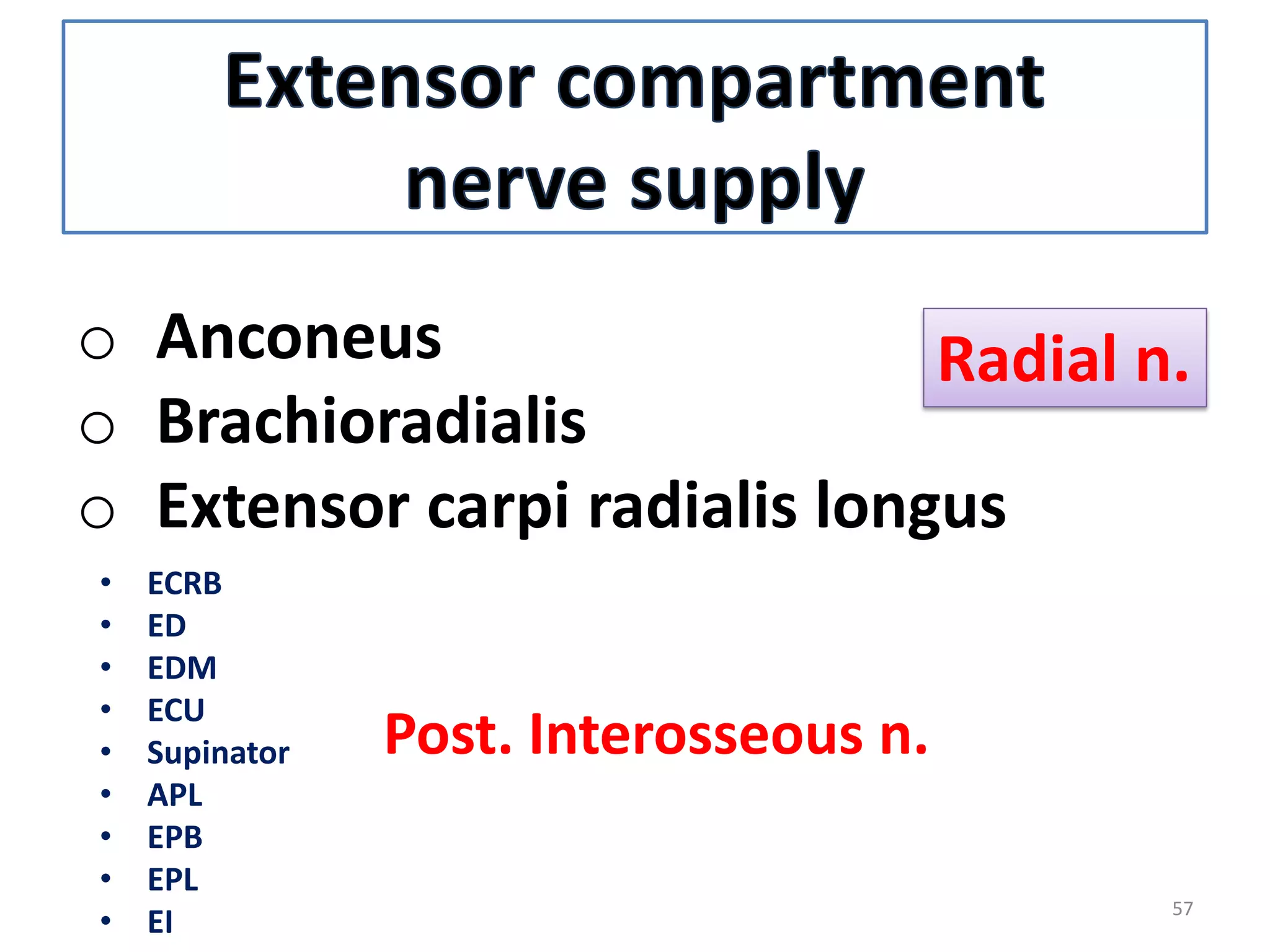
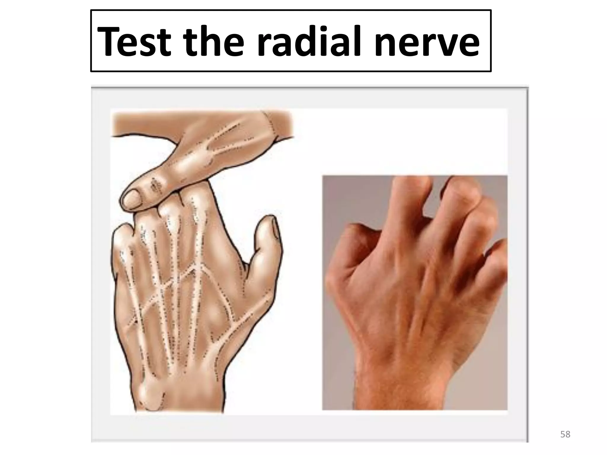
Faculty Of Medicine
Anatomy Department
1
Good morning
2
• deep group of muscles of the back
of forearm
a) Attachment
b) Action
c) N. supply
• Posterior interosseous nerve.
3
4
radius
ulna
Inter-osseous
membrane
Olecranon
coronoid
Trochlear notch
Styloid process
head
neck
tuberosity
Styloid process
5
6
12 muscles
1. Supinator
2. Abductor pollicis
longus
3. Extensor pollicis
brevis
4. Extensor pollicis
longus
5. Extensor indices
1. Anconeus
2. Brachioradialis
3. Extensor carpi radialis
longus
4. Extensor carpi radialis
brevis
5. Extensor digitorum
6. Extensor digiti minimi
7. Extensor carpi ulnaris
7
Muscles of back of forearm
Superficial
group (7)
Deep group
(5)
1
2
3
4
5
6
7
1. Brachioradialis
2. Extensor carpi radialis longus
3. Extensor carpi radialis brevis
4. Extensor digitorum
5. Extensor digiti minimi
6. Extensor carpi ulnaris
7. anconeus
8
Abductor
pollicis longus
Extensor
pollicis brevis
supinator
Extensor pollicis
longus
Extensor indices
9
- 3 of the deep muscles:-
1. abductor pollicis longus,
2. extensor pollicis brevis, and
3. extensor pollicis longus-
- Emerge from between tendons of :
 extensor digitorum &
 extensor carpi radialis brevis
- And pass into the thumb.
10
All superficial muscles cross the elbow joint
(arising from: Tip of lat. Epicondyle)
Deep muscles (except supinator)
 don’t cross the elbow
 Arise from:
 2 bones of forearm & interosseous membrane
 & pass to: thumb and index
While
11
o Anconeus
o Brachioradialis
o Extensor carpi radialis longus
Radial n.
• ECRB
• ED
• EDM
• ECU
• Supinator
• APL
• EPB
• EPL
• EI
Post. Interosseous n.
12
For any muscle
origin
insertion
action
n. supply
13
Ant. view
Post. view
Lat. view
supinator
14


15
Superficial head
1.lat. Epicondyle
2.Radial collateral lig.
3.Annular lig.
trochlea
capitulum
ulna
16
Supinator crest &
fossa
wide shallow
depression at the
lateral surface of
ulna, distal to the
tuberosity of ulna.
Deep head
17
lateral surface of the radius
(superior to: anterior oblique line & to insertion of
pronator teres muscle)
18
Supinator action:
Supinate extended forearm & hand
Supination
• Supination
(lat. Rotation forearm)
1- Thumb directed lateraly
2- palm faces anteriorly
19
Muscles of supination
•In flexed forearm
Biceps
brachii
•In extended
forearm
supinator
20
Pronation
1-Thumb directed medially
2-Palm faces backwards
3- body of radius lies obliquely
across ulna
21
Med. Rotation forearm
22
supination pronation
23
24
1. Abductor
pollicis longus
25
1)Ulna (post. S.)
2)Radius (post. S.)
3)Interosseous membrane.
1st metacarpal bone
(lat. Side of base)
26
1)Abduct &
2)Extend thumb
T
1
27
2. Extensor
pollicis brevis
28
1) Radius (post. S.)
2) Interosseous membrane
(post. S.)
proximal phalanx
thumb (base, post. S.)
29
Extension
proximal
phalanx thumb
30
3. Extensor
pollicis longus
31
1) Ulna (post. S.)
2) Interosseous membrane
(post. S.)
Distal phalanx thumb
(base, post. S.)
32
Extension
distal
phalanx thumb
4. Extensor
indices
33
34
1) Ulna (post. S.)
2) Interosseous membrane
(post. S.)
Extensor expansion
Of index finger
35
Assit extension
all joints
of index finger
2
36
Tendon
Extensor
indices
Tendon
of
extensor
digitorum
37
38
radius ulna
39
40
Radial nerve
(C5,6,7,8 and T1)
 largest branch of the
brachial plexus.
 It is continuation
of the posterior
cord of the plexus
C 5
6
7
8
T1
41
Leave the axilla by passing in lower
Triangular space to back of forearm
Run in
spiral
groove
with
brofunda
brachii a.
42
Pierce lat.
Intermuscular
septum
To reach ant.
Compartment again
43
44
Run in groove
bet. brachialis &
brachioradialis ,
extensor carpi
radialis longus
45
TERMINATION:
divides in front of the lateral epicondyle
into superficial & deep terminal branches
46
47
descends in the upper 2/3
of forearm
under cover of the
brachioradialis.
In the lower 1/3 of the forearm
it passes posteriorly
deep to tendon of
brachioradialis to reach the
dorsum of the hand .
Superficial terminal branch of radial n.
48
Supply
skin of the lateral 2/3 of the dorsum of the
hand
and the dorsal s. of the lateral 3 ½ fingers as
far as the second phalanges.
49
Deep terminal branch
Posterior interosseous nerve
 One of 2 terminal br. Of
radial n.
 Infront of lat.
Epicondyle humerus
50
● winds backwards passing between
superficial and deep fibers of the
supinator (piercing it) to reach the
back of the forearm.
● In the back
descends between the superficial
and deep m.
accompanied by:
1) the post. interosseous a. in the upper
part.
2)terminal part of ant. interosseous a. in
the lower 1/3 of the forearm
51
52
53
BRANCHES OF THE
POSTERIOR INTEROSSEOUS NERVE
Before
piercing supinator
Inside
supinator
After emerging
From supinator
1.Extensor
carpi radialis
brevis
2.Supinaor
supinator
1.Extensor digitorum .
2.Extensor digiti minimi.
3.Extensor carpi ulnaris.
4.Abductor pollicis
longus.
5. Extensor pollicis
brevis.
6. Extensor pollicis
longus.
7. Extensor indicis.
54
N.B.
Post. Interoseous n.
supplies all muscles on
Lat. side & dorsal S. of the forearm.
except
 anconeus
 brachioradialis &
 extensor carpi radialis longus.
55
The deep terminal branch with
anterior interosseous a.
Passes deep to
extensor retinaculum
(in the 4th compartment)
to the back of the carpus to supply joints .
56
radius ulna
4
1
57
o Anconeus
o Brachioradialis
o Extensor carpi radialis longus
Radial n.
• ECRB
• ED
• EDM
• ECU
• Supinator
• APL
• EPB
• EPL
• EI
Post. Interosseous n.
58
Test the radial nerve
59
Thank you
60
61

Back forearm