
Recommended
More Related Content
What's hot
What's hot (20)
Similar to Virus detection identification
Similar to Virus detection identification (20)
Recently uploaded
Recently uploaded (20)
Virus detection identification
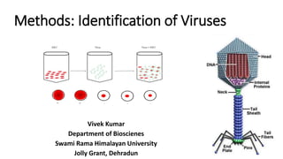
- 1. Methods: Identification of Viruses Vivek Kumar Department of Bioscienes Swami Rama Himalayan University Jolly Grant, Dehradun
- 2. Background • The first methods for detection of bacterial infections were available around 1880. • After staining, bacterial pathogens were recognized in the light microscope because of their size and could be cultivated in culture media. • Viruses evaded this approach, as they are significantly smaller, and as obligate parasites are not able to multiply in cell culture media. • Some viral infections could be associated with specific cellular changes. • Eg. certain depositions in the infected tissue - Negri inclusion bodies in nerve cells during rabies.
- 3. Introduction • In 1954, J. F. Enders was first to propose the adoption of a cell culture-based system to classify animal viruses into four distinct categories consisting • viruses that caused cell degeneration, • those that caused formation of inclusion bodies • and cell degeneration, • those that caused the formation of multinucleated cells (syncytia), • and those that caused no cytopathic effect or cytopathogenic effect (CPE).
- 4. • Since then, cell cultures have been successfully tested routinely for in vitro isolation of viruses. • Presumptive identification of virus types can be made by observing morphological changes produced in host cells (CPE), caused by cytopathogenic viruses. • Common damage to the host cells includes rounding of cells, a change in texture (granular or hyaline—glassy), and formation of a syncytium (fusion of infected cells).
- 5. • The extent of visible damage to cells caused by viral infection varies with strain, • type of host cells, • and multiplicity of infection (MOI); • Consequently CPEs becomes evident after as little as two days to as much as four weeks.
- 6. • Although most laboratories combine traditional and advanced laboratory approaches to optimize viral diagnostics. • Virus isolation from cell cultures still remains a primary method, particularly when viable and nonviable virus need to be differentiated. • If a viable virus has to be isolated, and when infection is not characteristic of any single virus.
- 7. Detection of a Virus • Regardless of the method of cultivation, once a virus has been introduced into a whole host organism, embryo, or tissue-culture cell, a sample can be prepared from the infected host, embryo, or cell line for further analysis under a brightfield, electron, or fluorescent microscope. • Cytopathic effects (CPEs) are distinct observable cell abnormalities due to viral infection. • CPEs can include loss of adherence to the surface of the container, changes in cell shape from flat to round, shrinkage of the nucleus, vacuoles in the cytoplasm, fusion of cytoplasmic membranes and the formation of multinucleated syncytia, inclusion bodies in the nucleus or cytoplasm, and complete cell lysis.
- 8. Paramyxovirus Syncytium and faint basophilic cytoplasmic inclusion bodies Poxyvirus Pink eosinophilic cytoplasmic inclusion bodies (arrows) and cell swelling
- 9. Herpesvirus Cytoplasmic stranding (arrows) and nuclear inclusion bodies (dashed arrow) Adenovirus Cell enlargement, rounding, and distinctive grape-like clusters
- 10. Hemagglutination Assay • A serological assay is used to detect the presence of certain types of viruses in patient serum. • Serum is the straw-colored liquid fraction of blood plasma from which clotting factors have been removed. • Serum can be used in a direct assay called a hemagglutination assay to detect specific types of viruses in the patient’s sample. • Hemagglutination is the agglutination (clumping) together of erythrocytes (red blood cells).
- 11. • Many viruses produce surface proteins or spikes called hemagglutinins that can bind to receptors on the membranes of erythrocytes and cause the cells to agglutinate. • Hemagglutination is observable without using the microscope, but this method does not always differentiate between infectious and noninfectious viral particles, since both can agglutinate erythrocytes.
- 12. • To identify a specific pathogenic virus using hemagglutination, we must use an indirect approach. • Proteins called antibodies, generated by the patient’s immune system to fight a specific virus, can be used to bind to components such as hemagglutinins that are uniquely associated with specific types of viruses. • The binding of the antibodies with the hemagglutinins found on the virus subsequently prevent erythrocytes from directly interacting with the virus.
- 13. • So when erythrocytes are added to the antibody-coated viruses, there is no appearance of agglutination; agglutination has been inhibited. • We call these types of indirect assays for virus-specific antibodies hemagglutination inhibition (HAI) assays. • HAI can be used to detect the presence of antibodies specific to many types of viruses that may be causing or have caused an infection in a patient even months or years after infection.
- 16. Nucleic Acid Amplification Test • Nucleic acid amplification tests (NAAT) are used in molecular biology to detect unique nucleic acid sequences of viruses in patient samples. • Polymerase chain reaction (PCR) is an NAAT used to detect the presence of viral DNA in a patient’s tissue or body fluid sample. • PCR is a technique that amplifies (i.e., synthesizes many copies) of a viral DNA segment of interest. • Using PCR, short nucleotide sequences called primers bind to specific sequences of viral DNA, enabling identification of the virus.
- 17. • Reverse transcriptase-PCR (RT-PCR) is an NAAT used to detect the presence of RNA viruses. • RT-PCR differs from PCR in that the enzyme reverse transcriptase (RT) is used to make a cDNA from the small amount of viral RNA in the specimen. • The cDNA can then be amplified by PCR. • Both PCR and RT-PCR are used to detect and confirm the presence of the viral nucleic acid in patient specimens.
- 18. Enzyme Immunoassay • Enzyme immunoassays (EIAs) rely on the ability of antibodies to detect and attach to specific biomolecules called antigens. • The detecting antibody attaches to the target antigen with a high degree of specificity in what might be a complex mixture of biomolecules. • Also included in this type of assay is a colorless enzyme attached to the detecting antibody. • The enzyme acts as a tag on the detecting antibody and can interact with a colorless substrate, leading to the production of a colored end product.
- 19. • EIAs often rely on layers of antibodies to capture and react with antigens, all of which are attached to a membrane filter. • EIAs for viral antigens are often used as preliminary screening tests. • If the results are positive, further confirmation will require tests with even greater sensitivity, such as a western blot or an NAAT.
- 23. Immunofluorescence (IF) Assay • Immunofluorescence (IF) technique is widely used for rapid detection of virus infections by identifying virus antigens in clinical specimens. • IF staining is usually considered very rapid (about 1 to 2 hr) and overall gives a sensitive and specific viral identification. • Unfortunately, IF technique may not able to confirm the identity of all virus strains, for instance viruses of the “enterovirus” group.
- 24. • Since most monoclonal antibodies (MAbs) for enteroviral identification have been shown to lack sensitivity, while cross-reactivity with rhinoviruses is extremely common. • In contrast, IF has been successfully used for better management of influenza virus infection and surveillance of influenza virus activity.
- 25. Molecular Methods • The development of molecular methods for the direct identification of a specific viral genome from the clinical sample is one of the greatest achievements of the 21st century. • Clearly nucleic acid amplification techniques including PCR, nucleic acid sequence-based amplification (NASBA) and Lawrence Livermore Microbial Detection Array (LMDA) are proven technology leaders for rapid detection and molecular identification for most known human viruses. • As such, PCR allows the in vitro amplification of a specific region of DNA sequences by a factor of 106 and thus considered as an extremely sensitive detection technique.
- 26. Viral Plaque Assay • Viral plaque assay is one of the most widely used methods in virology to purify a clonal population of virus or to determine viral titer. • Renato Dulbecco was first to develop this procedure in 1952 to calculate the infectivity of bacteriophages stocks. • A plaque usually is the result of infection of the cell by a single virion on the host cell monolayer. • As such, to perform a plaque assay, susceptible host cell monolayer is infected with 10-fold serial dilutions of virus.
- 27. Viral Plaque Assay Determination
- 28. • After an incubation period, monolayers are then covered with a semi-solid nutrient medium, (most commonly agar) to prevent the virus from spreading from host cells to other nearby uninfected cells. • As a result, each infectious particle produces a small circular zone in the monolayer called a plaque, while uninfected cells surround the plaques. • Eventually the plaque becomes large enough to be visible to the naked eye or with light microscopy.
- 29. Quantal Assays - TCID50, LD50, EID50 • Although plaque assay is an extremely useful method for determining viral titers, however there are several virus types which do not form plaques in culture. • Alternative procedures such as • TCID50, (Tissue culture infectious dose) • LD50, (Lethal dose) • EID50 (Embryo Infectious Dose )
- 30. • Assays are being used to determine the infectious titer of any such virus types, which can cause cytopathic effects (CPE) in tissue culture over a reasonable period of 5 to 20 days while cells in culture remain viable.
- 31. • However, since not all virus types cause CPE in tissue culture, and therefore cell line and virus must be carefully matched in order to see a cytopathic effect. • TCID50 is the tissue culture infectious dose defined as that dilution of virus required to infect 50% of the cell monolayers. • The Infectivity Titre is established by carrying out a titration.
- 32. • The unit of measurement of infectivity of avirulent Newcastle disease virus is the 50 percent Embryo Infectious Dose or EID50. • One EID50 unit is the amount of virus that will infect 50 percent of inoculated eggs. • *Lethal Dose, 50%" or median lethal dose, it is the amount of the substance required to kill 50% of the test population.
- 33. Immunofluorescence Foci Assay (IFA) • The IFA (also known as fluorescent foci assay FFA) is a rapid method of virus titration. • This technique allows the quantification of virus in cell lines, which does not support plaque formation or do not exhibit detectable CPE (to perform TCID50). • For example, hepatitis A virus (HAV) does not produce visible CPE without multiple passages in cell culture.
- 34. • In contrast to other infectivity- based assays, IFA is not dependent on the ability of induction of cell death, but it utilizes an antibody- based staining methods to detect virally infected cells as early as two days. • Overall IFA is considered more sensitive and faster than traditional plaque assays or TCID50, however it sometimes could be quite expensive, due to the cost of antibodies used.
- 35. X-Ray Crystallography • A method that is used to find out the arrangement of atoms within a crystal is known as X-ray crystallography. • A beam of X-rays scatters into many directions when it strikes a crystal. • According to the angles and intensities of these scattered beams, a three dimensional picture of the density of electrons within a crystal is produced from a crystallography. • The mean positions of the atoms in the crystal, their chemical bonding, their disorder and other information can be determined from the electron density.
- 37. Summary • A general method for obtaining and culturing cells. • Primary cultures contain cells obtained from an animal and they have a limited lifespan. • A basic method for virus purification using centrifugation. • Methods to count infectious virus particles (plaque assays and endpoint dilution). • The basic features of common techniques used to detect viruses and antibodies.