
Recommended
More Related Content
Similar to Presentation virology.pptx
Similar to Presentation virology.pptx (20)
More from ChinjuJoseSajith
More from ChinjuJoseSajith (20)
Recently uploaded
Recently uploaded (20)
Presentation virology.pptx
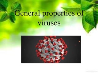
- 2. VIRUSES Viruses are the smallest obligate intracellular infective agents containing only one type of nucleic acid (DNA or RNA) as their genome. They do not possess a cellular organization and lack the enzymes necessary for protein and nucleic acid synthesis, They are resistant differences to antibiotics.
- 3. I. MORPHOLOGY OF VIRUSES. Size Viruses are much smaller than other organisms. The extracellular infectious virus particle is called the virion. The size of viruses ranges from 20 to 300 nm in diameter. The largest virus is the smallpox virus (300 nm) and the smallest is the parvovirus (20 nm)
- 5. Structure and Symmetry 1. STRUCTURE The virion consists of a nucleic acid core (genome) surrounded by a protein coat, the capsid. The capsid together with the enclosed nucleic acid is known as the nucleocapsid. The capsid is composed of a c large number of protein subunits (polypeptides) which are known as capsomers. Certain viruses also contain e envelope that surrounds the nucleic acid. It is lipoprotein in nature.
- 7. SYMMETRY Three types of symmetry are determined by the arrangement of capsid around the nucleic acid core. (i) Icosahedral (cubical) symmetry (ii) Helical symmetry (iii) Complex symmetry SHAPE The overall shape of virus particles varies in different groups . Pox virus is brick-shaped and rabies virus is bullet shaped.
- 9. CHEMICAL PROPERTIES NUCLEIC ACID Viruses contain only one kind of nucleic acid, either single or double stranded DNA or RNA. VIRAL PROTEIN AND LIPIDS Viruses contain protein which makes up the capsid. In case of enveloped viruses, they contain lipids (present in envelope).
- 10. CULTIVATION OF VIRUSES As viruses multiply only in living cells, they cannot be grown on any of the inanimate culture medium. Three methods are employed for the cultivation of viruses: A. Animal inoculation. B .Embryonated egg inoculation C. Cell culture Parvovirus
- 11. • A. ANIMAL INOCULATION Infant (suckling) mice are used in the isolation of arboviruses and coxsackie viruses, many of which do not grow in any other system. After inoculation, animals are observed for signs of disease or death. Later on, they are sacrificed and tissues are tested for the presence of virus.
- 13. B. Embryonated Egg Inoculation • Embryonated hen's eggs (7 to 12 days old) are inoculated by one of the several routes such as amniotic sac yolk sac and allantoic cavity
- 14. C. CELL CULTURE/TISSUE CULTURE This is the type of culture routinely employed for diagnostic virology. Cell cultures are classified into three different types (i) Primary cell culture (ii) Diploid cell strains (iii)Continuous cell lines
- 16. • (i) Primary cell cultures These are normal cells freshly taken from the organs of animal or human being and cultured. They are capable of very limited growth in culture, perhaps 5- 10 divisions at the most. (ii) Diploid cell strains They can be subcultured for a limited number. After about 50 serial subcultures they undergo senescence' and the cell strain is lost.
- 17. (iii) Continuous cell lines These are cells of a single type that are capable of indefinite growth in vitro. They are usually derived from cancerous tissue. They can be serially cultivated indefinitely, therefore, they are termed continuous cell lines.
- 18. LABORATORY DIAGNOSIS OFVIRAL INFECTIONS • A. Direct demonstration of virus and its components • B. Isolation of virus • C. Detection of the specific antibodies DIRECT DEMONSTRATION OF VIRUSAND ITS COMPONENTS 1. Electron Microscopy The detection of virus by electron microscopy (EM) is being used increasingly especially for viruses that are difficult to culture. Clinical applications of electron microscopy include detection of rotavirus and hepatitis
- 19. 2. Fluorescent Microscopy 3. Light Microscopy Inclusion bodies in tissue sections may be detected by light microscopy. Demonstration of inclusion bodies helps in diagnosis of some viral infections.
- 20. 4.Viral Antigens These may be detected by enzyme linked immunosorbent assay (ELISA) and latex agglutination.
- 21. 5. Polymerase Chain Reaction (PCR) With a PCR technique, a target DNA sequence can be amplified to the point where it can readily be identified using labelled probes in a hybridisation assay. Thus viral DNA extracted from a very small number of virions or infected cells can be detected. The technique can be used for the diagnosis of infections caused by HIV-1, HIV-2, hepatitis B virus, and Epstein-Barr virus.
- 22. • C. DETECTION OF SPECIFIC ANTIBODIES The demonstration of a rise in titre of antiviral antibodies during the course of a disease is strong evidence that it is the aetiological agent. For this, paired sera should be collected from the patient, the acute sample collected early in the course of the disease and the convalescent sample collected ten to fourteen days later. Examination of a single sample of serum is meaningful when IgM specific antibodies are detected.
- 23. CLASSIFICATION OF VIRUSES • Viruses are classified on the basis of physical and chemical biological, properties. Viruses are broadly classified into DNA and RNA viruses
- 24. PRIONS Prions are infectious proteins without any detectable nucleic acid. They are highly resistant to physical and chemical agents. They are resistant to heat (90°C for three minutes), UV rays and nucleases and sensitive to proteases. They produce slow infections with long incubation period (in years).