Immunohistochemistry Antibody Validation Report for Anti-FoxO1 Antibody (STJ93123)

Transcription factor that is the main target of insulin signaling and regulates metabolic homeostasis in response to oxidative stress. Binds to the insulin response element (IRE) with consensus sequence 5'-TT[G/A]TTTTG-3' and the related Daf-16 family binding element (DBE) with consensus sequence 5'-TT[G/A]TTTAC-3'. Activity suppressed by insulin. Main regulator of redox balance and osteoblast numbers and controls bone mass. Orchestrates the endocrine function of the skeleton in regulating glucose metabolism. Acts synergistically with ATF4 to suppress osteocalcin/BGLAP activity, increasing glucose levels and triggering glucose intolerance and insulin insensitivity. Also suppresses the transcriptional activity of RUNX2, an upstream activator of osteocalcin/BGLAP. In hepatocytes, promotes gluconeogenesis by acting together with PPARGC1A and CEBPA to activate the expression of genes such as IGFBP1, G6PC and PCK1. Important regulator of cell death acting downstream of CDK1, PKB/AKT1 and SKT4/MST1. Promotes neural cell death. Mediates insulin action on adipose tissue. Regulates the expression of adipogenic genes such as PPARG during preadipocyte differentiation and, adipocyte size and adipose tissue-specific gene expression in response to excessive calorie intake. Regulates the transcriptional activity of GADD45A and repair of nitric oxide-damaged DNA in beta-cells. Required for the autophagic cell death induction in response to starvation or oxidative stress in a transcription-independent Anti-FoxO1-http://www.stjohnslabs.com/foxo1-antibody-p-92348 Join our Antibody Validation Project - http://www.stjohnslabs.com/services/antibody-validation

Recommended
Recommended
More Related Content
What's hot
What's hot (20)
Viewers also liked
Viewers also liked (20)
Similar to Immunohistochemistry Antibody Validation Report for Anti-FoxO1 Antibody (STJ93123)
Similar to Immunohistochemistry Antibody Validation Report for Anti-FoxO1 Antibody (STJ93123) (13)
More from St John's Laboratory Ltd
More from St John's Laboratory Ltd (20)
Recently uploaded
Recently uploaded (20)
Immunohistochemistry Antibody Validation Report for Anti-FoxO1 Antibody (STJ93123)
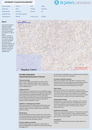
- 1. Figure: Immunohistochemical analysis of paraffin embedded Human uterus tissue. 1: FoxO1 Polyclonal Antibody was diluted at 1:200 (4 degree Celsius,overnight). 2: Sodium citrate pH 6.0 was used for antibody retrieval (>98 degree Celsius,20min). 3: Secondary antibody was diluted at 1:200 (room temperature, 30min). Negative control was used by secondary antibody only. Report Number 93123-a Host Rabbit Application IHC-P Clonality Polyclonal Model Number STJ93123 Clone ID NA Antibody Name Anti-FoxO1 antibody Testing Species HUMAN Testing Tissue UTERUS ANTIBODY VALIDATION REPORT b. (A small amount of distilled water was added into the incubation box to prevent evaporation of antibody). 18. Secondary antibody incubation a. Slides were washed 3 times, with PBS on a shaker for 5min. Shortly after the slides were dried the corresponding secondary antibody solution was added (HRP labelled), covering the tissues, and incubated at room temperature for 30min. b. 19. DAB staining a. Slides were washed 3 times, with PBS on a shaker for 5min. b. Shortly after, the slides were dried and fresh DAB staining buffer was added inside the circles. The staining time was adjusted under a microscope. Yellow-brown colour represented a positive result. Slides were washed with water to stop the staining. c. 20. Haematoxylin staining a. Haematoxylin was used to counter-staining for 1min, and then the slides were washed with water. 1% Hydrochloric acid and alcohol was added for several seconds and then washed with water. Ammonia was used to reveal blue colour, and then flushed with water. b. 21. Desolation and Clearing i. Slides were incubated sequentially into: 75% alcohol 5min, 85% alcohol 5min, Anhydrous ethanol - 5min, Anhydrous ethanol - 5min & Xylene - 5min. Shortly after slides were dried and neutral gum was used to seal the slides. ii. 22. Visualization a. Results were validated with microscope, and the slides were scanned. Paraffin-Embedded Immunohistochemistry Protocol 12. 13. Tissue processing a. Slides were incubated sequentially into Xylene; 15min – Xylene, 15min - Anhydrous ethanol, 5min - Anhydrous ethanol, 5min - 85% alcohol, 5min - 75% alcohol & 5min – wash in distilled water. b. 14. Antigen retrieval a. Tissue slides were incubated with citric acid (PH6.0) antigen retrieval buffer and microwaved for antigen retrieval (heated until boiled and then stopped heating) for 8min. Slides were then heated with medium power for 7min. During this process slides were kept from drying out. After cooling down at room temperature, slides were washed with PBS on shaker for 5min, repeated for 3 times. b. 15. Inhibition of endogenous peroxidase a. Slides were placed in 3% Hydrogen peroxide solution, and incubated for 10 min at room temperature without light exposure. Slides were then washed 3 times with PBS on a shaker for 5mins. b. 16. BSA Blocking a. Shortly after slides were dried, a PAP pen was used to draw circles around the tissue sections (and to prevent draining of the antibody solution). Inside the circles, BSA was used to cover the tissue evenly, blocking for 30min. b. 17. Primary antibody incubation After blocking solution was removed a 1:200 solution of primary antibody/PBS was added on the slide, and incubated overnight at 4°C. St John's Laboratory Ltd. www.stjohnslabs.com
- 2. Figure: Immunohistochemical analysis of paraffin embedded Human uterus cancer tissue. 1: FoxO1 Polyclonal Antibody was diluted at 1:200 (4 degree Celsius,overnight). 2: Sodium citrate pH 6.0 was used for antibody retrieval (>98 degree Celsius,20min). 3: Secondary antibody was diluted at 1:200 (room temperature, 30min). Negative control was used by secondary antibody only. Report Number 93123-b Host Rabbit Application IHC-P Clonality Polyclonal Model Number STJ93123 Clone ID NA Antibody Name Anti-FoxO1 antibody Testing Species HUMAN Testing Tissue UTERUS CANCER ANTIBODY VALIDATION REPORT b. (A small amount of distilled water was added into the incubation box to prevent evaporation of antibody). 7. Secondary antibody incubation a. Slides were washed 3 times, with PBS on a shaker for 5min. Shortly after the slides were dried the corresponding secondary antibody solution was added (HRP labelled), covering the tissues, and incubated at room temperature for 30min. b. 8. DAB staining a. Slides were washed 3 times, with PBS on a shaker for 5min. b. Shortly after, the slides were dried and fresh DAB staining buffer was added inside the circles. The staining time was adjusted under a microscope. Yellow-brown colour represented a positive result. Slides were washed with water to stop the staining. c. 9. Haematoxylin staining a. Haematoxylin was used to counter-staining for 1min, and then the slides were washed with water. 1% Hydrochloric acid and alcohol was added for several seconds and then washed with water. Ammonia was used to reveal blue colour, and then flushed with water. b. 10. Desolation and Clearing i. Slides were incubated sequentially into: 75% alcohol 5min, 85% alcohol 5min, Anhydrous ethanol - 5min, Anhydrous ethanol - 5min & Xylene - 5min. Shortly after slides were dried and neutral gum was used to seal the slides. ii. 11. Visualization a. Results were validated with microscope, and the slides were scanned. Paraffin-Embedded Immunohistochemistry Protocol 1. 2. Tissue processing a. Slides were incubated sequentially into Xylene; 15min – Xylene, 15min - Anhydrous ethanol, 5min - Anhydrous ethanol, 5min - 85% alcohol, 5min - 75% alcohol & 5min – wash in distilled water. b. 3. Antigen retrieval a. Tissue slides were incubated with citric acid (PH6.0) antigen retrieval buffer and microwaved for antigen retrieval (heated until boiled and then stopped heating) for 8min. Slides were then heated with medium power for 7min. During this process slides were kept from drying out. After cooling down at room temperature, slides were washed with PBS on shaker for 5min, repeated for 3 times. b. 4. Inhibition of endogenous peroxidase a. Slides were placed in 3% Hydrogen peroxide solution, and incubated for 10 min at room temperature without light exposure. Slides were then washed 3 times with PBS on a shaker for 5mins. b. 5. BSA Blocking a. Shortly after slides were dried, a PAP pen was used to draw circles around the tissue sections (and to prevent draining of the antibody solution). Inside the circles, BSA was used to cover the tissue evenly, blocking for 30min. b. 6. Primary antibody incubation After blocking solution was removed a 1:200 solution of primary antibody/PBS was added on the slide, and incubated overnight at 4°C. St John's Laboratory Ltd. www.stjohnslabs.com
- 3. Figure: Immunohistochemical analysis of paraffin embedded Rat kidney tissue. 1: FoxO1 Polyclonal Antibody was diluted at 1:200 (4 degree Celsius,overnight). 2: Sodium citrate pH 6.0 was used for antibody retrieval (>98 degree Celsius,20min). 3: Secondary antibody was diluted at 1:200 (room temperature, 30min). Negative control was used by secondary antibody only. Report Number 93123-c Host Rabbit Application IHC-P Clonality Polyclonal Model Number STJ93123 Clone ID NA Antibody Name Anti-FoxO1 antibody Testing Species RAT Testing Tissue KIDNEY ANTIBODY VALIDATION REPORT b. (A small amount of distilled water was added into the incubation box to prevent evaporation of antibody). 23. Secondary antibody incubation a. Slides were washed 3 times, with PBS on a shaker for 5min. Shortly after the slides were dried the corresponding secondary antibody solution was added (HRP labelled), covering the tissues, and incubated at room temperature for 30min. b. 24. DAB staining a. Slides were washed 3 times, with PBS on a shaker for 5min. b. Shortly after, the slides were dried and fresh DAB staining buffer was added inside the circles. The staining time was adjusted under a microscope. Yellow-brown colour represented a positive result. Slides were washed with water to stop the staining. c. 25. Haematoxylin staining a. Haematoxylin was used to counter-staining for 1min, and then the slides were washed with water. 1% Hydrochloric acid and alcohol was added for several seconds and then washed with water. Ammonia was used to reveal blue colour, and then flushed with water. b. 26. Desolation and Clearing i. Slides were incubated sequentially into: 75% alcohol 5min, 85% alcohol 5min, Anhydrous ethanol - 5min, Anhydrous ethanol - 5min & Xylene - 5min. Shortly after slides were dried and neutral gum was used to seal the slides. ii. 27. Visualization a. Results were validated with microscope, and the slides were scanned. Paraffin-Embedded Immunohistochemistry Protocol 28. 29. Tissue processing a. Slides were incubated sequentially into Xylene; 15min – Xylene, 15min - Anhydrous ethanol, 5min - Anhydrous ethanol, 5min - 85% alcohol, 5min - 75% alcohol & 5min – wash in distilled water. b. 30. Antigen retrieval a. Tissue slides were incubated with citric acid (PH6.0) antigen retrieval buffer and microwaved for antigen retrieval (heated until boiled and then stopped heating) for 8min. Slides were then heated with medium power for 7min. During this process slides were kept from drying out. After cooling down at room temperature, slides were washed with PBS on shaker for 5min, repeated for 3 times. b. 31. Inhibition of endogenous peroxidase a. Slides were placed in 3% Hydrogen peroxide solution, and incubated for 10 min at room temperature without light exposure. Slides were then washed 3 times with PBS on a shaker for 5mins. b. 32. BSA Blocking a. Shortly after slides were dried, a PAP pen was used to draw circles around the tissue sections (and to prevent draining of the antibody solution). Inside the circles, BSA was used to cover the tissue evenly, blocking for 30min. b. 33. Primary antibody incubation After blocking solution was removed a 1:200 solution of primary antibody/PBS was added on the slide, and incubated overnight at 4°C. St John's Laboratory Ltd. www.stjohnslabs.com
- 4. Figure: Immunohistochemical analysis of paraffin embedded Rat brain tissue. 1: FoxO1 Polyclonal Antibody was diluted at 1:200 (4 degree Celsius,overnight). 2: Sodium citrate pH 6.0 was used for antibody retrieval (>98 degree Celsius,20min). 3: Secondary antibody was diluted at 1:200 (room temperature, 30min). Negative control was used by secondary antibody only. Report Number 93123-d Host Rabbit Application IHC-P Clonality Polyclonal Model Number STJ93123 Clone ID NA Antibody Name Anti-FoxO1 antibody Testing Species RAT Testing Tissue BRAIN ANTIBODY VALIDATION REPORT b. (A small amount of distilled water was added into the incubation box to prevent evaporation of antibody). 34. Secondary antibody incubation a. Slides were washed 3 times, with PBS on a shaker for 5min. Shortly after the slides were dried the corresponding secondary antibody solution was added (HRP labelled), covering the tissues, and incubated at room temperature for 30min. b. 35. DAB staining a. Slides were washed 3 times, with PBS on a shaker for 5min. b. Shortly after, the slides were dried and fresh DAB staining buffer was added inside the circles. The staining time was adjusted under a microscope. Yellow-brown colour represented a positive result. Slides were washed with water to stop the staining. c. 36. Haematoxylin staining a. Haematoxylin was used to counter-staining for 1min, and then the slides were washed with water. 1% Hydrochloric acid and alcohol was added for several seconds and then washed with water. Ammonia was used to reveal blue colour, and then flushed with water. b. 37. Desolation and Clearing i. Slides were incubated sequentially into: 75% alcohol 5min, 85% alcohol 5min, Anhydrous ethanol - 5min, Anhydrous ethanol - 5min & Xylene - 5min. Shortly after slides were dried and neutral gum was used to seal the slides. ii. 38. Visualization a. Results were validated with microscope, and the slides were scanned. Paraffin-Embedded Immunohistochemistry Protocol 39. 40. Tissue processing a. Slides were incubated sequentially into Xylene; 15min – Xylene, 15min - Anhydrous ethanol, 5min - Anhydrous ethanol, 5min - 85% alcohol, 5min - 75% alcohol & 5min – wash in distilled water. b. 41. Antigen retrieval a. Tissue slides were incubated with citric acid (PH6.0) antigen retrieval buffer and microwaved for antigen retrieval (heated until boiled and then stopped heating) for 8min. Slides were then heated with medium power for 7min. During this process slides were kept from drying out. After cooling down at room temperature, slides were washed with PBS on shaker for 5min, repeated for 3 times. b. 42. Inhibition of endogenous peroxidase a. Slides were placed in 3% Hydrogen peroxide solution, and incubated for 10 min at room temperature without light exposure. Slides were then washed 3 times with PBS on a shaker for 5mins. b. 43. BSA Blocking a. Shortly after slides were dried, a PAP pen was used to draw circles around the tissue sections (and to prevent draining of the antibody solution). Inside the circles, BSA was used to cover the tissue evenly, blocking for 30min. b. 44. Primary antibody incubation After blocking solution was removed a 1:200 solution of primary antibody/PBS was added on the slide, and incubated overnight at 4°C. St John's Laboratory Ltd. www.stjohnslabs.com
- 5. Figure: Immunohistochemical analysis of paraffin embedded Mouse heart tissue. 1: FoxO1 Polyclonal Antibody was diluted at 1:200 (4 degree Celsius,overnight). 2: Sodium citrate pH 6.0 was used for antibody retrieval (>98 degree Celsius,20min). 3: Secondary antibody was diluted at 1:200 (room temperature, 30min). Negative control was used by secondary antibody only. Report Number 93123-e Host Rabbit Application IHC-P Clonality Polyclonal Model Number STJ93123 Clone ID NA Antibody Name Anti-FoxO1 antibody Testing Species MOUSE Testing Tissue HEART ANTIBODY VALIDATION REPORT b. (A small amount of distilled water was added into the incubation box to prevent evaporation of antibody). 45. Secondary antibody incubation a. Slides were washed 3 times, with PBS on a shaker for 5min. Shortly after the slides were dried the corresponding secondary antibody solution was added (HRP labelled), covering the tissues, and incubated at room temperature for 30min. b. 46. DAB staining a. Slides were washed 3 times, with PBS on a shaker for 5min. b. Shortly after, the slides were dried and fresh DAB staining buffer was added inside the circles. The staining time was adjusted under a microscope. Yellow-brown colour represented a positive result. Slides were washed with water to stop the staining. c. 47. Haematoxylin staining a. Haematoxylin was used to counter-staining for 1min, and then the slides were washed with water. 1% Hydrochloric acid and alcohol was added for several seconds and then washed with water. Ammonia was used to reveal blue colour, and then flushed with water. b. 48. Desolation and Clearing i. Slides were incubated sequentially into: 75% alcohol 5min, 85% alcohol 5min, Anhydrous ethanol - 5min, Anhydrous ethanol - 5min & Xylene - 5min. Shortly after slides were dried and neutral gum was used to seal the slides. ii. 49. Visualization a. Results were validated with microscope, and the slides were scanned. Paraffin-Embedded Immunohistochemistry Protocol 50. 51. Tissue processing a. Slides were incubated sequentially into Xylene; 15min – Xylene, 15min - Anhydrous ethanol, 5min - Anhydrous ethanol, 5min - 85% alcohol, 5min - 75% alcohol & 5min – wash in distilled water. b. 52. Antigen retrieval a. Tissue slides were incubated with citric acid (PH6.0) antigen retrieval buffer and microwaved for antigen retrieval (heated until boiled and then stopped heating) for 8min. Slides were then heated with medium power for 7min. During this process slides were kept from drying out. After cooling down at room temperature, slides were washed with PBS on shaker for 5min, repeated for 3 times. b. 53. Inhibition of endogenous peroxidase a. Slides were placed in 3% Hydrogen peroxide solution, and incubated for 10 min at room temperature without light exposure. Slides were then washed 3 times with PBS on a shaker for 5mins. b. 54. BSA Blocking a. Shortly after slides were dried, a PAP pen was used to draw circles around the tissue sections (and to prevent draining of the antibody solution). Inside the circles, BSA was used to cover the tissue evenly, blocking for 30min. b. 55. Primary antibody incubation After blocking solution was removed a 1:200 solution of primary antibody/PBS was added on the slide, and incubated overnight at 4°C. St John's Laboratory Ltd. www.stjohnslabs.com
- 6. Figure: Immunohistochemical analysis of paraffin embedded Mouse liver tissue. 1: FoxO1 Polyclonal Antibody was diluted at 1:200 (4 degree Celsius,overnight). 2: Sodium citrate pH 6.0 was used for antibody retrieval (>98 degree Celsius,20min). 3: Secondary antibody was diluted at 1:200 (room temperature, 30min). Negative control was used by secondary antibody only. Report Number 93123-f Host Rabbit Application IHC-P Clonality Polyclonal Model Number STJ93123 Clone ID NA Antibody Name Anti-FoxO1 antibody Testing Species MOUSE Testing Tissue LIVER ANTIBODY VALIDATION REPORT b. (A small amount of distilled water was added into the incubation box to prevent evaporation of antibody). 56. Secondary antibody incubation a. Slides were washed 3 times, with PBS on a shaker for 5min. Shortly after the slides were dried the corresponding secondary antibody solution was added (HRP labelled), covering the tissues, and incubated at room temperature for 30min. b. 57. DAB staining a. Slides were washed 3 times, with PBS on a shaker for 5min. b. Shortly after, the slides were dried and fresh DAB staining buffer was added inside the circles. The staining time was adjusted under a microscope. Yellow-brown colour represented a positive result. Slides were washed with water to stop the staining. c. 58. Haematoxylin staining a. Haematoxylin was used to counter-staining for 1min, and then the slides were washed with water. 1% Hydrochloric acid and alcohol was added for several seconds and then washed with water. Ammonia was used to reveal blue colour, and then flushed with water. b. 59. Desolation and Clearing i. Slides were incubated sequentially into: 75% alcohol 5min, 85% alcohol 5min, Anhydrous ethanol - 5min, Anhydrous ethanol - 5min & Xylene - 5min. Shortly after slides were dried and neutral gum was used to seal the slides. ii. 60. Visualization a. Results were validated with microscope, and the slides were scanned. Paraffin-Embedded Immunohistochemistry Protocol 61. 62. Tissue processing a. Slides were incubated sequentially into Xylene; 15min – Xylene, 15min - Anhydrous ethanol, 5min - Anhydrous ethanol, 5min - 85% alcohol, 5min - 75% alcohol & 5min – wash in distilled water. b. 63. Antigen retrieval a. Tissue slides were incubated with citric acid (PH6.0) antigen retrieval buffer and microwaved for antigen retrieval (heated until boiled and then stopped heating) for 8min. Slides were then heated with medium power for 7min. During this process slides were kept from drying out. After cooling down at room temperature, slides were washed with PBS on shaker for 5min, repeated for 3 times. b. 64. Inhibition of endogenous peroxidase a. Slides were placed in 3% Hydrogen peroxide solution, and incubated for 10 min at room temperature without light exposure. Slides were then washed 3 times with PBS on a shaker for 5mins. b. 65. BSA Blocking a. Shortly after slides were dried, a PAP pen was used to draw circles around the tissue sections (and to prevent draining of the antibody solution). Inside the circles, BSA was used to cover the tissue evenly, blocking for 30min. b. 66. Primary antibody incubation After blocking solution was removed a 1:200 solution of primary antibody/PBS was added on the slide, and incubated overnight at 4°C. St John's Laboratory Ltd. www.stjohnslabs.com
- 7. Figure: Immunohistochemical analysis of paraffin embedded Mouse lung tissue. 1: FoxO1 Polyclonal Antibody was diluted at 1:200 (4 degree Celsius,overnight). 2: Sodium citrate pH 6.0 was used for antibody retrieval (>98 degree Celsius,20min). 3: Secondary antibody was diluted at 1:200 (room temperature, 30min). Negative control was used by secondary antibody only. Report Number 93123-g Host Rabbit Application IHC-P Clonality Polyclonal Model Number STJ93123 Clone ID NA Antibody Name Anti-FoxO1 antibody Testing Species MOUSE Testing Tissue LUNG ANTIBODY VALIDATION REPORT b. (A small amount of distilled water was added into the incubation box to prevent evaporation of antibody). 67. Secondary antibody incubation a. Slides were washed 3 times, with PBS on a shaker for 5min. Shortly after the slides were dried the corresponding secondary antibody solution was added (HRP labelled), covering the tissues, and incubated at room temperature for 30min. b. 68. DAB staining a. Slides were washed 3 times, with PBS on a shaker for 5min. b. Shortly after, the slides were dried and fresh DAB staining buffer was added inside the circles. The staining time was adjusted under a microscope. Yellow-brown colour represented a positive result. Slides were washed with water to stop the staining. c. 69. Haematoxylin staining a. Haematoxylin was used to counter-staining for 1min, and then the slides were washed with water. 1% Hydrochloric acid and alcohol was added for several seconds and then washed with water. Ammonia was used to reveal blue colour, and then flushed with water. b. 70. Desolation and Clearing i. Slides were incubated sequentially into: 75% alcohol 5min, 85% alcohol 5min, Anhydrous ethanol - 5min, Anhydrous ethanol - 5min & Xylene - 5min. Shortly after slides were dried and neutral gum was used to seal the slides. ii. 71. Visualization a. Results were validated with microscope, and the slides were scanned. Paraffin-Embedded Immunohistochemistry Protocol 72. 73. Tissue processing a. Slides were incubated sequentially into Xylene; 15min – Xylene, 15min - Anhydrous ethanol, 5min - Anhydrous ethanol, 5min - 85% alcohol, 5min - 75% alcohol & 5min – wash in distilled water. b. 74. Antigen retrieval a. Tissue slides were incubated with citric acid (PH6.0) antigen retrieval buffer and microwaved for antigen retrieval (heated until boiled and then stopped heating) for 8min. Slides were then heated with medium power for 7min. During this process slides were kept from drying out. After cooling down at room temperature, slides were washed with PBS on shaker for 5min, repeated for 3 times. b. 75. Inhibition of endogenous peroxidase a. Slides were placed in 3% Hydrogen peroxide solution, and incubated for 10 min at room temperature without light exposure. Slides were then washed 3 times with PBS on a shaker for 5mins. b. 76. BSA Blocking a. Shortly after slides were dried, a PAP pen was used to draw circles around the tissue sections (and to prevent draining of the antibody solution). Inside the circles, BSA was used to cover the tissue evenly, blocking for 30min. b. 77. Primary antibody incubation After blocking solution was removed a 1:200 solution of primary antibody/PBS was added on the slide, and incubated overnight at 4°C. St John's Laboratory Ltd. www.stjohnslabs.com