Immunohistochemistry Antibody Validation Report for Anti-Phospho-FAK (Y397) Antibody (STJ90899)

Non-receptor protein-tyrosine kinase that plays an essential role in regulating cell migration, adhesion, spreading, reorganization of the actin cytoskeleton, formation and disassembly of focal adhesions and cell protrusions, cell cycle progression, cell proliferation and apoptosis. Required for early embryonic development and placenta development. Required for embryonic angiogenesis, normal cardiomyocyte migration and proliferation, and normal heart development. Regulates axon growth and neuronal cell migration, axon branching and synapse formation; required for normal development of the nervous system. Plays a role in osteogenesis and differentiation of osteoblasts. Functions in integrin signal transduction, but also in signaling downstream of numerous growth factor receptors, G-protein coupled receptors (GPCR), EPHA2, netrin receptors and LDL receptors. Anti-Phospho-FAK (Y397)-http://www.stjohnslabs.com/phospho-fak-y397-antibody-p-90511 Join our Antibody Validation Project - http://www.stjohnslabs.com/services/antibody-validation

Recommended
Recommended
More Related Content
What's hot
What's hot (20)
Viewers also liked
Viewers also liked (19)
Similar to Immunohistochemistry Antibody Validation Report for Anti-Phospho-FAK (Y397) Antibody (STJ90899)
Similar to Immunohistochemistry Antibody Validation Report for Anti-Phospho-FAK (Y397) Antibody (STJ90899) (18)
More from St John's Laboratory Ltd
More from St John's Laboratory Ltd (20)
Recently uploaded
Recently uploaded (20)
Immunohistochemistry Antibody Validation Report for Anti-Phospho-FAK (Y397) Antibody (STJ90899)
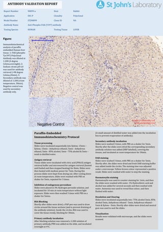
- 1. Figure: Immunohistochemical analysis of paraffin embedded Human liver tissue. 1: FAK (phospho Tyr397) Polyclonal Antibody was diluted at 1:200 (4 degree Celsius,overnight). 2: Sodium citrate pH 6.0 was used for antibody retrieval (>98 degree Celsius,20min). 3: Secondary antibody was diluted at 1:200 (room temperature, 30min). Negative control was used by secondary antibody only. Report Number 90899-a Host Rabbit Application IHC-P Clonality Polyclonal Model Number STJ90899 Clone ID NA Antibody Name Anti-Phospho-FAK (Y397) antibody Testing Species HUMAN Testing Tissue LIVER ANTIBODY VALIDATION REPORT b. (A small amount of distilled water was added into the incubation box to prevent evaporation of antibody). 30. Secondary antibody incubation a. Slides were washed 3 times, with PBS on a shaker for 5min. Shortly after the slides were dried the corresponding secondary antibody solution was added (HRP labelled), covering the tissues, and incubated at room temperature for 30min. b. 31. DAB staining a. Slides were washed 3 times, with PBS on a shaker for 5min. b. Shortly after, the slides were dried and fresh DAB staining buffer was added inside the circles. The staining time was adjusted under a microscope. Yellow-brown colour represented a positive result. Slides were washed with water to stop the staining. c. 32. Haematoxylin staining a. Haematoxylin was used to counter-staining for 1min, and then the slides were washed with water. 1% Hydrochloric acid and alcohol was added for several seconds and then washed with water. Ammonia was used to reveal blue colour, and then flushed with water. b. 33. Desolation and Clearing i. Slides were incubated sequentially into: 75% alcohol 5min, 85% alcohol 5min, Anhydrous ethanol - 5min, Anhydrous ethanol - 5min & Xylene - 5min. Shortly after slides were dried and neutral gum was used to seal the slides. ii. 34. Visualization a. Results were validated with microscope, and the slides were scanned. Paraffin-Embedded Immunohistochemistry Protocol 24. 25. Tissue processing a. Slides were incubated sequentially into Xylene; 15min – Xylene, 15min - Anhydrous ethanol, 5min - Anhydrous ethanol, 5min - 85% alcohol, 5min - 75% alcohol & 5min – wash in distilled water. b. 26. Antigen retrieval a. Tissue slides were incubated with citric acid (PH6.0) antigen retrieval buffer and microwaved for antigen retrieval (heated until boiled and then stopped heating) for 8min. Slides were then heated with medium power for 7min. During this process slides were kept from drying out. After cooling down at room temperature, slides were washed with PBS on shaker for 5min, repeated for 3 times. b. 27. Inhibition of endogenous peroxidase a. Slides were placed in 3% Hydrogen peroxide solution, and incubated for 10 min at room temperature without light exposure. Slides were then washed 3 times with PBS on a shaker for 5mins. b. 28. BSA Blocking a. Shortly after slides were dried, a PAP pen was used to draw circles around the tissue sections (and to prevent draining of the antibody solution). Inside the circles, BSA was used to cover the tissue evenly, blocking for 30min. b. 29. Primary antibody incubation After blocking solution was removed a 1:200 solution of primary antibody/PBS was added on the slide, and incubated overnight at 4°C. St John's Laboratory Ltd. www.stjohnslabs.com
- 2. Figure: Immunohistochemical analysis of paraffin embedded Human stomach cancer tissue. 1: FAK (phospho Tyr397) Polyclonal Antibody was diluted at 1:200 (4 degree Celsius,overnight). 2: Sodium citrate pH 6.0 was used for antibody retrieval (>98 degree Celsius,20min). 3: Secondary antibody was diluted at 1:200 (room temperature, 30min). Negative control was used by secondary antibody only. Report Number 90899-b Host Rabbit Application IHC-P Clonality Polyclonal Model Number STJ90899 Clone ID NA Antibody Name Anti-Phospho-FAK (Y397) antibody Testing Species HUMAN Testing Tissue STOMACH CANCER ANTIBODY VALIDATION REPORT b. (A small amount of distilled water was added into the incubation box to prevent evaporation of antibody). 19. Secondary antibody incubation a. Slides were washed 3 times, with PBS on a shaker for 5min. Shortly after the slides were dried the corresponding secondary antibody solution was added (HRP labelled), covering the tissues, and incubated at room temperature for 30min. b. 20. DAB staining a. Slides were washed 3 times, with PBS on a shaker for 5min. b. Shortly after, the slides were dried and fresh DAB staining buffer was added inside the circles. The staining time was adjusted under a microscope. Yellow-brown colour represented a positive result. Slides were washed with water to stop the staining. c. 21. Haematoxylin staining a. Haematoxylin was used to counter-staining for 1min, and then the slides were washed with water. 1% Hydrochloric acid and alcohol was added for several seconds and then washed with water. Ammonia was used to reveal blue colour, and then flushed with water. b. 22. Desolation and Clearing i. Slides were incubated sequentially into: 75% alcohol 5min, 85% alcohol 5min, Anhydrous ethanol - 5min, Anhydrous ethanol - 5min & Xylene - 5min. Shortly after slides were dried and neutral gum was used to seal the slides. ii. 23. Visualization a. Results were validated with microscope, and the slides were scanned. Paraffin-Embedded Immunohistochemistry Protocol 13. 14. Tissue processing a. Slides were incubated sequentially into Xylene; 15min – Xylene, 15min - Anhydrous ethanol, 5min - Anhydrous ethanol, 5min - 85% alcohol, 5min - 75% alcohol & 5min – wash in distilled water. b. 15. Antigen retrieval a. Tissue slides were incubated with citric acid (PH6.0) antigen retrieval buffer and microwaved for antigen retrieval (heated until boiled and then stopped heating) for 8min. Slides were then heated with medium power for 7min. During this process slides were kept from drying out. After cooling down at room temperature, slides were washed with PBS on shaker for 5min, repeated for 3 times. b. 16. Inhibition of endogenous peroxidase a. Slides were placed in 3% Hydrogen peroxide solution, and incubated for 10 min at room temperature without light exposure. Slides were then washed 3 times with PBS on a shaker for 5mins. b. 17. BSA Blocking a. Shortly after slides were dried, a PAP pen was used to draw circles around the tissue sections (and to prevent draining of the antibody solution). Inside the circles, BSA was used to cover the tissue evenly, blocking for 30min. b. 18. Primary antibody incubation After blocking solution was removed a 1:200 solution of primary antibody/PBS was added on the slide, and incubated overnight at 4°C. St John's Laboratory Ltd. www.stjohnslabs.com
- 3. Figure: Immunohistochemical analysis of paraffin embedded Mouse brain tissue. 1: FAK (phospho Tyr397) Polyclonal Antibody was diluted at 1:200 (4 degree Celsius,overnight). 2: Sodium citrate pH 6.0 was used for antibody retrieval (>98 degree Celsius,20min). 3: Secondary antibody was diluted at 1:200 (room temperature, 30min). Negative control was used by secondary antibody only. Report Number 90899-c Host Rabbit Application IHC-P Clonality Polyclonal Model Number STJ90899 Clone ID NA Antibody Name Anti-Phospho-FAK (Y397) antibody Testing Species MOUSE Testing Tissue BRAIN ANTIBODY VALIDATION REPORT b. (A small amount of distilled water was added into the incubation box to prevent evaporation of antibody). 8. Secondary antibody incubation a. Slides were washed 3 times, with PBS on a shaker for 5min. Shortly after the slides were dried the corresponding secondary antibody solution was added (HRP labelled), covering the tissues, and incubated at room temperature for 30min. b. 9. DAB staining a. Slides were washed 3 times, with PBS on a shaker for 5min. b. Shortly after, the slides were dried and fresh DAB staining buffer was added inside the circles. The staining time was adjusted under a microscope. Yellow-brown colour represented a positive result. Slides were washed with water to stop the staining. c. 10. Haematoxylin staining a. Haematoxylin was used to counter-staining for 1min, and then the slides were washed with water. 1% Hydrochloric acid and alcohol was added for several seconds and then washed with water. Ammonia was used to reveal blue colour, and then flushed with water. b. 11. Desolation and Clearing i. Slides were incubated sequentially into: 75% alcohol 5min, 85% alcohol 5min, Anhydrous ethanol - 5min, Anhydrous ethanol - 5min & Xylene - 5min. Shortly after slides were dried and neutral gum was used to seal the slides. ii. 12. Visualization a. Results were validated with microscope, and the slides were scanned. Paraffin-Embedded Immunohistochemistry Protocol 2. 3. Tissue processing a. Slides were incubated sequentially into Xylene; 15min – Xylene, 15min - Anhydrous ethanol, 5min - Anhydrous ethanol, 5min - 85% alcohol, 5min - 75% alcohol & 5min – wash in distilled water. b. 4. Antigen retrieval a. Tissue slides were incubated with citric acid (PH6.0) antigen retrieval buffer and microwaved for antigen retrieval (heated until boiled and then stopped heating) for 8min. Slides were then heated with medium power for 7min. During this process slides were kept from drying out. After cooling down at room temperature, slides were washed with PBS on shaker for 5min, repeated for 3 times. b. 5. Inhibition of endogenous peroxidase a. Slides were placed in 3% Hydrogen peroxide solution, and incubated for 10 min at room temperature without light exposure. Slides were then washed 3 times with PBS on a shaker for 5mins. b. 6. BSA Blocking a. Shortly after slides were dried, a PAP pen was used to draw circles around the tissue sections (and to prevent draining of the antibody solution). Inside the circles, BSA was used to cover the tissue evenly, blocking for 30min. b. 7. Primary antibody incubation After blocking solution was removed a 1:200 solution of primary antibody/PBS was added on the slide, and incubated overnight at 4°C. St John's Laboratory Ltd. www.stjohnslabs.com