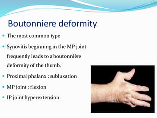
Ashraf Abdelaziz MD
Lecturer of orthopedic surgery
Hand and reconstructive surgery
Alzhraa University Hospital
Al-Azhar university
2016
Introduction
 Rheumatoid arthritis (RA) is the most common
cause of chronic inflammatory joint disease.
 Most typical features are a
 Symmetrical polyarthritis
 Tenosynovitis
 Morning stiffness,
 Elevation of the erythrocyte sedimentation rate (ESR)
 Autoantibodies that target immunoglobulins
(rheumatoid factors) in the serum
Stages of RA
1. Pre Clinical
2. Synovitis
3. Destruction
4. Deformity
Development of Deformity
The persistent inflammation causes;
 Erosion of the articular cartilage, tenosynovitis &
eventually rupture of the tendons occurs.
 Progressive process leads to joint instabilty &
deformity.
 Stretch the supporting structures of a joint, causing a
flexion, extension, or lateral deformity
 Disruption of the normal thumb biomechanics leads
to significant loss of ability to carry out daily living
activities
Thumb Deformity (Nalebuff) Classification
Boutonniere deformity
 The most common type
 Synovitis beginning in the MP joint
frequently leads to a boutonnière
deformity of the thumb.
 Proximal phalanx : subluxation
 MP joint : flexion
 IP joint hyperextension
 EPB tendon insertion become attenuated
 EPL tendon displaces ulnarly
 The patient loses the ability to actively extend the MP
joint
 Articular erosion and ligament laxity occur to varying
degrees.
Treatment Options
Mild stage; both MP and IP joints are
correctable passively
 Synovectomy of the MP joint and
reconstruction of the extensor mechanism
 Nalebuff procedure
EPL-rerouting procedure through the
dorsal capsule of the joint to provide
additional extensor force
Moderate stage; a fixed MP joint is present
with or without intra-articular joint
destruction
 Most patients are seen at this stage.
Arthrodesis is recommended for the MP
joint, if adjacent joints minimally affected(15
degrees flexion, abd, and pronation)
 Arthroplasty if the a adjacent joints affected
Sever stage ;
 Both a fixed MP joint flexion deformity and a fixed
IP joint hyperextension deformity.
 Surgery depends on the severity of the deformity,
the status of the articular surfaces.
Synovectomy, Arthrodesis, or Arthroplasty.
Treatment Options for (Boutonniýre Deformity)
Stage MP IP
Mild Synovectomy Synovectomy
EPL rerouting Restore FPL function
Flexor tenodesis
Moderate Fusion Joint release
Arthroplasty
Sever Arthroplasty Fusion
Joint release
Swan Neck Deformity
 The second most common thumb deformity
 Synovitis begins in the CMC joint
 Deformity:
 Dorsal subluxation of the metacarpal base
 Hyperextension of the metacarpophalangeal
joint (swan-neck deformity).
 Metacarpal adduction
Operative Treatment
Mild stage, a painful CMC joint, weak pinch
1. Conservative therapy (splinting or injection)
2. Resection arthroplasty and tendon interposition
Moderate stage show varying degrees of CMC joint
deformity and passively correctable MP joint
hyperextension
 Resection arthroplasty
 + MP joint fusion
Sever stage ; complete CMC j
dislocation, fixed adduction and
fixed hyperextension of the MP
joint
 CMC resection arthroplasty with
ligament reconstruction and MP
joint fusion
Game Keeper’s Thumb
 Type IV deformity
 Destruction of the
capsuloligamentous supports
on the ulnar side of MP joint
 Laxity of the ulnar collateral
ligament of the MP joint
Operative Treatment
 MP joint synovectomy and collateral
ligament reconstruction are performed.
 In advanced cases, MP joint arthrodesis is
done.
 Adductor fascia release
Arthritis Mutilans (Opera Glass Hand)
 Arthritis Mutilans of Hand
 Shortening of fingers due to
destruction of phalanges and
joints.
 Excess skin gets folded
transversely resembling ‘opera
glass’
Operative Treatment
 Arthrodesis is the procedure of choice
 Bone grafts will be required to restore length and to
allow fusion to occur
Tenosynovitis
 RA is a disease of the synovium and Tendon sheath.
 Presentation :
 Pain
 Tendon dysfunction (e.g. trigger finger)
 Tendon rupture
Rupture of the Extensor Pollicis Longus
 Tenosynovitis leads to tendon rupture
 Frequently ruptures at the Lister tubercle,
where it enclosed in tunnel.
 Incomplete extension of the IP joint
 The intrinsics alone can extend IP to
neutral
 Rupture of EPL loses extension of the MP
joint because the EPB is not strong
enough to extend this joint
 If the tendon is ruptured and the functional loss is
significant, EPL function should be restored.
 Tendon transfer; The two most commonly used are
EIP or ECRL
 The most common flexor tendon rupture in RA
patients.
 Erosion of the capsule and ligaments over radial
osteophytes contribute to flexor pollicis longus
rupture in the carpal tunnel (Mannerfelt lesion).
Rupture of the Flexor Pollicis Longus
Surgical options
 Tendon graft,
 Tendon transfer; FDS of Middle finger is usually used
for transfer.
Take a home massage
 RA is the most common cause of chronic inflammatory joint
disease.
 Don’t forget to mange the disease
 Boutonniere deformity and Swan Neck Deformity the common
types of thumb deformity
 Concern to any joint affected and deformity to plane for the
treatment
 Physiotherapy and splinting prevent progress of the deformity
Thank You

Thumb deformity

  • 1.
    Ashraf Abdelaziz MD Lecturerof orthopedic surgery Hand and reconstructive surgery Alzhraa University Hospital Al-Azhar university 2016
  • 2.
    Introduction  Rheumatoid arthritis(RA) is the most common cause of chronic inflammatory joint disease.  Most typical features are a  Symmetrical polyarthritis  Tenosynovitis  Morning stiffness,  Elevation of the erythrocyte sedimentation rate (ESR)  Autoantibodies that target immunoglobulins (rheumatoid factors) in the serum
  • 3.
    Stages of RA 1.Pre Clinical 2. Synovitis 3. Destruction 4. Deformity
  • 4.
    Development of Deformity Thepersistent inflammation causes;  Erosion of the articular cartilage, tenosynovitis & eventually rupture of the tendons occurs.  Progressive process leads to joint instabilty & deformity.
  • 5.
     Stretch thesupporting structures of a joint, causing a flexion, extension, or lateral deformity  Disruption of the normal thumb biomechanics leads to significant loss of ability to carry out daily living activities
  • 6.
  • 7.
    Boutonniere deformity  Themost common type  Synovitis beginning in the MP joint frequently leads to a boutonnière deformity of the thumb.  Proximal phalanx : subluxation  MP joint : flexion  IP joint hyperextension
  • 8.
     EPB tendoninsertion become attenuated  EPL tendon displaces ulnarly  The patient loses the ability to actively extend the MP joint  Articular erosion and ligament laxity occur to varying degrees.
  • 9.
    Treatment Options Mild stage;both MP and IP joints are correctable passively  Synovectomy of the MP joint and reconstruction of the extensor mechanism  Nalebuff procedure EPL-rerouting procedure through the dorsal capsule of the joint to provide additional extensor force
  • 10.
    Moderate stage; afixed MP joint is present with or without intra-articular joint destruction  Most patients are seen at this stage. Arthrodesis is recommended for the MP joint, if adjacent joints minimally affected(15 degrees flexion, abd, and pronation)  Arthroplasty if the a adjacent joints affected
  • 11.
    Sever stage ; Both a fixed MP joint flexion deformity and a fixed IP joint hyperextension deformity.  Surgery depends on the severity of the deformity, the status of the articular surfaces. Synovectomy, Arthrodesis, or Arthroplasty.
  • 12.
    Treatment Options for(Boutonniýre Deformity) Stage MP IP Mild Synovectomy Synovectomy EPL rerouting Restore FPL function Flexor tenodesis Moderate Fusion Joint release Arthroplasty Sever Arthroplasty Fusion Joint release
  • 13.
    Swan Neck Deformity The second most common thumb deformity  Synovitis begins in the CMC joint  Deformity:  Dorsal subluxation of the metacarpal base  Hyperextension of the metacarpophalangeal joint (swan-neck deformity).  Metacarpal adduction
  • 14.
    Operative Treatment Mild stage,a painful CMC joint, weak pinch 1. Conservative therapy (splinting or injection) 2. Resection arthroplasty and tendon interposition Moderate stage show varying degrees of CMC joint deformity and passively correctable MP joint hyperextension  Resection arthroplasty  + MP joint fusion
  • 15.
    Sever stage ;complete CMC j dislocation, fixed adduction and fixed hyperextension of the MP joint  CMC resection arthroplasty with ligament reconstruction and MP joint fusion
  • 16.
    Game Keeper’s Thumb Type IV deformity  Destruction of the capsuloligamentous supports on the ulnar side of MP joint  Laxity of the ulnar collateral ligament of the MP joint
  • 17.
    Operative Treatment  MPjoint synovectomy and collateral ligament reconstruction are performed.  In advanced cases, MP joint arthrodesis is done.  Adductor fascia release
  • 18.
    Arthritis Mutilans (OperaGlass Hand)  Arthritis Mutilans of Hand  Shortening of fingers due to destruction of phalanges and joints.  Excess skin gets folded transversely resembling ‘opera glass’
  • 19.
    Operative Treatment  Arthrodesisis the procedure of choice  Bone grafts will be required to restore length and to allow fusion to occur
  • 20.
    Tenosynovitis  RA isa disease of the synovium and Tendon sheath.  Presentation :  Pain  Tendon dysfunction (e.g. trigger finger)  Tendon rupture
  • 21.
    Rupture of theExtensor Pollicis Longus  Tenosynovitis leads to tendon rupture  Frequently ruptures at the Lister tubercle, where it enclosed in tunnel.  Incomplete extension of the IP joint  The intrinsics alone can extend IP to neutral  Rupture of EPL loses extension of the MP joint because the EPB is not strong enough to extend this joint
  • 22.
     If thetendon is ruptured and the functional loss is significant, EPL function should be restored.  Tendon transfer; The two most commonly used are EIP or ECRL
  • 23.
     The mostcommon flexor tendon rupture in RA patients.  Erosion of the capsule and ligaments over radial osteophytes contribute to flexor pollicis longus rupture in the carpal tunnel (Mannerfelt lesion). Rupture of the Flexor Pollicis Longus
  • 24.
    Surgical options  Tendongraft,  Tendon transfer; FDS of Middle finger is usually used for transfer.
  • 25.
    Take a homemassage  RA is the most common cause of chronic inflammatory joint disease.  Don’t forget to mange the disease  Boutonniere deformity and Swan Neck Deformity the common types of thumb deformity  Concern to any joint affected and deformity to plane for the treatment  Physiotherapy and splinting prevent progress of the deformity
  • 26.