Heart part 1 ll SOHAN PATEL
•Download as PPT, PDF•
0 likes•64 views

Basic introduction of heart

Recommended
More Related Content
What's hot
What's hot (20)
Similar to Heart part 1 ll SOHAN PATEL
Similar to Heart part 1 ll SOHAN PATEL (20)
Recently uploaded
Recently uploaded (20)
Heart part 1 ll SOHAN PATEL
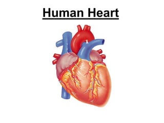
- 1. Human Heart
- 2. 2 Introduction • Hollow, muscular organ • The heart is a complex muscular pump. • 300 grams (size of a fist) • It maintains blood pressure of the body. • The heart pumps about 1,00,000 times • moves 7200 liters (1900 gallons) of blood every day.
- 4. 4 HEART ANATOMY 1. Location of heart 2. Covering layers of heart 3. Layers of heart 4. Chambers of heart 5. valves
- 5. Location of the heart • The heart is located between the lungs. • behind the sternum • above the diaphragm. • Its centre is located about 1.5 cm to the left of the midsagittal plane.
- 6. 6 Heart’s position in thorax
- 7. SOHAN A. PATEL ASSISTANT PROFESSOR, DEPT. OF PHARMACOLOGY,
- 8. 6. Heart Valves Function- • prevent blood from flowing backwards Two types of valves in heart 1. Atrioventricular valves (AV) 2. Semi-lunar valves (SL)
- 9. 1. Atrioventricular Valves Located between Atrium and ventricles. Right AV valve (Tricuspid) • Located between Right Atrium and ventricle. • Contains 3 cusps Left AV valve (Mitral or Bicuspid) • Located between left Atrium and ventricle. •Contains 2 cusps
- 10. 2. Semilunar valves • Located at exit of ventricles, originate from endothelial lining of veins Heart contains two semilunar valves • Pulmonic valve • Aortic valve
- 13. Internal structure of Heart
- 15. The Heart These are arteries. They carry blood away from the heart. This is a vein. It brings blood from the body, except the lungs. Coronary arteries, the hearts own blood supply The heart has four chambers 2 atria 2 ventricles now lets look inside the heart
- 16. The Heart Left Ventricle Left Atrium Right Atrium Right Ventricle valve Vein from Lungs Artery to Head and BodyArtery to Lungs Vein from Head and Body valve
- 17. How does the Heart work? blood from the body blood from the lungs The heart beat begins when the heart muscles relax and blood flows into the atria. STEP ONE
- 18. The atria then contract and the valves open to allow blood into the ventricles. How does the Heart work? STEP TWO
- 19. How does the Heart work? The valves close to stop blood flowing backwards. The ventricles contract forcing the blood to leave the heart. At the same time, the atria are relaxing and once again filling with blood. The cycle then repeats itself. STEP THREE
- 20. 3 Kinds of Circulation: • Pulmonary circulation • Coronary circulation • Systemic circulation
- 21. Pulmonary Circulation Movement of blood from the heart, to the lungs, and back to the heart again
- 23. Coronary Circulation Movement of blood through the tissues of the heart
- 25. Systemic Circulation Supplies nourishment to all of the tissue located throughout the body , except for the heart and lungs
- 28. Hollow tubes that circulate your blood There are 3 types of blood vessels a. ARTERY b. VEIN c. CAPILLARY Blood Vessels
- 29. Arteries • Carry blood AWAY from the heart • Main artery called the aorta • Aorta divides and branches and become Many smaller arteries • Each region of your body has system of arteries supplying it with fresh, oxygen-rich blood.
- 30. The ARTERY thick muscle and elastic fibres Arteries carry blood away from the heart. the elastic fibres allow the artery to stretch under pressure the thick muscle can contract to push the blood along.
- 33. Veins • Carry blood to the heart • Receive blood from the capillaries • Transport waste-rich/ oxygen-poor blood back to the heart • Allow blood to move in one direction
- 34. The VEIN Veins carry blood towards from the heart. thin muscle and elastic fibres veins have valves which act to stop the blood from going in the wrong direction. body muscles surround the veins so that when they contract to move the body, they also squeeze the veins and push the blood along the vessel.
- 35. Capillaries • Very thin • Only one cell thick • Connect arteries & veins
- 36. Capillaries • Food and oxygen released to the body cells • Carbon dioxide and other waste products returned to the bloodstream
- 37. The CAPILLARY Capillaries link Arteries with Veins the wall of a capillary is only one cell thick they exchange materials between the blood and other body cells. The exchange of materials between the blood and the body can only occur through capillaries.
- 38. artery vein capillaries body cell The CAPILLARY A collection of capillaries is known as a capillary bed.
- 48. Properties of Cardiac muscle:- 1.Excitability or irritability 2.Contractility 3.Automaticity 4.Rhythmicity 5.Refractory period 6.Tonicity
- 49. Coordination of chamber contraction, relaxation
- 50. I. Excitability (Irritability): = the ability of cardiac muscle to respond to adequate stimuli by generating an action potential followed by a mechanical contraction.
- 54. Electrophysiology of the cardiac muscle cell
- 55. The Conduction System • Electrical signal begins in the sinoatrial (SA) node: "natural pacemaker." – causes the atria to contract. • The signal then passes through the atrioventricular (AV) node. – sends the signal to the ventricles via the “bundle of His” – causes the ventricles to contract.
- 58. Abnormalities of Heart:- 1. Hypertension 2. CHF (Conjunctive heart failure) 3. Hyperlipidemic 4. Arraythmias 5. Angina pectories