SlideShare a Scribd company logo
1 of 43
Y chromosome Profiling and Gender Typing
 Human Y chromosome Genome
Pseudo-Autosomal Region
Male-Specific Y Region
Polymorphic Sequences
 Profiling Systems
Y-STR
Core Y-STR Loci
Multiplex Y-STR
 Gender Typing
Amelogenin Locus
AMELY Null Mutations
Y Chromosome Profiling and Gender Typing
• The Y chromosome is inherited from the father, unique to males and
passed on to all male offspring.
• The chromosome encodes dozens of genes required for male-specific
functions, including sex determination and spermatogenesis.
• Y chromosome loci are very important for forensic DNA profiling.
• For instance, the Y chromosome STR (Y-STR) used in forensic DNA
testing is male-specific (for humans and certain higher primates) and is
thus useful in investigations of sexual assault cases involving male
suspects.
• The evidence gathered in such cases usually consists of mixtures of
high levels of female DNA and low levels of male DNA.
• The Y chromosome-specific loci can be examined without interference
from large amounts of female DNA; differential extraction of sperm
and nonsperm cells may not be needed.
• Furthermore, the Y-STR system is useful for determining numbers of
male criminals in sexual assault cases involving more than one male.
• The Y-STR loci used for forensic applications are located in the non-
recombining section of the Y chromosome so that paternal lineages can
be established.
• The technique can be used for paternity testing and identification of
missing persons.
• The major disadvantage of Y chromosome loci is that their
discriminating power is low compared to the discriminating power of
autosomal loci.
• Because Y chromosome loci are linked, the product rule for statistical
calculations for profile probability does not apply.
• Also, the Y chromosome loci test cannot distinguish individuals with
the same paternal lineage.
Human Y Chromosome Genome
• The human Y chromosome genome contains approximately 60 million
bp and the chromosome can be divided into two regions: the pseudo-
autosomal region (PAR) and the male-specific Y (MSY) region.
1.Pseudo-Autosomal Region
 Approximately 5% of the Y chromosome sequence is located at the
telomeres of the chromosome.
 In particular, PAR1 is located on the tip of the short arm and PAR2 is
located at the tip of the long arm (Figure 1).
 This region undergoes recombination with homologous regions on the
X chromosome during meiosis in males.
Figure 1. Human Y-STR chromosome structure. PAR = pseudo-
autosomal region. MSY = male-specific Y region. Yp = short arm of Y
chromosome. Yq = long arm of Y chromosome.
2.Male-Specific Y Region
• The remainder of the Y chromosome is known as the MSY region.
• It was previously called the non-recombining Y (NRY) region (Figure
1 above) or It does not participate in homologous recombination.
• However, certain sections involve intra chromosomal gene conversion.
• About 40 megabases (Mb) within the MSY region are
heterochromatic (highly repetitive sequences) including the
centromeric region and the bulk of the distal long arm.
• The euchromatic region is about 23 Mb and most of it has
been sequenced.
• Certain sections of the euchromatic region share some
homology with the X chromosome.
• For instance, X-transposed sequences of the Y chromosome
are 99% identical to sequences within Xq21 (a band in the
long arm of the X chromosome).
• Additionally, dozens of genes located in the euchromatic
region share 60% to 96% homology with their X
chromosome counterparts.
• These X-homologous regions should be avoided when
selecting Y chromosome-specific markers for forensic DNA
profiling.
3 Polymorphic Sequences
• The Y chromosome contains an abundance of repetitive
elements, namely STRs, Alu, and LINE elements.
• Many of these are highly polymorphic.
• To date Y-STRs are usually used for Y chromosome DNA
testing.
• Single nucleotide polymorphisms (SNPs) at the Y
chromosome are also useful for forensic applications.
Profiling Systems
1. Y-STR
• More than 400 STR loci have been identified in the Y chromosome
genome.
• The precise locations of these loci have been sequentially mapped
using human genome sequencing data.
• The distribution of Y-STR loci at the Y chromosome has also been
analyzed.
• Most Y-STR loci, approximately 60% of the 400 identified, are located
at the long arm of the chromosome; about 22% are located at the short
arm and a few are found in the centromeric region.
• Y-STRs in the telomeric region have yet to be identified.
• Only about 5% of Y-STRs are located within 5′ untranslated or intron
regions of protein coding genes.
• The repeat unit length of identified Y-STRs have been analyzed.
• Among the 400 Y-STRs, 6% are dimeric repeats, 39% are trimeric,
45% are tetrameric, 9% are pentameric, and 1% are hexameric.
• Fewer than half the STRs have been characterized. Some loci are
polymorphic and are useful for forensic applications and developing
new Y-STR multiplex systems.
• The STR loci at the Y chromosome are usually referred to as
haplotypes.
• A haplotype is a collection of alleles that are usually linked (inherited
together) since homologous recombination does not occur on the
majority of the Y chromosome.
2. Core Y-STR Loci
• In 1997, the European minimal haplotype (EMH) locus set was
recommended by the International Y-STR User Group for forensic
applications.
• This haplotype set includes a core set of nine Y-STR loci: DYS19,
DYS385 a and b, DYS389I, DYS389II, DYS390, DYS391, DYS392,
and DYS393.
• In 2003, the U.S. haplotype loci were recommended by the Scientific
Working Group on DNAAnalysis Methods (SWGDAM) for forensic
DNA analysis.
• The U.S. haplotype loci includes the EMH loci set plus two additional
loci, DYS438 and DYS439.
• DYS385 and DYS389 are multi-local Y-STR loci (MLL).
• The MLL designation refers to a presence of a particular STR at more
than one site on the Y chromosome DNA due to duplication.
• To date, about 50 such MLL Y-STRs have been identified. Further
MLL subdivisions are designated bi-local, trilocal, etc. DYS385 and
DYS389 are bi-local.
• The DYS385 locus has two inverted duplicated clusters and is
separated by a 4 × 10⁴ bp interstitial region (Figure 19.5).
• It can be amplified by a single set of primers. One allele is observed if
the duplicates are the same length.
• If the duplicated clusters have different lengths, they can generate two
different alleles when amplified.
• The smaller sized allele is designated “a” and the larger sized allele is
designated “b.”
• The DYS389 locus has two duplicated clusters with the same
orientation.
• In a single set of PCR primers, there are two binding sites for the same
forward primer at each 5′ flanking sequence of the core repeat region
of DYS389.
• These binding sites between DYS389I and DYS389II are about 120 bp
apart.
• Therefore, two amplicons are produced.
• DYS389I is designed for the smaller allele and DYS389II is
designated for the larger allele.
• The average mutation rate for the core Y-STR loci is approximately
10–3 per generation—similar to the mutation rate of autosomal STR
loci. Mutations can exert major impacts on the interpretation of
paternity test results.
3 Multiplex Y-STR
 The application Y-STR for forensic casework was initiated in Europe.
 In the U.S., the laboratory of the Office of Chief Medical Examiner in
New York City was the first to perform Y-STR testing of four loci
(DYS19, DYS390, DYS389I and II) for casework.
 The use of Y-STR loci has been facilitated by various commercially
available PCR amplification kits in multiplex systems.
 ReliaGene Technologies developed the first commercial multiplex Y-
STR system, the Y-PLEXTM6.
 The kit includes DYS19, DYS385a and b, DYS389II, DYS390,
DYS391, and DYS393.
 Additional commercially available kits with more Y-STR loci are now
available and have been validated for forensic use.
 To improve discriminating power, multiplex systems including new Y-
STR loci are desired.
 Many new Y-STR loci are being characterized for developing new
multiplex systems.
Gender Typing
• Gender typing of a biological sample is useful in forensic
investigation, for example, for victim identification in disaster cases
and suspect identification in sexual assaults.
• One commonly used gender typing marker is the amelogenin (AMEL)
locus.
1 Amelogenin Locus
• This region encodes extracellular matrix proteins involved in tooth
enamel formation.
• Mutations in the AMEL gene can lead to an enamel defect known as
amelogenesis imperfecta.
• The AMEL locus has two homologous genes: AMELX (Xp22.1–
Xp22.3) is located on the human X chromosome and AMELY
(Yp11.2) is located on the human Y chromosome.
• Although the genes constitute a homologous pair, they differ in size
and sequence.
• Gender typing can be performed using various primers designed
specifically for the sequences of the homologous region on these
genes, followed by amplification. Different sizes of amplicons are
obtained.
• The most commonly used gender typing method at the AMEL locus is
the detection of a 6-bp deletion at intron 1 of AMELX.
• This deletion is not present in AMELY.
• Primer sets were developed to amplify both alleles in a single PCR by
Forensic Science Service in the United Kingdom in 1993.
• The amplicons generated from AMELY and AMELX are separated by
electrophoresis.
• The observation of the AMELX fragment alone indicates a female,
whereas the observation of both AMELX and AMELY indicates a
male.
• Nevertheless, primate and some rudiment DNA can be amplified as
well but the amplicon sizes vary.
• The AMEL locus has been co-amplified with other markers to provide
a combined gender and identity test.
• Such combined tests have been used in D1S80 AFLP and various STR
multiplex analyses.
2 AMELY Null Mutations
• Several cases of AMELY null mutations have been reported.
• Only the AMELX fragment was detected in these AMELY null males.
• Many of them are phenotypically normal but present the AMEL gender
types of females.
• Various interstitial deletions at the Y chromosome short arm have been
identified as the cause of some AMELY null gender typing.
• The frequency of AMELY null males is rare, but is higher in Sri Lanka
and India.
Mitochondrial DNA Profiling
• Forensic mitochondrial DNA (mtDNA) analysis is a useful tool for
human identification.
• Because mtDNA is maternally inherited, it is especially useful for
identifying victims.
• Additionally, the mtDNA genome produces much higher numbers of
copies per cell than the nuclear genome.
• Thus, mtDNA testing is frequently used when nuclear DNA in samples
is insufficient.
• For example, hair shafts, bones, and decomposed samples may be
tested with mtDNA analysis.
Human Mitochondrial Genome
• Mitochondria are subcellular organelles that serve as the energy-generating
components of cells (Figure 1).
• Each cell contains hundreds of mitochondria that have their own
extrachromosomal genomes separate from the nuclear genome.
• Although each human mitochondrion contains several copies of the mtDNA
genome, the exact copy number varies for each cell.
• However, it is estimated that hundreds of copies of mtDNA genome exist in
most cell types.
• Recombination has not been observed in mtDNA.
• Thus, the mtDNA type, also referred to as the mitotype, is considered a
haplotype treated as a single locus.
• The mitochondrial genome has a higher mutation rate (up to 10 times higher)
than its nuclear counterpart.
1 Genetic Contents of Mitochondrial Organelle Genomes
• Organelle genomes are usually much smaller than their nuclear
counterparts.
• The much smaller mitochondrial genome has been sequenced and is
known as the Cambridge reference sequence.
• It was established in the early 1980s, later modified, and is now known
as the revised Cambridge reference sequence (rCRS) that is presently
used as the standard for sequence comparisons.
• The human mitochondrial genome is a circular DNA molecule
consisting of 16,569 bp containing 37 genes (Figure 1.).
• Thirteen of these genes code for proteins involved in the respiratory
complex, a main energy-generating component in mitochondria.
Figure 1 Mitochondrion. mtDNA = mitochondrial DNA.
• The other 24 specify noncoding RNA molecules required for
expression of the mitochondrial genome.
• The genes in the human mitochondrial genome are much more closely
packed than in the nuclear genome and contain no introns.
• A control region, also known as a displacement loop (D loop),
contains the origin of replication for one of the mtDNA strands but
does not code for any gene products (Figure 2).
• An asymmetric distribution of nucleotides gives rise to light (L) and
heavy (H) strands when mtDNA molecules are separated in alkaline
CsCl gradients.
• The H strand that contains a greater number of guanine nucleotides
has a higher molecular weight in comparison to the L strand.
 Figure 2. Human circular mitochondrial genome.
 The transcription direction for the H (heavy) and L (light) strands are
indicated by arrows (PH, PL).
 The origins of replication are labeled OH for heavy strand and OL for light
strand, respectively.
 The mitochondrial DNA genome encodes genes. ND = NADH coenzyme Q
oxidoreductase complex. CO = cytochrome c oxidase complex. Cytb =
cytochrome b. ATP = ATP synthase. rRNA, ribosomal RNA.
 Transfer RNA genes are shown as indicated.
2 Maternal Inheritance of mtDNA
• Maternal inheritance is typically observed for the mtDNA genome.
• mtDNA is inherited differently from nuclear genes; it does not obey
the rules of Mendelian inheritance and is thus called non Mendelian
inheritance.
• The mitochondria of the spermatozoa are located at the mid pieces of
spermatozoa.
• At conception, only the head portion of a spermatozoon (containing a
nucleus but no mitochondria) enters the egg.
• The fertilized egg contains the maternal mitochondria which is
transmitted to progeny.
• The mtDNA sequence is identical for relatives within the same
maternal lineage (Figure 3).
• This characteristic of maternal inheritance is useful for identifying
samples by comparing them with samples from maternal relatives.
mtDNA Polymorphic Regions
1. Hypervariable Regions
2. Heteroplasmy
Hypervariable Regions
• The most polymorphic region of mtDNA is located within the D loop
(Figure 4).
• The three hypervariable regions in the D-loop region are designated
HV1 (16024–16365; 342 bp), HV2 (73–340; 268 bp), and HV3
(438–574; 137 bp).
• The most common polymorphic regions of the human mtDNA
genome analyzed for forensic purposes are the two
hypervariable regions within the D loop known as
hypervariable region I (HV1) and hypervariable region II
(HV2).
Figure 4 Hypervariable regions of
the D loop in mtDNA (with
nucleotide positions).
Figure 3 Pedigree of a human family showing inheritance of mtDNA
Females and males are denoted by circles and squares, respectively.
Red symbols indicate individuals who inherited the same mtDNA.
Heteroplasmy
• Heteroplasmy occurs when an individual carries more than one
mtDNA haplotype. Occurs due to non disjunction during meiosis.
• Heteroplasmy may be observed with one type of tissue and be absent in
other tissue types; for example, it is commonly observed in hair
samples.
• Several instances of heteroplasmy may be observed in different tissue
types.
• An individual may exhibit one mitotype in one tissue and a different
mitotype in another.
• Thus, it is necessary to obtain and process additional samples to
confirm the heteroplasmy when it is observed in a questioned sample
but not in a known sample or vice versa.
• The two types of Heteroplasmy are sequence and length
heteroplasmies.
Sequence Heteroplasmy
• Sequence heteroplasmy is defined as the presence of two nucleotides
at a single position shown as overlapping peaks in a sequence
electropherogram (Figure 5).
• Heteroplasmy usually occurs at one position, but on rare occasions can
be observed at more than one position.
• Heteroplasmy may complicate the interpretation of mtDNA results, but
its presence can also improve the strength of a match
Length Heteroplasmy
• Both HV1 and HV2 of the human mtDNA D-loop region contain
homopolymeric cytosine sequences known as C stretches.
• The HVI region contains a C stretch between positions 16184 and 16193,
interrupted by a thymine at position 16189.
• If a base transition from T to C occurs at position 16189 (a variant present in
approximately 20% of the population), it results in an uninterrupted C
stretch.
• A similar C stretch resides between positions 303 and 315 of the HV2 region.
• Length heteroplasmies are often observed at the uninterrupted C stretches,
which create serious problems with sequence analysis downstream from the
homopolymeric regions (Figure 6).
• It is not clear whether the length heteroplasmy is due to replication slippage
at the C stretches or results from a mixture of length variants in the cells.
• If length heteroplasmy occurs, alternative sequencing primers can be used to
obtain the downstream sequences of the C stretches.
Figure .5 Electropherogram showing mtDNA sequence heteroplasmy at
position 234 R (A/G) as indicated by arrow. N = unresolved sequence.
Figure .6 Electropherogram showing mtDNA length heteroplamy at
C stretch of HV1 region where position 16189 is a C as indicated
by arrow. N = unresolved sequence.
Forensic mtDNA Testing
1 General Considerations
• mtDNA analysis is often used on samples derived from skeletal or
decomposed remains.
• The surface of the sample should be cleaned to remove any adhering
debris or contaminants.
• Bones and teeth are pulverized to facilitate extraction of the mtDNA.
• Duplicate extractions (e.g., two sections of a single hair) are
recommended if sufficient sample material is available.
• mtDNA is extracted using a similar method to nuclear DNA (nuclear
DNA is coextracted with mtDNA).
• The amount of mtDNA can therefore be estimated from the quantity
of nuclear DNA obtained.
 For mtDNA sequencing, analysis of both strands of the mtDNA in a
given region must be performed to ensure accuracy.
 Due to the high sensitivity of mtDNA analysis, it is essential to
minimize risks of contamination during the procedure.
 Contamination must be strictly monitored using proper controls such
as reagent blanks and negative controls (samples containing all
reagents except DNA template).
 Finally, a positive control must also be used to monitor the success of
the analysis.
 It should be introduced at the amplification step and remain through
the sequencing process.
 A positive control consists of a DNA template of known sequence such
as DNA purified from an HL60 cell line.
mtDNA Sequencing
• To sequence a specific region of mtDNA, a combination of PCR
amplification and DNA sequencing techniques is employed that reduce
the time and labor needed to obtain DNA sequences from genomic
DNA templates.
• mtDNA sequencing usually consists of
1) PCR amplification,
2) DNA sequencing reactions,
3) separation using electrophoresis, and
4) data collection and sequence analysis .
PCR Amplification
• The extracted DNA samples must be amplified to yield sufficient
quantities of template for sequencing reactions.
• PCR amplification of all or a part of the D loop region can be carried
out with various primer sets.
• If a sample contains high quality and high copy number mtDNA, the
HV1, and HV2 regions can be amplified as two amplicons, each of
about 350 to 400 bp in length.
• If a sample is degraded or contains low copy number mtDNA, the
hypervariable regions can be amplified as smaller PCR products. PCR
amplification of mtDNA is usually done in 34 to 38 cycles.
• Protocols for highly degraded DNA specimens sometimes require 42
cycles.
• The use of higher PCR cycle numbers can improve the yield of the amplicon.
• Following mtDNA amplification, a purification step is necessary to remove
excess primers and deoxynucleotide triphosphates (dNTPs).
• This step can be performed by using filtration devices such as a MicroconR
to remove small molecules from the sample or by using nuclease digestion
with shrimp alkaline phosphatase or exonuclease I to degrade remaining
primers and dNTPs.
• The concentration of the PCR product is important for an optimal sequencing
reaction in the next phase of mtDNA sequencing.
• The quality and quantity of the mtDNA amplicon must be evaluated to
confirm the presence or absence of PCR products and their concentrations.
• This can be done using an agarose yield gel to visualize the PCR products of
the sample or via capillary electrophoresis, a more informative method, for
quantifying PCR products.
DNA Sequencing Reactions
• The best known DNA sequencing techniques are the chain
termination method and the chemical degradation method developed,
respectively, by Sanger and Gilbert.
• Over the years, the chain termination method became more common
because it was applicable for automation and did not require the toxic
chemicals necessary for the chemical degradation method.
Electrophoresis and Sequence Analysis
• The cycle sequencing products can be separated using electrophoresis
in a 4% polyacrylamide denatured gel or a POP-6 polymer (Applied
Biosystems) as the matrix for capillary electrophoresis.
• Following data collection, sequence data analysis can be performed
with the Sequencher™software (Gene Codes Corporation, Ann Arbor,
MI, USA).
Interpretation of mtDNA Profiling Results
• Interpretation guidelines are used when the evaluation of sequencing
results from evidence and reference samples are necessary.
• General guidelines were set forth by the Scientific Working Group on
DNAAnalysis Methods (SWGDAM) and the DNA Commission of the
International Society of Forensic Genetics (ISFG).
• The limitations of mtDNA technology should be taken into account as
should the higher mutation rates found with the mtDNA genome than
found with the nuclear genome.
• Mutations seem to be more common in certain tissues.
• For that reason the sources of the tissues investigated should be taken
into consideration as well.
• In reporting mtDNA profiling results, the most common categories of
conclusions are: cannot exclude, exclusion, and inconclusive result.
• Exclusion — If the sequences are different, then the samples can be
excluded as originating from the same source.
• Additionally, the SWGDAM’s guidelines define that this conclusion
can be made if there are two or more nucleotide differences between
the questioned and known samples.
• Cannot exclude — If the sequences are the same, the reference
sample and evidence cannot be excluded as potentially arising from
the same source.
• When a mtDNA profile cannot be excluded, it is desirable to evaluate
the weight of the evidence.
• In cases where the same heteroplasmy is observed in both questioned
and known samples, its presence increases the strength of the
evidence.
• However, if heteroplasmy is observed in a questioned sample but not
in a known sample or vice versa, a common maternal lineage still
cannot be excluded.
• Inconclusive result — If the questioned and known samples differ by
a single nucleotide, and no evidence of heteroplasmy is present, the
interpretation may be that the results are inconclusive.

More Related Content

What's hot

What's hot (20)

Introduction to Forensic Biology
Introduction to Forensic BiologyIntroduction to Forensic Biology
Introduction to Forensic Biology
 
Forensic significance of DNA Profiling (Forensic biology)
 Forensic significance of DNA Profiling (Forensic biology)  Forensic significance of DNA Profiling (Forensic biology)
Forensic significance of DNA Profiling (Forensic biology)
 
Dna typing methods
Dna typing methodsDna typing methods
Dna typing methods
 
Role of toxicology findings and drug Recognition expert
Role of toxicology findings and drug Recognition expertRole of toxicology findings and drug Recognition expert
Role of toxicology findings and drug Recognition expert
 
voice recognition
voice recognition voice recognition
voice recognition
 
Forensic Biology - Biological evidences
Forensic Biology - Biological evidencesForensic Biology - Biological evidences
Forensic Biology - Biological evidences
 
Botanical evidences
Botanical evidencesBotanical evidences
Botanical evidences
 
Mt DNA
Mt DNAMt DNA
Mt DNA
 
FORENSIC DNA PROFILING: Strengths and Limitations
FORENSIC DNA PROFILING: Strengths and LimitationsFORENSIC DNA PROFILING: Strengths and Limitations
FORENSIC DNA PROFILING: Strengths and Limitations
 
Quantitation of DNA
Quantitation of DNAQuantitation of DNA
Quantitation of DNA
 
Paternity index
Paternity indexPaternity index
Paternity index
 
DNA fingerprinting
DNA fingerprintingDNA fingerprinting
DNA fingerprinting
 
Automated Fingerprint Identification System (AFIS)
Automated Fingerprint Identification System (AFIS)Automated Fingerprint Identification System (AFIS)
Automated Fingerprint Identification System (AFIS)
 
Paints as evidence
Paints as evidencePaints as evidence
Paints as evidence
 
Snp
SnpSnp
Snp
 
Introduction to Forensic science labs in India
Introduction to Forensic science labs in IndiaIntroduction to Forensic science labs in India
Introduction to Forensic science labs in India
 
DNA Profiling
DNA ProfilingDNA Profiling
DNA Profiling
 
Dna fingerprinting
Dna fingerprintingDna fingerprinting
Dna fingerprinting
 
Forensic DNA Typing-M. Asif
Forensic DNA Typing-M. AsifForensic DNA Typing-M. Asif
Forensic DNA Typing-M. Asif
 
Forensic analysis of soil
Forensic analysis of soilForensic analysis of soil
Forensic analysis of soil
 

Similar to The role of Y chromosome and mitochondrial DNA in forensic science

Y_Workshop_WI_planz (1).ppt123457800kjbvc
Y_Workshop_WI_planz (1).ppt123457800kjbvcY_Workshop_WI_planz (1).ppt123457800kjbvc
Y_Workshop_WI_planz (1).ppt123457800kjbvc
alizain9604
 
Y_Workshop_WI_planz (3).ppt12345789999987543
Y_Workshop_WI_planz (3).ppt12345789999987543Y_Workshop_WI_planz (3).ppt12345789999987543
Y_Workshop_WI_planz (3).ppt12345789999987543
alizain9604
 
Amelogenin test abnormalities revealed in Belaruisan population during forens...
Amelogenin test abnormalities revealed in Belaruisan population during forens...Amelogenin test abnormalities revealed in Belaruisan population during forens...
Amelogenin test abnormalities revealed in Belaruisan population during forens...
Alena Shyla
 
Introductiontokaryotyping.pptx
Introductiontokaryotyping.pptxIntroductiontokaryotyping.pptx
Introductiontokaryotyping.pptx
Arumugam90
 
Chromosomes 221009
Chromosomes 221009Chromosomes 221009
Chromosomes 221009
marudhan
 
The sex chromosomes and their abnormalities
The sex chromosomes and their abnormalitiesThe sex chromosomes and their abnormalities
The sex chromosomes and their abnormalities
mohammad bahrami
 

Similar to The role of Y chromosome and mitochondrial DNA in forensic science (20)

Y_Workshop_WI_planz (1).ppt123457800kjbvc
Y_Workshop_WI_planz (1).ppt123457800kjbvcY_Workshop_WI_planz (1).ppt123457800kjbvc
Y_Workshop_WI_planz (1).ppt123457800kjbvc
 
Y_Workshop_WI_planz (3).ppt12345789999987543
Y_Workshop_WI_planz (3).ppt12345789999987543Y_Workshop_WI_planz (3).ppt12345789999987543
Y_Workshop_WI_planz (3).ppt12345789999987543
 
Unusual struc of y chromosome
Unusual struc of y chromosomeUnusual struc of y chromosome
Unusual struc of y chromosome
 
Human y chromosome
Human y chromosomeHuman y chromosome
Human y chromosome
 
Chromosomes /certified fixed orthodontic courses by Indian dental academy
Chromosomes /certified fixed orthodontic courses by Indian dental academy Chromosomes /certified fixed orthodontic courses by Indian dental academy
Chromosomes /certified fixed orthodontic courses by Indian dental academy
 
Seminar /certified fixed orthodontic courses by Indian dental academy
Seminar /certified fixed orthodontic courses by Indian dental academy Seminar /certified fixed orthodontic courses by Indian dental academy
Seminar /certified fixed orthodontic courses by Indian dental academy
 
Chromosome: A Complete Overview
Chromosome: A Complete OverviewChromosome: A Complete Overview
Chromosome: A Complete Overview
 
Amelogenin test abnormalities revealed in Belaruisan population during forens...
Amelogenin test abnormalities revealed in Belaruisan population during forens...Amelogenin test abnormalities revealed in Belaruisan population during forens...
Amelogenin test abnormalities revealed in Belaruisan population during forens...
 
Introductiontokaryotyping.pptx
Introductiontokaryotyping.pptxIntroductiontokaryotyping.pptx
Introductiontokaryotyping.pptx
 
Introduction to karyotyping examination.
Introduction to karyotyping examination.Introduction to karyotyping examination.
Introduction to karyotyping examination.
 
Forensic dna typing by John M Butler
Forensic dna typing by John M ButlerForensic dna typing by John M Butler
Forensic dna typing by John M Butler
 
D.N.A and genetics /certified fixed orthodontic courses by Indian dental acad...
D.N.A and genetics /certified fixed orthodontic courses by Indian dental acad...D.N.A and genetics /certified fixed orthodontic courses by Indian dental acad...
D.N.A and genetics /certified fixed orthodontic courses by Indian dental acad...
 
Introduction to Basic Cytogenetics by Tanzeel Rehman
Introduction to Basic Cytogenetics by Tanzeel RehmanIntroduction to Basic Cytogenetics by Tanzeel Rehman
Introduction to Basic Cytogenetics by Tanzeel Rehman
 
dr.archana_rani_chromosomes.pdf
dr.archana_rani_chromosomes.pdfdr.archana_rani_chromosomes.pdf
dr.archana_rani_chromosomes.pdf
 
Chromosomes 221009
Chromosomes 221009Chromosomes 221009
Chromosomes 221009
 
The sex chromosomes and their abnormalities
The sex chromosomes and their abnormalitiesThe sex chromosomes and their abnormalities
The sex chromosomes and their abnormalities
 
Karyotyping
Karyotyping Karyotyping
Karyotyping
 
Y Chromosome & DNA Testing in Forensic Science.pptx
Y Chromosome & DNA Testing in Forensic Science.pptxY Chromosome & DNA Testing in Forensic Science.pptx
Y Chromosome & DNA Testing in Forensic Science.pptx
 
Forensic Anthropology الاستعراف
Forensic Anthropology الاستعراف Forensic Anthropology الاستعراف
Forensic Anthropology الاستعراف
 
Chromosome Gene.pptx
Chromosome Gene.pptxChromosome Gene.pptx
Chromosome Gene.pptx
 

Recently uploaded

Activity 01 - Artificial Culture (1).pdf
Activity 01 - Artificial Culture (1).pdfActivity 01 - Artificial Culture (1).pdf
Activity 01 - Artificial Culture (1).pdf
ciinovamais
 
BASLIQ CURRENT LOOKBOOK LOOKBOOK(1) (1).pdf
BASLIQ CURRENT LOOKBOOK  LOOKBOOK(1) (1).pdfBASLIQ CURRENT LOOKBOOK  LOOKBOOK(1) (1).pdf
BASLIQ CURRENT LOOKBOOK LOOKBOOK(1) (1).pdf
SoniaTolstoy
 
1029-Danh muc Sach Giao Khoa khoi 6.pdf
1029-Danh muc Sach Giao Khoa khoi  6.pdf1029-Danh muc Sach Giao Khoa khoi  6.pdf
1029-Danh muc Sach Giao Khoa khoi 6.pdf
QucHHunhnh
 
Kisan Call Centre - To harness potential of ICT in Agriculture by answer farm...
Kisan Call Centre - To harness potential of ICT in Agriculture by answer farm...Kisan Call Centre - To harness potential of ICT in Agriculture by answer farm...
Kisan Call Centre - To harness potential of ICT in Agriculture by answer farm...
Krashi Coaching
 
1029 - Danh muc Sach Giao Khoa 10 . pdf
1029 -  Danh muc Sach Giao Khoa 10 . pdf1029 -  Danh muc Sach Giao Khoa 10 . pdf
1029 - Danh muc Sach Giao Khoa 10 . pdf
QucHHunhnh
 

Recently uploaded (20)

Measures of Dispersion and Variability: Range, QD, AD and SD
Measures of Dispersion and Variability: Range, QD, AD and SDMeasures of Dispersion and Variability: Range, QD, AD and SD
Measures of Dispersion and Variability: Range, QD, AD and SD
 
Activity 01 - Artificial Culture (1).pdf
Activity 01 - Artificial Culture (1).pdfActivity 01 - Artificial Culture (1).pdf
Activity 01 - Artificial Culture (1).pdf
 
Key note speaker Neum_Admir Softic_ENG.pdf
Key note speaker Neum_Admir Softic_ENG.pdfKey note speaker Neum_Admir Softic_ENG.pdf
Key note speaker Neum_Admir Softic_ENG.pdf
 
social pharmacy d-pharm 1st year by Pragati K. Mahajan
social pharmacy d-pharm 1st year by Pragati K. Mahajansocial pharmacy d-pharm 1st year by Pragati K. Mahajan
social pharmacy d-pharm 1st year by Pragati K. Mahajan
 
Holdier Curriculum Vitae (April 2024).pdf
Holdier Curriculum Vitae (April 2024).pdfHoldier Curriculum Vitae (April 2024).pdf
Holdier Curriculum Vitae (April 2024).pdf
 
Arihant handbook biology for class 11 .pdf
Arihant handbook biology for class 11 .pdfArihant handbook biology for class 11 .pdf
Arihant handbook biology for class 11 .pdf
 
microwave assisted reaction. General introduction
microwave assisted reaction. General introductionmicrowave assisted reaction. General introduction
microwave assisted reaction. General introduction
 
Paris 2024 Olympic Geographies - an activity
Paris 2024 Olympic Geographies - an activityParis 2024 Olympic Geographies - an activity
Paris 2024 Olympic Geographies - an activity
 
BASLIQ CURRENT LOOKBOOK LOOKBOOK(1) (1).pdf
BASLIQ CURRENT LOOKBOOK  LOOKBOOK(1) (1).pdfBASLIQ CURRENT LOOKBOOK  LOOKBOOK(1) (1).pdf
BASLIQ CURRENT LOOKBOOK LOOKBOOK(1) (1).pdf
 
Nutritional Needs Presentation - HLTH 104
Nutritional Needs Presentation - HLTH 104Nutritional Needs Presentation - HLTH 104
Nutritional Needs Presentation - HLTH 104
 
Measures of Central Tendency: Mean, Median and Mode
Measures of Central Tendency: Mean, Median and ModeMeasures of Central Tendency: Mean, Median and Mode
Measures of Central Tendency: Mean, Median and Mode
 
fourth grading exam for kindergarten in writing
fourth grading exam for kindergarten in writingfourth grading exam for kindergarten in writing
fourth grading exam for kindergarten in writing
 
1029-Danh muc Sach Giao Khoa khoi 6.pdf
1029-Danh muc Sach Giao Khoa khoi  6.pdf1029-Danh muc Sach Giao Khoa khoi  6.pdf
1029-Danh muc Sach Giao Khoa khoi 6.pdf
 
Kisan Call Centre - To harness potential of ICT in Agriculture by answer farm...
Kisan Call Centre - To harness potential of ICT in Agriculture by answer farm...Kisan Call Centre - To harness potential of ICT in Agriculture by answer farm...
Kisan Call Centre - To harness potential of ICT in Agriculture by answer farm...
 
Z Score,T Score, Percential Rank and Box Plot Graph
Z Score,T Score, Percential Rank and Box Plot GraphZ Score,T Score, Percential Rank and Box Plot Graph
Z Score,T Score, Percential Rank and Box Plot Graph
 
1029 - Danh muc Sach Giao Khoa 10 . pdf
1029 -  Danh muc Sach Giao Khoa 10 . pdf1029 -  Danh muc Sach Giao Khoa 10 . pdf
1029 - Danh muc Sach Giao Khoa 10 . pdf
 
IGNOU MSCCFT and PGDCFT Exam Question Pattern: MCFT003 Counselling and Family...
IGNOU MSCCFT and PGDCFT Exam Question Pattern: MCFT003 Counselling and Family...IGNOU MSCCFT and PGDCFT Exam Question Pattern: MCFT003 Counselling and Family...
IGNOU MSCCFT and PGDCFT Exam Question Pattern: MCFT003 Counselling and Family...
 
APM Welcome, APM North West Network Conference, Synergies Across Sectors
APM Welcome, APM North West Network Conference, Synergies Across SectorsAPM Welcome, APM North West Network Conference, Synergies Across Sectors
APM Welcome, APM North West Network Conference, Synergies Across Sectors
 
Accessible design: Minimum effort, maximum impact
Accessible design: Minimum effort, maximum impactAccessible design: Minimum effort, maximum impact
Accessible design: Minimum effort, maximum impact
 
Mattingly "AI & Prompt Design: Structured Data, Assistants, & RAG"
Mattingly "AI & Prompt Design: Structured Data, Assistants, & RAG"Mattingly "AI & Prompt Design: Structured Data, Assistants, & RAG"
Mattingly "AI & Prompt Design: Structured Data, Assistants, & RAG"
 

The role of Y chromosome and mitochondrial DNA in forensic science

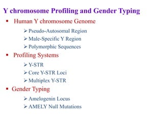
  • 1. Y chromosome Profiling and Gender Typing  Human Y chromosome Genome Pseudo-Autosomal Region Male-Specific Y Region Polymorphic Sequences  Profiling Systems Y-STR Core Y-STR Loci Multiplex Y-STR  Gender Typing Amelogenin Locus AMELY Null Mutations
  • 2. Y Chromosome Profiling and Gender Typing • The Y chromosome is inherited from the father, unique to males and passed on to all male offspring. • The chromosome encodes dozens of genes required for male-specific functions, including sex determination and spermatogenesis. • Y chromosome loci are very important for forensic DNA profiling. • For instance, the Y chromosome STR (Y-STR) used in forensic DNA testing is male-specific (for humans and certain higher primates) and is thus useful in investigations of sexual assault cases involving male suspects.
  • 3. • The evidence gathered in such cases usually consists of mixtures of high levels of female DNA and low levels of male DNA. • The Y chromosome-specific loci can be examined without interference from large amounts of female DNA; differential extraction of sperm and nonsperm cells may not be needed. • Furthermore, the Y-STR system is useful for determining numbers of male criminals in sexual assault cases involving more than one male. • The Y-STR loci used for forensic applications are located in the non- recombining section of the Y chromosome so that paternal lineages can be established. • The technique can be used for paternity testing and identification of missing persons.
  • 4. • The major disadvantage of Y chromosome loci is that their discriminating power is low compared to the discriminating power of autosomal loci. • Because Y chromosome loci are linked, the product rule for statistical calculations for profile probability does not apply. • Also, the Y chromosome loci test cannot distinguish individuals with the same paternal lineage.
  • 5. Human Y Chromosome Genome • The human Y chromosome genome contains approximately 60 million bp and the chromosome can be divided into two regions: the pseudo- autosomal region (PAR) and the male-specific Y (MSY) region. 1.Pseudo-Autosomal Region  Approximately 5% of the Y chromosome sequence is located at the telomeres of the chromosome.  In particular, PAR1 is located on the tip of the short arm and PAR2 is located at the tip of the long arm (Figure 1).  This region undergoes recombination with homologous regions on the X chromosome during meiosis in males.
  • 6. Figure 1. Human Y-STR chromosome structure. PAR = pseudo- autosomal region. MSY = male-specific Y region. Yp = short arm of Y chromosome. Yq = long arm of Y chromosome.
  • 7. 2.Male-Specific Y Region • The remainder of the Y chromosome is known as the MSY region. • It was previously called the non-recombining Y (NRY) region (Figure 1 above) or It does not participate in homologous recombination. • However, certain sections involve intra chromosomal gene conversion. • About 40 megabases (Mb) within the MSY region are heterochromatic (highly repetitive sequences) including the centromeric region and the bulk of the distal long arm.
  • 8. • The euchromatic region is about 23 Mb and most of it has been sequenced. • Certain sections of the euchromatic region share some homology with the X chromosome. • For instance, X-transposed sequences of the Y chromosome are 99% identical to sequences within Xq21 (a band in the long arm of the X chromosome). • Additionally, dozens of genes located in the euchromatic region share 60% to 96% homology with their X chromosome counterparts. • These X-homologous regions should be avoided when selecting Y chromosome-specific markers for forensic DNA profiling.
  • 9. 3 Polymorphic Sequences • The Y chromosome contains an abundance of repetitive elements, namely STRs, Alu, and LINE elements. • Many of these are highly polymorphic. • To date Y-STRs are usually used for Y chromosome DNA testing. • Single nucleotide polymorphisms (SNPs) at the Y chromosome are also useful for forensic applications.
  • 10. Profiling Systems 1. Y-STR • More than 400 STR loci have been identified in the Y chromosome genome. • The precise locations of these loci have been sequentially mapped using human genome sequencing data. • The distribution of Y-STR loci at the Y chromosome has also been analyzed. • Most Y-STR loci, approximately 60% of the 400 identified, are located at the long arm of the chromosome; about 22% are located at the short arm and a few are found in the centromeric region. • Y-STRs in the telomeric region have yet to be identified. • Only about 5% of Y-STRs are located within 5′ untranslated or intron regions of protein coding genes.
  • 11. • The repeat unit length of identified Y-STRs have been analyzed. • Among the 400 Y-STRs, 6% are dimeric repeats, 39% are trimeric, 45% are tetrameric, 9% are pentameric, and 1% are hexameric. • Fewer than half the STRs have been characterized. Some loci are polymorphic and are useful for forensic applications and developing new Y-STR multiplex systems. • The STR loci at the Y chromosome are usually referred to as haplotypes. • A haplotype is a collection of alleles that are usually linked (inherited together) since homologous recombination does not occur on the majority of the Y chromosome.
  • 12. 2. Core Y-STR Loci • In 1997, the European minimal haplotype (EMH) locus set was recommended by the International Y-STR User Group for forensic applications. • This haplotype set includes a core set of nine Y-STR loci: DYS19, DYS385 a and b, DYS389I, DYS389II, DYS390, DYS391, DYS392, and DYS393. • In 2003, the U.S. haplotype loci were recommended by the Scientific Working Group on DNAAnalysis Methods (SWGDAM) for forensic DNA analysis. • The U.S. haplotype loci includes the EMH loci set plus two additional loci, DYS438 and DYS439.
  • 13. • DYS385 and DYS389 are multi-local Y-STR loci (MLL). • The MLL designation refers to a presence of a particular STR at more than one site on the Y chromosome DNA due to duplication. • To date, about 50 such MLL Y-STRs have been identified. Further MLL subdivisions are designated bi-local, trilocal, etc. DYS385 and DYS389 are bi-local. • The DYS385 locus has two inverted duplicated clusters and is separated by a 4 × 10⁴ bp interstitial region (Figure 19.5). • It can be amplified by a single set of primers. One allele is observed if the duplicates are the same length. • If the duplicated clusters have different lengths, they can generate two different alleles when amplified. • The smaller sized allele is designated “a” and the larger sized allele is designated “b.”
  • 14. • The DYS389 locus has two duplicated clusters with the same orientation. • In a single set of PCR primers, there are two binding sites for the same forward primer at each 5′ flanking sequence of the core repeat region of DYS389. • These binding sites between DYS389I and DYS389II are about 120 bp apart. • Therefore, two amplicons are produced. • DYS389I is designed for the smaller allele and DYS389II is designated for the larger allele. • The average mutation rate for the core Y-STR loci is approximately 10–3 per generation—similar to the mutation rate of autosomal STR loci. Mutations can exert major impacts on the interpretation of paternity test results.
  • 15. 3 Multiplex Y-STR  The application Y-STR for forensic casework was initiated in Europe.  In the U.S., the laboratory of the Office of Chief Medical Examiner in New York City was the first to perform Y-STR testing of four loci (DYS19, DYS390, DYS389I and II) for casework.  The use of Y-STR loci has been facilitated by various commercially available PCR amplification kits in multiplex systems.  ReliaGene Technologies developed the first commercial multiplex Y- STR system, the Y-PLEXTM6.  The kit includes DYS19, DYS385a and b, DYS389II, DYS390, DYS391, and DYS393.
  • 16.  Additional commercially available kits with more Y-STR loci are now available and have been validated for forensic use.  To improve discriminating power, multiplex systems including new Y- STR loci are desired.  Many new Y-STR loci are being characterized for developing new multiplex systems.
  • 17. Gender Typing • Gender typing of a biological sample is useful in forensic investigation, for example, for victim identification in disaster cases and suspect identification in sexual assaults. • One commonly used gender typing marker is the amelogenin (AMEL) locus. 1 Amelogenin Locus • This region encodes extracellular matrix proteins involved in tooth enamel formation. • Mutations in the AMEL gene can lead to an enamel defect known as amelogenesis imperfecta.
  • 18. • The AMEL locus has two homologous genes: AMELX (Xp22.1– Xp22.3) is located on the human X chromosome and AMELY (Yp11.2) is located on the human Y chromosome. • Although the genes constitute a homologous pair, they differ in size and sequence. • Gender typing can be performed using various primers designed specifically for the sequences of the homologous region on these genes, followed by amplification. Different sizes of amplicons are obtained. • The most commonly used gender typing method at the AMEL locus is the detection of a 6-bp deletion at intron 1 of AMELX. • This deletion is not present in AMELY.
  • 19. • Primer sets were developed to amplify both alleles in a single PCR by Forensic Science Service in the United Kingdom in 1993. • The amplicons generated from AMELY and AMELX are separated by electrophoresis. • The observation of the AMELX fragment alone indicates a female, whereas the observation of both AMELX and AMELY indicates a male. • Nevertheless, primate and some rudiment DNA can be amplified as well but the amplicon sizes vary. • The AMEL locus has been co-amplified with other markers to provide a combined gender and identity test. • Such combined tests have been used in D1S80 AFLP and various STR multiplex analyses.
  • 20. 2 AMELY Null Mutations • Several cases of AMELY null mutations have been reported. • Only the AMELX fragment was detected in these AMELY null males. • Many of them are phenotypically normal but present the AMEL gender types of females. • Various interstitial deletions at the Y chromosome short arm have been identified as the cause of some AMELY null gender typing. • The frequency of AMELY null males is rare, but is higher in Sri Lanka and India.
  • 21. Mitochondrial DNA Profiling • Forensic mitochondrial DNA (mtDNA) analysis is a useful tool for human identification. • Because mtDNA is maternally inherited, it is especially useful for identifying victims. • Additionally, the mtDNA genome produces much higher numbers of copies per cell than the nuclear genome. • Thus, mtDNA testing is frequently used when nuclear DNA in samples is insufficient. • For example, hair shafts, bones, and decomposed samples may be tested with mtDNA analysis.
  • 22. Human Mitochondrial Genome • Mitochondria are subcellular organelles that serve as the energy-generating components of cells (Figure 1). • Each cell contains hundreds of mitochondria that have their own extrachromosomal genomes separate from the nuclear genome. • Although each human mitochondrion contains several copies of the mtDNA genome, the exact copy number varies for each cell. • However, it is estimated that hundreds of copies of mtDNA genome exist in most cell types. • Recombination has not been observed in mtDNA. • Thus, the mtDNA type, also referred to as the mitotype, is considered a haplotype treated as a single locus. • The mitochondrial genome has a higher mutation rate (up to 10 times higher) than its nuclear counterpart.
  • 23. 1 Genetic Contents of Mitochondrial Organelle Genomes • Organelle genomes are usually much smaller than their nuclear counterparts. • The much smaller mitochondrial genome has been sequenced and is known as the Cambridge reference sequence. • It was established in the early 1980s, later modified, and is now known as the revised Cambridge reference sequence (rCRS) that is presently used as the standard for sequence comparisons. • The human mitochondrial genome is a circular DNA molecule consisting of 16,569 bp containing 37 genes (Figure 1.). • Thirteen of these genes code for proteins involved in the respiratory complex, a main energy-generating component in mitochondria.
  • 24. Figure 1 Mitochondrion. mtDNA = mitochondrial DNA.
  • 25. • The other 24 specify noncoding RNA molecules required for expression of the mitochondrial genome. • The genes in the human mitochondrial genome are much more closely packed than in the nuclear genome and contain no introns. • A control region, also known as a displacement loop (D loop), contains the origin of replication for one of the mtDNA strands but does not code for any gene products (Figure 2). • An asymmetric distribution of nucleotides gives rise to light (L) and heavy (H) strands when mtDNA molecules are separated in alkaline CsCl gradients. • The H strand that contains a greater number of guanine nucleotides has a higher molecular weight in comparison to the L strand.
  • 26.  Figure 2. Human circular mitochondrial genome.  The transcription direction for the H (heavy) and L (light) strands are indicated by arrows (PH, PL).  The origins of replication are labeled OH for heavy strand and OL for light strand, respectively.  The mitochondrial DNA genome encodes genes. ND = NADH coenzyme Q oxidoreductase complex. CO = cytochrome c oxidase complex. Cytb = cytochrome b. ATP = ATP synthase. rRNA, ribosomal RNA.  Transfer RNA genes are shown as indicated.
  • 27. 2 Maternal Inheritance of mtDNA • Maternal inheritance is typically observed for the mtDNA genome. • mtDNA is inherited differently from nuclear genes; it does not obey the rules of Mendelian inheritance and is thus called non Mendelian inheritance. • The mitochondria of the spermatozoa are located at the mid pieces of spermatozoa. • At conception, only the head portion of a spermatozoon (containing a nucleus but no mitochondria) enters the egg. • The fertilized egg contains the maternal mitochondria which is transmitted to progeny. • The mtDNA sequence is identical for relatives within the same maternal lineage (Figure 3). • This characteristic of maternal inheritance is useful for identifying samples by comparing them with samples from maternal relatives.
  • 28. mtDNA Polymorphic Regions 1. Hypervariable Regions 2. Heteroplasmy Hypervariable Regions • The most polymorphic region of mtDNA is located within the D loop (Figure 4). • The three hypervariable regions in the D-loop region are designated HV1 (16024–16365; 342 bp), HV2 (73–340; 268 bp), and HV3 (438–574; 137 bp). • The most common polymorphic regions of the human mtDNA genome analyzed for forensic purposes are the two hypervariable regions within the D loop known as hypervariable region I (HV1) and hypervariable region II (HV2).
  • 29. Figure 4 Hypervariable regions of the D loop in mtDNA (with nucleotide positions). Figure 3 Pedigree of a human family showing inheritance of mtDNA Females and males are denoted by circles and squares, respectively. Red symbols indicate individuals who inherited the same mtDNA.
  • 30. Heteroplasmy • Heteroplasmy occurs when an individual carries more than one mtDNA haplotype. Occurs due to non disjunction during meiosis. • Heteroplasmy may be observed with one type of tissue and be absent in other tissue types; for example, it is commonly observed in hair samples. • Several instances of heteroplasmy may be observed in different tissue types. • An individual may exhibit one mitotype in one tissue and a different mitotype in another. • Thus, it is necessary to obtain and process additional samples to confirm the heteroplasmy when it is observed in a questioned sample but not in a known sample or vice versa. • The two types of Heteroplasmy are sequence and length heteroplasmies.
  • 31. Sequence Heteroplasmy • Sequence heteroplasmy is defined as the presence of two nucleotides at a single position shown as overlapping peaks in a sequence electropherogram (Figure 5). • Heteroplasmy usually occurs at one position, but on rare occasions can be observed at more than one position. • Heteroplasmy may complicate the interpretation of mtDNA results, but its presence can also improve the strength of a match
  • 32. Length Heteroplasmy • Both HV1 and HV2 of the human mtDNA D-loop region contain homopolymeric cytosine sequences known as C stretches. • The HVI region contains a C stretch between positions 16184 and 16193, interrupted by a thymine at position 16189. • If a base transition from T to C occurs at position 16189 (a variant present in approximately 20% of the population), it results in an uninterrupted C stretch. • A similar C stretch resides between positions 303 and 315 of the HV2 region. • Length heteroplasmies are often observed at the uninterrupted C stretches, which create serious problems with sequence analysis downstream from the homopolymeric regions (Figure 6). • It is not clear whether the length heteroplasmy is due to replication slippage at the C stretches or results from a mixture of length variants in the cells. • If length heteroplasmy occurs, alternative sequencing primers can be used to obtain the downstream sequences of the C stretches.
  • 33. Figure .5 Electropherogram showing mtDNA sequence heteroplasmy at position 234 R (A/G) as indicated by arrow. N = unresolved sequence. Figure .6 Electropherogram showing mtDNA length heteroplamy at C stretch of HV1 region where position 16189 is a C as indicated by arrow. N = unresolved sequence.
  • 34. Forensic mtDNA Testing 1 General Considerations • mtDNA analysis is often used on samples derived from skeletal or decomposed remains. • The surface of the sample should be cleaned to remove any adhering debris or contaminants. • Bones and teeth are pulverized to facilitate extraction of the mtDNA. • Duplicate extractions (e.g., two sections of a single hair) are recommended if sufficient sample material is available. • mtDNA is extracted using a similar method to nuclear DNA (nuclear DNA is coextracted with mtDNA). • The amount of mtDNA can therefore be estimated from the quantity of nuclear DNA obtained.
  • 35.  For mtDNA sequencing, analysis of both strands of the mtDNA in a given region must be performed to ensure accuracy.  Due to the high sensitivity of mtDNA analysis, it is essential to minimize risks of contamination during the procedure.  Contamination must be strictly monitored using proper controls such as reagent blanks and negative controls (samples containing all reagents except DNA template).  Finally, a positive control must also be used to monitor the success of the analysis.  It should be introduced at the amplification step and remain through the sequencing process.  A positive control consists of a DNA template of known sequence such as DNA purified from an HL60 cell line.
  • 36. mtDNA Sequencing • To sequence a specific region of mtDNA, a combination of PCR amplification and DNA sequencing techniques is employed that reduce the time and labor needed to obtain DNA sequences from genomic DNA templates. • mtDNA sequencing usually consists of 1) PCR amplification, 2) DNA sequencing reactions, 3) separation using electrophoresis, and 4) data collection and sequence analysis .
  • 37. PCR Amplification • The extracted DNA samples must be amplified to yield sufficient quantities of template for sequencing reactions. • PCR amplification of all or a part of the D loop region can be carried out with various primer sets. • If a sample contains high quality and high copy number mtDNA, the HV1, and HV2 regions can be amplified as two amplicons, each of about 350 to 400 bp in length. • If a sample is degraded or contains low copy number mtDNA, the hypervariable regions can be amplified as smaller PCR products. PCR amplification of mtDNA is usually done in 34 to 38 cycles. • Protocols for highly degraded DNA specimens sometimes require 42 cycles.
  • 38. • The use of higher PCR cycle numbers can improve the yield of the amplicon. • Following mtDNA amplification, a purification step is necessary to remove excess primers and deoxynucleotide triphosphates (dNTPs). • This step can be performed by using filtration devices such as a MicroconR to remove small molecules from the sample or by using nuclease digestion with shrimp alkaline phosphatase or exonuclease I to degrade remaining primers and dNTPs. • The concentration of the PCR product is important for an optimal sequencing reaction in the next phase of mtDNA sequencing. • The quality and quantity of the mtDNA amplicon must be evaluated to confirm the presence or absence of PCR products and their concentrations. • This can be done using an agarose yield gel to visualize the PCR products of the sample or via capillary electrophoresis, a more informative method, for quantifying PCR products.
  • 39. DNA Sequencing Reactions • The best known DNA sequencing techniques are the chain termination method and the chemical degradation method developed, respectively, by Sanger and Gilbert. • Over the years, the chain termination method became more common because it was applicable for automation and did not require the toxic chemicals necessary for the chemical degradation method.
  • 40. Electrophoresis and Sequence Analysis • The cycle sequencing products can be separated using electrophoresis in a 4% polyacrylamide denatured gel or a POP-6 polymer (Applied Biosystems) as the matrix for capillary electrophoresis. • Following data collection, sequence data analysis can be performed with the Sequencher™software (Gene Codes Corporation, Ann Arbor, MI, USA).
  • 41. Interpretation of mtDNA Profiling Results • Interpretation guidelines are used when the evaluation of sequencing results from evidence and reference samples are necessary. • General guidelines were set forth by the Scientific Working Group on DNAAnalysis Methods (SWGDAM) and the DNA Commission of the International Society of Forensic Genetics (ISFG). • The limitations of mtDNA technology should be taken into account as should the higher mutation rates found with the mtDNA genome than found with the nuclear genome. • Mutations seem to be more common in certain tissues. • For that reason the sources of the tissues investigated should be taken into consideration as well. • In reporting mtDNA profiling results, the most common categories of conclusions are: cannot exclude, exclusion, and inconclusive result.
  • 42. • Exclusion — If the sequences are different, then the samples can be excluded as originating from the same source. • Additionally, the SWGDAM’s guidelines define that this conclusion can be made if there are two or more nucleotide differences between the questioned and known samples. • Cannot exclude — If the sequences are the same, the reference sample and evidence cannot be excluded as potentially arising from the same source. • When a mtDNA profile cannot be excluded, it is desirable to evaluate the weight of the evidence. • In cases where the same heteroplasmy is observed in both questioned and known samples, its presence increases the strength of the evidence.
  • 43. • However, if heteroplasmy is observed in a questioned sample but not in a known sample or vice versa, a common maternal lineage still cannot be excluded. • Inconclusive result — If the questioned and known samples differ by a single nucleotide, and no evidence of heteroplasmy is present, the interpretation may be that the results are inconclusive.