X ray machine
•Download as ODP, PDF•
5 likes•2,921 views
Our expert team with years of experience in their respective fields have enabled us become a leading x-ray manufacturer India by manufacturing world-class diagnostic equipment. http://www.allengers.com
Report
Share
Report
Share

Recommended
x ray machine ppt
An x-ray machine uses x-rays to generate images of structures inside the body. It is a complex device used for purposes like airport security screening and medical imaging. Maintenance of x-ray machines includes repairs, preventative maintenance, and scheduled inspections to increase availability and reliability. Troubleshooting involves testing different components like transformers, cables, and tubes to diagnose issues like failed power supply or exposure problems.
Digital radiography
working and usage of digital radiography. types of digital radiography its advantages and disadvantages. care and maintenance of systems
Modern x-ray tube
The document provides details on the history and development of the modern X-ray tube. It discusses early experiments with evacuated glass tubes in the 18th and 19th centuries. Key developments include Roentgen's discovery of X-rays in 1895 using a Crookes tube, and Coolidge's invention of the hot cathode tube with a tungsten filament in 1913. The modern X-ray tube consists of a cathode that emits a focused electron beam, a rotating or stationary anode that produces X-rays upon electron impact, and components to dissipate heat and maintain the vacuum within the glass envelope.
Qc in xray
This document discusses quality control and quality assurance for x-ray machines. It outlines various tests that should be conducted, including central beam alignment, focal spot size, tube voltage, and timer checks. These tests help ensure the machine is functioning properly and producing high quality images. Acceptable tolerances and testing frequencies are provided. The roles of the quality assurance committee, including the medical physicist, radiologist, biomedical engineer, and technicians are described.
Automatic processing of X-ray film.pptx
Automatic processing of X-ray film involves passing exposed film through different chemical solutions - developer, fixer, wash, and dryer - inside an automatic processor. The processor uses rollers to transport the film between temperature-controlled solutions over a total processing time of around 90 seconds. This ensures consistent film quality and faster processing compared to manual methods. While automatic processors produce dry films immediately, they can potentially cause artifacts and require more expensive maintenance than manual processing.
Mobile & portable radiography
This document discusses portable and mobile x-ray machines. Portable x-rays can be carried by one person and used in hospitals, distant locations, or patients' homes to image in-patients or guide surgeons. Mobile x-rays are larger wheeled units that can be motorized or pushed. They have components like a base, generator, control panel, and supported x-ray tube. Mobile x-rays are classified by power source like capacitor discharge or batteries, and by output like low, average, or high power. Capacitor discharge units use a charged capacitor as the power source, while battery powered units use rechargeable batteries. Safety precautions for portable and mobile x-rays include long exposure cables and lead protection
soft tissue radiography
This document discusses techniques for visualizing soft tissues in radiography. Soft tissues have less differential attenuation compared to bones, making contrast reduced. Special techniques are needed to improve contrast and demonstrate soft tissues clearly. These include adjusting the kVp and adding filters to change image contrast. Using a normal or low kVp can help visualize certain soft tissues like adenoid and effusions more clearly. High kVp is useful for exams like BA enemas where thicker tissues are involved. Digital technology also helps improve soft tissue visibility compared to conventional radiography. Proper technique selection is important to optimize contrast and sharpness while reducing artifacts.
Viewing and storage of x ray film
This document discusses the viewing and storage of x-ray films. It describes x-ray viewers as boxes with light lamps used to observe radiograph films. X-ray viewers come in single or multiple formats. Films should be viewed in darkrooms using different light intensities depending on the film density. The document also outlines proper storage methods to protect films, such as wrapping, boxes, and digital formats. Films need storage in low temperature, humidity, and light conditions to avoid artifacts like fogging, strikes, or static marks.
Distortion
This document discusses two types of distortion that can occur in radiography: size distortion and shape distortion. Size distortion refers to unequal magnification and is influenced by the object-to-film distance and film-to-focus distance. Shape distortion occurs when there is elongation or foreshortening due to the central ray-part-film alignment. Magnification radiography can be used intentionally to visualize small structures and comes at the cost of increased patient dose. Proper technique such as parallel part-film alignment and perpendicular central ray direction can minimize distortion.
Quality Assurance on Diagnostic Equipment/Accessories
This document provides an overview of quality assurance procedures for diagnostic radiography equipment and accessories. It discusses the importance of quality assurance and quality control in maintaining high standards, reducing radiation dose and costs from repeated tests. It then details specific tests and inspection criteria for various components of radiographic systems, including generators, computers, viewers and monitors. Frequency of testing and corrective actions are provided. Procedures are described for testing lead aprons, light field-radiation field alignment, and other equipment. References for additional information are listed at the end.
compiter radiography and digital radiography
This document discusses computed radiography (CR) and digital radiography (DR). CR uses reusable imaging plates instead of film, which are read by a laser scanner. DR uses a digital detector incorporated into x-ray equipment to provide direct digital output. Both have greater exposure latitude than screen-film and allow computer post-processing to enhance images. Technologists must monitor exposure indices to avoid overexposure with CR and DR systems. The document also covers digital fluoroscopy techniques like frame averaging.
Paediatric radiography
Paediatric radiography requires special techniques and considerations due to children's developing cognitive abilities and need for trust and comfort. The technologist's friendly introduction and clear explanation of the procedure helps build rapport. Immobilization devices like boards and straps help reduce motion blur while sandbags, tape and towels can also immobilize. Technologists minimize radiation dose through high mA, short exposures and shielding gonads when possible. Clear communication between technologist and parents helps each understand their role in supporting the child.
Tomographic equipment
Tomographic equipment allows for the production of sharp images by moving the x-ray tube and detector in opposite directions during exposure. There are two main types - attachments that connect to existing equipment using linkage mechanisms, pivot units, and drives, and specialized tomography tables. Tables are categorized into three groups based on their motion capabilities. All tomographic equipment aims to focus the anatomy of interest while blurring surrounding structures through controlled tube movement during x-ray exposure.
Faults in x ray tube and its care
This document discusses common faults in x-ray tubes, their causes, and remedies. It outlines faults that can occur in the tube housing, glass/metal envelope, filament, and anode. Examples of faults include cracking of the housing, loss of vacuum, vaporization or breakage of the filament, and kinking or roughening of the anode surface. The document also provides tips for proper care of x-ray tubes, such as following rating charts and limiting operation to prevent overheating. Overall, the document provides an overview of potential issues that can arise in x-ray tubes and how to address them.
Radiographic cassettes
The document is a presentation about radiographic cassettes by Sudil Paudyal. It discusses the functions and features of radiographic cassettes, how they are constructed, the materials used and different types available including single screen, double screen, curved, gridded, multi-section, vacuum, and computed radiography cassettes. It also covers how cassettes should be loaded, unloaded, and cared for to maximize the life of the intensifying screens.
Spect technology
SPECT involves injecting a radiopharmaceutical that emits gamma rays. Detectors rotate around the body to acquire data from multiple angles and produce 3D images. It allows visualization of organ function. A gamma camera detects gamma rays and includes a collimator, scintillation detector, photomultiplier tubes, and computer. SPECT is used for heart, brain, and tumor imaging. It has lower resolution than PET but is commonly used to detect coronary artery disease.
Components of X-Ray Machine
This document summarizes the key components of an X-ray system, including the operating console, high frequency generator, and X-ray tube. It describes the internal parts of the X-ray tube, such as the cathode and anode. Additional parts that help form X-ray images like the collimator, grid, and bucky are also outlined. Finally, some common medical uses of X-rays like detecting broken bones or cancer are mentioned.
X ray tube
The document provides information about X-ray tubes, including their history, components, and developments over time. It discusses:
- The key components of an X-ray tube including the cathode, filament, focusing cup, and anode. Electrons are emitted from the filament and accelerated toward the anode to produce X-rays.
- The development of X-ray tubes from the original Crookes tube to modern Coolidge tubes. Coolidge tubes introduced thermionic emission to produce electrons instead of relying on residual gas ionization.
- Advances over time including rotating anodes, improved cooling methods, and different target materials to produce more intense and focused X-rays for various medical and industrial applications
Ct Generations
1. The first generation of CT used a single narrow x-ray beam and detector that rotated around the patient in a translate-rotate motion. It took 5-6 minutes to complete a scan.
2. The second generation used multiple narrow beams and detectors, reducing scan time by a factor equal to the number of detectors by collecting multiple views simultaneously. Scan times were reduced to 20 seconds.
3. The third generation eliminated translation motion by using a fan-beam of x-rays and multiple stationary detectors arranged in a ring. Only rotational motion was needed, simplifying the mechanics. This further reduced scan times.
Beam restricted device and filter used in x ray
This document discusses various beam restricting devices and filters used in radiography to reduce radiation exposure. It describes common beam restricting devices like diaphragms, cones, cylinders and collimators which are used to limit the size of the primary x-ray beam and reduce scatter radiation. It also discusses different types of filters like inherent, aluminum, compound and molybdenum filters which absorb low energy photons and improve image quality. Maintaining proper collimation and use of appropriate filters helps achieve the ALARA principle of keeping radiation exposure As Low As Reasonably Achievable.
OT theatre radiography.ppt
The document discusses the role and responsibilities of a radiographer in the operating theatre. It outlines the key tasks of preparing equipment, ensuring patient details are entered correctly, and using protective equipment. The radiographer aids surgical procedures by producing diagnostic images to visualize anatomy and equipment placement. Key responsibilities include minimizing radiation dose, maintaining sterilization, effective communication with the surgical team, and working collaboratively to improve imaging techniques.
Radiographic exposure and image quality
The document discusses various radiographic exposure factors and how they influence the quantity and quality of x-radiation exposure to patients. It describes how factors like kVp, mA, and exposure time determine the radiation dose and beam quality. It also discusses how the design of the x-ray machine like focal spot size, filtration, and high voltage generation impact technical settings. Film factors like sensitometry, contrast, and processing also influence radiographic image quality.
Emergency radiography
emergency radiography has emerged as one of the newest and fastest growing radiology subspecialties .and to easy radiographer and technologist
CT Generation (Generation of CT)
This document discusses the history and evolution of different generations of computed tomography (CT) technology. It describes the key limitations and innovations of each generation from the first generation CT scanner created in 1971, which took 5 minutes to produce an image, to modern multi-slice CT scanners. The higher the generation number, the faster imaging times and more slices that could be acquired simultaneously. However, a higher generation does not always indicate a higher performance system.
Mammographic equipment and its advancement
This document discusses the advancement of mammographic equipment. It begins by introducing the components and purpose of mammography equipment. Key components discussed in detail include the x-ray tube, compressor, anti-scatter grid, cassette holder, and digital detectors. The document then covers recent advancements, such as digital mammography technologies like computed radiography, full-field digital mammography, and digital breast tomosynthesis, which uses 3D imaging to improve cancer detection rates.
Dark room procedures
The document discusses procedures for a dark room used in medical radiography. It provides details on the layout, equipment, and setup of a dark room. Key points include:
- A dark room must be lightproof and have adequate space for a dry bench, wet bench, and sink. Special lighting and filters are used to allow safe viewing of films.
- Equipment includes cassettes, hangers, a film bin, and either a manual or automatic film processor. Chemicals are needed for developing, fixing, and washing films.
- Proper development involves immersing films in tanks containing developer, fixer, and rinse solutions. Strict temperature control and timing is required to reveal the latent image on
Quality assurance
This document discusses quality assurance and quality control tests for diagnostic x-ray equipment. It defines quality assurance as maintaining high quality imaging through personnel training and evaluation, while quality control refers to evaluating radiographic equipment and identifying issues. Regular quality control tests check parameters like radiation and optical field alignment, focal spot size, tube voltage accuracy, exposure timer accuracy, total filtration, and radiation leakage. Performing these tests ensures optimal image quality, minimum radiation exposure, and cost effectiveness of diagnostic x-ray equipment.
Dark room and film processing techniques rv
This document provides information on dark room and film processing techniques. It discusses the key components and functions of a dark room for handling radiographic films without light exposure. It also describes the various stages of film processing including development, fixing, washing and drying. Both manual and automatic processing techniques are covered, outlining the different steps, equipment, chemical solutions and factors involved in each method. Automatic processors provide controlled, consistent processing using chemical tanks and a transport system to move films through development, fixation, washing and drying cycles.
Portable n mobile unit
Portable and mobile radiographic units can be either portable or mobile. Portable units are small enough to be carried by one person for use outside of a radiology department. Mobile units are larger and mounted on wheels, able to be moved throughout a hospital. Both use an X-ray tube, generator, and control unit to produce radiographic images. Newer units are using high frequency generators, computed radiography, or direct radiography for more efficient and higher quality imaging. Mobile C-arm units are important for fluoroscopy in operating rooms.
Introduction to the parts of x ray machine
The document describes the key components of an X-ray machine:
1. The X-ray tube which produces X-rays and contains a cathode and anode in a vacuum tube housed in a protective casing.
2. The operating console which controls the voltage, current, and exposure time of the X-ray tube.
3. Additional components include a high voltage transformer, collimator to control the beam, patient table, grid to reduce scattering, and Bucky device which holds the X-ray film.
More Related Content
What's hot
Distortion
This document discusses two types of distortion that can occur in radiography: size distortion and shape distortion. Size distortion refers to unequal magnification and is influenced by the object-to-film distance and film-to-focus distance. Shape distortion occurs when there is elongation or foreshortening due to the central ray-part-film alignment. Magnification radiography can be used intentionally to visualize small structures and comes at the cost of increased patient dose. Proper technique such as parallel part-film alignment and perpendicular central ray direction can minimize distortion.
Quality Assurance on Diagnostic Equipment/Accessories
This document provides an overview of quality assurance procedures for diagnostic radiography equipment and accessories. It discusses the importance of quality assurance and quality control in maintaining high standards, reducing radiation dose and costs from repeated tests. It then details specific tests and inspection criteria for various components of radiographic systems, including generators, computers, viewers and monitors. Frequency of testing and corrective actions are provided. Procedures are described for testing lead aprons, light field-radiation field alignment, and other equipment. References for additional information are listed at the end.
compiter radiography and digital radiography
This document discusses computed radiography (CR) and digital radiography (DR). CR uses reusable imaging plates instead of film, which are read by a laser scanner. DR uses a digital detector incorporated into x-ray equipment to provide direct digital output. Both have greater exposure latitude than screen-film and allow computer post-processing to enhance images. Technologists must monitor exposure indices to avoid overexposure with CR and DR systems. The document also covers digital fluoroscopy techniques like frame averaging.
Paediatric radiography
Paediatric radiography requires special techniques and considerations due to children's developing cognitive abilities and need for trust and comfort. The technologist's friendly introduction and clear explanation of the procedure helps build rapport. Immobilization devices like boards and straps help reduce motion blur while sandbags, tape and towels can also immobilize. Technologists minimize radiation dose through high mA, short exposures and shielding gonads when possible. Clear communication between technologist and parents helps each understand their role in supporting the child.
Tomographic equipment
Tomographic equipment allows for the production of sharp images by moving the x-ray tube and detector in opposite directions during exposure. There are two main types - attachments that connect to existing equipment using linkage mechanisms, pivot units, and drives, and specialized tomography tables. Tables are categorized into three groups based on their motion capabilities. All tomographic equipment aims to focus the anatomy of interest while blurring surrounding structures through controlled tube movement during x-ray exposure.
Faults in x ray tube and its care
This document discusses common faults in x-ray tubes, their causes, and remedies. It outlines faults that can occur in the tube housing, glass/metal envelope, filament, and anode. Examples of faults include cracking of the housing, loss of vacuum, vaporization or breakage of the filament, and kinking or roughening of the anode surface. The document also provides tips for proper care of x-ray tubes, such as following rating charts and limiting operation to prevent overheating. Overall, the document provides an overview of potential issues that can arise in x-ray tubes and how to address them.
Radiographic cassettes
The document is a presentation about radiographic cassettes by Sudil Paudyal. It discusses the functions and features of radiographic cassettes, how they are constructed, the materials used and different types available including single screen, double screen, curved, gridded, multi-section, vacuum, and computed radiography cassettes. It also covers how cassettes should be loaded, unloaded, and cared for to maximize the life of the intensifying screens.
Spect technology
SPECT involves injecting a radiopharmaceutical that emits gamma rays. Detectors rotate around the body to acquire data from multiple angles and produce 3D images. It allows visualization of organ function. A gamma camera detects gamma rays and includes a collimator, scintillation detector, photomultiplier tubes, and computer. SPECT is used for heart, brain, and tumor imaging. It has lower resolution than PET but is commonly used to detect coronary artery disease.
Components of X-Ray Machine
This document summarizes the key components of an X-ray system, including the operating console, high frequency generator, and X-ray tube. It describes the internal parts of the X-ray tube, such as the cathode and anode. Additional parts that help form X-ray images like the collimator, grid, and bucky are also outlined. Finally, some common medical uses of X-rays like detecting broken bones or cancer are mentioned.
X ray tube
The document provides information about X-ray tubes, including their history, components, and developments over time. It discusses:
- The key components of an X-ray tube including the cathode, filament, focusing cup, and anode. Electrons are emitted from the filament and accelerated toward the anode to produce X-rays.
- The development of X-ray tubes from the original Crookes tube to modern Coolidge tubes. Coolidge tubes introduced thermionic emission to produce electrons instead of relying on residual gas ionization.
- Advances over time including rotating anodes, improved cooling methods, and different target materials to produce more intense and focused X-rays for various medical and industrial applications
Ct Generations
1. The first generation of CT used a single narrow x-ray beam and detector that rotated around the patient in a translate-rotate motion. It took 5-6 minutes to complete a scan.
2. The second generation used multiple narrow beams and detectors, reducing scan time by a factor equal to the number of detectors by collecting multiple views simultaneously. Scan times were reduced to 20 seconds.
3. The third generation eliminated translation motion by using a fan-beam of x-rays and multiple stationary detectors arranged in a ring. Only rotational motion was needed, simplifying the mechanics. This further reduced scan times.
Beam restricted device and filter used in x ray
This document discusses various beam restricting devices and filters used in radiography to reduce radiation exposure. It describes common beam restricting devices like diaphragms, cones, cylinders and collimators which are used to limit the size of the primary x-ray beam and reduce scatter radiation. It also discusses different types of filters like inherent, aluminum, compound and molybdenum filters which absorb low energy photons and improve image quality. Maintaining proper collimation and use of appropriate filters helps achieve the ALARA principle of keeping radiation exposure As Low As Reasonably Achievable.
OT theatre radiography.ppt
The document discusses the role and responsibilities of a radiographer in the operating theatre. It outlines the key tasks of preparing equipment, ensuring patient details are entered correctly, and using protective equipment. The radiographer aids surgical procedures by producing diagnostic images to visualize anatomy and equipment placement. Key responsibilities include minimizing radiation dose, maintaining sterilization, effective communication with the surgical team, and working collaboratively to improve imaging techniques.
Radiographic exposure and image quality
The document discusses various radiographic exposure factors and how they influence the quantity and quality of x-radiation exposure to patients. It describes how factors like kVp, mA, and exposure time determine the radiation dose and beam quality. It also discusses how the design of the x-ray machine like focal spot size, filtration, and high voltage generation impact technical settings. Film factors like sensitometry, contrast, and processing also influence radiographic image quality.
Emergency radiography
emergency radiography has emerged as one of the newest and fastest growing radiology subspecialties .and to easy radiographer and technologist
CT Generation (Generation of CT)
This document discusses the history and evolution of different generations of computed tomography (CT) technology. It describes the key limitations and innovations of each generation from the first generation CT scanner created in 1971, which took 5 minutes to produce an image, to modern multi-slice CT scanners. The higher the generation number, the faster imaging times and more slices that could be acquired simultaneously. However, a higher generation does not always indicate a higher performance system.
Mammographic equipment and its advancement
This document discusses the advancement of mammographic equipment. It begins by introducing the components and purpose of mammography equipment. Key components discussed in detail include the x-ray tube, compressor, anti-scatter grid, cassette holder, and digital detectors. The document then covers recent advancements, such as digital mammography technologies like computed radiography, full-field digital mammography, and digital breast tomosynthesis, which uses 3D imaging to improve cancer detection rates.
Dark room procedures
The document discusses procedures for a dark room used in medical radiography. It provides details on the layout, equipment, and setup of a dark room. Key points include:
- A dark room must be lightproof and have adequate space for a dry bench, wet bench, and sink. Special lighting and filters are used to allow safe viewing of films.
- Equipment includes cassettes, hangers, a film bin, and either a manual or automatic film processor. Chemicals are needed for developing, fixing, and washing films.
- Proper development involves immersing films in tanks containing developer, fixer, and rinse solutions. Strict temperature control and timing is required to reveal the latent image on
Quality assurance
This document discusses quality assurance and quality control tests for diagnostic x-ray equipment. It defines quality assurance as maintaining high quality imaging through personnel training and evaluation, while quality control refers to evaluating radiographic equipment and identifying issues. Regular quality control tests check parameters like radiation and optical field alignment, focal spot size, tube voltage accuracy, exposure timer accuracy, total filtration, and radiation leakage. Performing these tests ensures optimal image quality, minimum radiation exposure, and cost effectiveness of diagnostic x-ray equipment.
Dark room and film processing techniques rv
This document provides information on dark room and film processing techniques. It discusses the key components and functions of a dark room for handling radiographic films without light exposure. It also describes the various stages of film processing including development, fixing, washing and drying. Both manual and automatic processing techniques are covered, outlining the different steps, equipment, chemical solutions and factors involved in each method. Automatic processors provide controlled, consistent processing using chemical tanks and a transport system to move films through development, fixation, washing and drying cycles.
What's hot (20)
Quality Assurance on Diagnostic Equipment/Accessories
Quality Assurance on Diagnostic Equipment/Accessories
Viewers also liked
Portable n mobile unit
Portable and mobile radiographic units can be either portable or mobile. Portable units are small enough to be carried by one person for use outside of a radiology department. Mobile units are larger and mounted on wheels, able to be moved throughout a hospital. Both use an X-ray tube, generator, and control unit to produce radiographic images. Newer units are using high frequency generators, computed radiography, or direct radiography for more efficient and higher quality imaging. Mobile C-arm units are important for fluoroscopy in operating rooms.
Introduction to the parts of x ray machine
The document describes the key components of an X-ray machine:
1. The X-ray tube which produces X-rays and contains a cathode and anode in a vacuum tube housed in a protective casing.
2. The operating console which controls the voltage, current, and exposure time of the X-ray tube.
3. Additional components include a high voltage transformer, collimator to control the beam, patient table, grid to reduce scattering, and Bucky device which holds the X-ray film.
x-ray machine
X-rays are a form of electromagnetic radiation that can pass through soft tissue but are partially or fully blocked by dense tissues like bone. Wilhelm Roentgen discovered x-rays in 1895 while experimenting with cathode ray tubes. X-rays are used in medical imaging to generate diagnostic images of internal structures in the body by passing x-ray beams through the body onto photographic film or digital sensors. The denser tissues absorb more of the x-ray beam, resulting in contrast between different internal structures visible on the radiograph. Common medical uses of x-rays include detection of fractures, tumors, dental decay, and foreign objects in the body.
Portable X-ray Radiography
The document summarizes a presentation given at an RPM working meeting at Bruce Power about using portable x-ray radiography equipment to conduct radiography tests. It discusses the goals of maintaining safety controls and reducing dose, costs, and exclusion zones while maintaining quality. It provides details on the portable x-ray tube and digital imaging equipment used, test setup, and results from a field application that showed significant savings in time, costs, and dose compared to traditional radiography.
03 xray film etc
The document discusses x-ray film, including its composition, how a latent image is formed, different film characteristics like density and contrast, common film types and sizes, intensifying screens, and manual and automatic film processing. Key points include that film is composed of silver halide crystals, screens decrease the radiation needed for images, and processing involves developing, fixing, washing and drying the film.
Setting up Radiology Diagnostic Centers
This document outlines the components needed to build a modern diagnostic center chain, including both an advanced central diagnostic center and several satellite imaging centers. It discusses key considerations for the location, layout, infrastructure, equipment, staffing, training, use of digital radiology systems, financial projections, and a franchise model for expanding the network of centers. The goal is to establish a central hub with advanced modalities connected to smaller satellite clinics via telemedicine to improve diagnostic access.
X rays
X-rays are a form of electromagnetic radiation that can penetrate materials and are used in medical imaging to see inside the body. Wilhelm Röntgen discovered X-rays in 1895 while experimenting with cathode ray tubes. He noticed a fluorescent screen glowing nearby even when covered, and realized the rays could pass through some materials but not others like bone. This allowed him to see the outline of bones in his hand, demonstrating their medical application. Today, X-rays are widely used in medicine, industry, and research due to their ability to penetrate soft tissue while being blocked by denser materials like bones and metals.
X Rays Ppt
The document discusses x-rays, how they produce images, and their risks and benefits. It explains that x-rays are electromagnetic waves that can pass through tissues at different levels, exposing film to create internal images of the body. German physicist Wilhelm Röntgen discovered x-rays in 1895 while experimenting with vacuum tubes. Lead shields are used during x-rays to block radiation and prevent long-term exposure, as x-rays can detect health problems. The document identifies different bones and joints visible in sample x-ray images.
X ray films
The document discusses x-ray films and the process of developing radiographic images. It describes the composition of intraoral x-ray films, including the film base, emulsion, and packet components. It then explains the processes of latent image formation when film is exposed to x-rays and the chemical steps to develop the latent image, including developing, fixing, washing and drying the film. Finally, it discusses factors that influence radiographic image quality such as density, contrast and sharpness.
Viewers also liked (9)
Similar to X ray machine
IJSRED-V2I2P32
The document describes a high frequency surgical C-arm with a stationary anode tube, 9-inch image intensifier, and computerized workstation manufactured by ADONIS Medical Systems. It provides an overview of the technical specifications and components of the mobile C-arm system. The C-arm features a 40 kHz generator, stationary anode x-ray tube, 9-inch image intensifier, 1/2-inch CCD camera, computerized workstation, and can be used for procedures in orthopedics, urology and other areas. It allows for fluoroscopy, radiography, image storage and processing capabilities. The system is designed to provide efficient, reliable and easy to use intraoperative imaging.
e_book_PDF diagnositic X ray system and X ray components.pdf
Description
Dongmun's another innovation, Portable,
X-Ray Units is Digital Technology High Frequency inverter
type to provide the high efficiency in x-ray equipments,
designed for uses in large hospitals, clinics, private
practices, dental clinics. bed-side diagnosis and
emergencies, veterinary.
Features
● Compact and light weight design.
● Digital LED display , reversible.
● 6 selection presetting anatomical memory mode.
● Line voltage compensation.
● Remote control hand switch. for exposure.
● halogen lamp collimator.
● Scale tape for SID measurement & laser pointer.
QMI 2014 Instrumentation Catalog
This document is a catalog from Quality Material Inspection, Inc. that describes their non-contact air-coupled ultrasonic NDT instrumentation and scanning systems. The company develops, manufactures, and markets the AIRSCAN line which uses air-coupled ultrasound to inspect materials without contamination. Their flagship product is the Sonda 007CX instrument, which is designed to replace conventional UT instruments for C-Scan applications. They also offer various scanning gantries and systems up to 6 feet by 3 feet in size to perform inspections with the AIRSCAN instrumentation.
Presentation Of Vikomed In English
Vikomed is a Vietnam-Korea joint venture medical equipment company established in 2007. It manufactures and sells advanced medical technologies including extracorporeal shock wave lithotripters, high frequency digital x-rays, and high-end surgical c-arms. Vikomed's strengths include over 15 years of experience in medical equipment, manufacturing its own detectors, installing over 100 systems, and focusing on prompt customer service.
Pixel cp operators manual
This document appears to be an operator's manual for an x-ray generator. It provides information on safety, specifications, controls, programming, error codes, exposure tables, maintenance, and x-ray tube data. The introduction describes the generator's features such as output power levels up to 80 kW, kV ranges from 125-150 depending on the model, and user-friendly controls. Safety precautions are outlined, and applicable standards are listed.
Introduction of imaging dept
The document discusses various medical imaging modalities and their specifications and uses. It provides information on radiography/fluoroscopy, computed tomography, ultrasound, and other modalities. For each modality, it describes the uses, specifications of the equipment, room requirements, electrical needs, HVAC requirements, and other pre-installation considerations. The document serves as a reference for properly installing and utilizing different medical imaging technologies.
Introduction of imaging dept
The document provides information on various medical imaging modalities and their specifications. It discusses radiography/fluoroscopy, computed tomography, ultrasound, magnetic resonance imaging, their uses, equipment details, room requirements, and electrical/HVAC specifications. The modalities described provide non-invasive imaging of the internal body structures for diagnostic and interventional procedures. Precautions are outlined for each to ensure patient and staff safety.
Catalogo Examion Produvet
This document provides an overview of EXAMION® Systems, a supplier of digital and conventional X-ray systems. It describes their portfolio of DR, CR, and portable X-ray generators. EXAMION® offers complete DR systems integrating an X-ray generator, detector, and acquisition software. They also provide CR digitizer systems that allow existing X-ray equipment to be used for digital radiography. A range of portable X-ray generators are presented for mobile and small animal use. The EXAMION® AQS software is highlighted as the heart of the digital systems, controlling image acquisition, processing, and archiving.
NextGuard_Brochure_WEB
The Thermo Scientific NextGuard x-ray detection system provides cost-effective and sensitive detection of foreign objects in packaged food products. It uses a unique non-linear x-ray detector design that eliminates inspection blind spots and ensures 100% detection for any product size or shape. The system offers options for customization, easy maintenance, and is designed for use in compliance with worldwide food safety standards.
Go 2 d cataloge
GO 2D is a new panoramic x-ray device from NewTom that offers precise 2D imaging with a user-friendly and reliable system. It produces high quality panoramic images with low radiation exposure using exclusive SafeBeam technology. The device installs quickly and is integrated with NNT software for enhanced workflow and diagnostics.
Metallurgical and mechanical testing machines in pune conation technologies
Metallurgical and mechanical testing machines in pune conation technologiesBizzporto Information and Marketting Services Private Limited
“Conation Technologies” are committed to offer quality control lab equipment like Metallurgical
and Mechanical Testing Machines to our customers with latest and best technologies at affordable prices.
Our product range includes Metallurgical Microscopes, Universal Microscopes, Portable Microscopes,
Stereozooms, Metallurgical Image Analyzer, Image Analyzers for welding analysis, fibre analysis,
clinker analysis etc., Cleanliness (Millipore Filter Paper) Analysis System, Microhardness Testers,Metallurgical Sample Preparation Machines, Mechanical Testing Machines,Jominy End Quench Test set-up, and Special Purpose Machines.TEST TRADING - Utilizarea generatorilor Rx portabili pentru scanarea autoturi...
TEST TRADING - Utilizarea generatorilor Rx portabili pentru scanarea autoturismelor si camionetelor - Dl. Emil Studineanu
Trans o sonic
Trans-O-Sonic manufactures ultrasonic cleaners and cleaning equipment. It was established in 1980 and develops new designs while enhancing product lifecycles and integrating new technologies. It provides ultrasonic cleaners, vapour degreasers, washing machines, and other equipment for industries like engineering, electronics, electroplating, and more. The document provides details on their material handling systems, multi-chamber cleaners, transducers, and other products, with contact information for the partner.
CO2 Accessories
The document describes various CO2 laser accessories for medical procedures, including:
- The SurgiTouch system which provides pre-set parameters and recommendations to guide laser surgery for different specialties. It allows automated scanning for precise incisions and tissue removal.
- Scanning accessories like the AcuBlade and OtoScan which enable precise, char-free incisions and ablation through fast laser beam scanning.
- Sets for procedures like bronchoscopy, treating the oral cavity and nasal passages, and colposcopy.
- The UltraScan computerized pattern generator for precise laser skin resurfacing.
Setting a new radiotherapy facility-steps involved
Setting a new radiotherapy facility-steps involved. Explains what all things to be done to setup new oncology centre.
Angstrom advanced scientific instrument introduction
As an leading supplier of Scientific Instruments, Angstrom Advanced has extensive experiences in designing, manufacturing, building and installing a variety of Scientific Instruments; especially Ellipsometer, AA2000 Atomic Force Microscope, AA5000 Multi-function SPM Systems, AA8000 Multi-function SEM System, Gas Generators, X-ray Diffractometer, etc
Portfolio_0215_5
The document provides an overview of Stryker's product catalog and marketing campaigns for 2011. Key highlights include:
1. The catalog carried Stryker's "quality of life" theme throughout and featured more visual content and hyperlinks to drive customers online.
2. Marketing campaigns promoted new products like the Crossfire integrated arthroscopy system and MiniLap instruments through brochures, posters, and tradeshow booths emphasizing resonant benefits.
3. Advertisements and a tradeshow booth introduced Stryker's expanded portfolio for single-site surgeries including longer instruments and a pneumatic scope holder.
Application of Automation in different sectors
This document discusses various applications of automation in different sectors such as medical, defense, mining, and agriculture. In the medical field, it describes how nanorobots and automated dispensing devices can help with diagnosis and targeted drug delivery. It also discusses robotic surgery tools like AESOP, Zeus, and da Vinci. In defense, it outlines automated systems for missiles, drones, and radar targeting. For mining, it explains technologies like seismic imaging, laser imaging, and automated drilling, hauling, and exploration equipment. Finally, it discusses sensors and automation for site-specific spraying and yield mapping in agriculture.
mobile image intensifier units.ppt
Image intensifiers, Fluoroscopy, Angiography, Angioplasty, Real-time imaging, Videocon, input phosphor, Photocathode, C-arm, TV monitors, Mini C-arm system, Operation theatres, Radiation protection devices, X-ray tube, stenting, interventional radiology
HeatSign Laser Marking Machines
Here you can see all HeatSign Laser Marking Machines include laser etching machine, fiber laser engraver, CO2 laser marking macine, industry laser marker, handheld portable laser engraving systems etc.
Similar to X ray machine (20)
e_book_PDF diagnositic X ray system and X ray components.pdf
e_book_PDF diagnositic X ray system and X ray components.pdf
Metallurgical and mechanical testing machines in pune conation technologies
Metallurgical and mechanical testing machines in pune conation technologies
TEST TRADING - Utilizarea generatorilor Rx portabili pentru scanarea autoturi...
TEST TRADING - Utilizarea generatorilor Rx portabili pentru scanarea autoturi...
Setting a new radiotherapy facility-steps involved
Setting a new radiotherapy facility-steps involved
Angstrom advanced scientific instrument introduction
Angstrom advanced scientific instrument introduction
Recently uploaded
Innovative Minds France's Most Impactful Healthcare Leaders.pdf
This edition features a handful of Innovative Minds: France's Most Impactful Healthcare Leaders that are leading us into a better future.
NEEDLE STICK INJURY - JOURNAL CLUB PRESENTATION - DR SHAMIN EABENSON
NEEDLE STICK INJURY - JOURNAL CLUB PRESENTATION - DR SHAMIN EABENSON
Luxurious Spa In Ajman Chandrima Massage Center
Chandrima Spa Ajman is one of the leading Massage Center in Ajman, which is open 24 hours exclusively for men. Being one of the most affordable Spa in Ajman, we offer Body to Body massage, Kerala Massage, Malayali Massage, Indian Massage, Pakistani Massage Russian massage, Thai massage, Swedish massage, Hot Stone Massage, Deep Tissue Massage, and many more. Indulge in the ultimate massage experience and book your appointment today. We are confident that you will leave our Massage spa feeling refreshed, rejuvenated, and ready to take on the world.
Visit : https://massagespaajman.com/
Call : 052 987 1315
Letter to MREC - application to conduct study
Application to conduct study on research title 'Awareness and knowledge of oral cancer and precancer among dental outpatient in Klinik Pergigian Merlimau, Melaka'
Let's Talk About It: Breast Cancer (What is Mindset and Does it Really Matter?)
Your mindset is the way you make sense of the world around you. This lens influences the way you think, the way you feel, and how you might behave in certain situations. Let's talk about mindset myths that can get us into trouble and ways to cultivate a mindset to support your cancer survivorship in authentic ways. Let’s Talk About It!
U Part Wigs_ A Natural Look with Minimal Effort Jokerwigs.in.pdf
Joker Wigs has been a one-stop-shop for hair products for over 26 years. We provide high-quality hair wigs, hair extensions, hair toppers, hair patch, and more for both men and women.
一比一原版(EUR毕业证)鹿特丹伊拉斯姆斯大学毕业证如何办理
EUR毕业证offer【微信95270640】《鹿特丹伊拉斯姆斯大学毕业证书原版制作EUR成绩单》【Q微信95270640】《仿制EUR毕业证成绩单鹿特丹伊拉斯姆斯大学学位证书pdf电子图》,A.为什么留学生需要操作留信认证?
留信认证全称全国留学生信息服务网认证,隶属于北京中科院。①留信认证门槛条件更低,费用更美丽,并且包过,完单周期短,效率高②留信认证虽然不能去国企,但是一般的公司都没有问题,因为国内很多公司连基本的留学生学历认证都不了解。这对于留学生来说,这就比自己光拿一个证书更有说服力,因为留学学历可以在留信网站上进行查询!
B.为什么我们提供的毕业证成绩单具有使用价值?
查询留服认证是国内鉴别留学生海外学历的唯一途径,但认证只是个体行为,不是所有留学生都操作,所以没有办理认证的留学生的学历在国内也是查询不到的,他们也仅仅只有一张文凭。所以这时候我们提供的和学校颁发的一模一样的毕业证成绩单,就有了使用价值。
实体公司,专业可靠,办理加拿大毕业证|办美国成绩单|做德国文凭学历认证|办新西兰学位证,办澳洲文凭认证,办留信网认证(网上可查,实体公司,专业可靠)铸就十年品质!信誉!实体公司!
办理国外鹿特丹伊拉斯姆斯大学毕业证书 #成绩单改成绩 #教育部学历学位认证 #毕业证认证 #留服认证 #使馆认证(留学回国人员证明) #(证)等
真实教育部认证教育部存档中国教育部留学服务中心认证(即教育部留服认证)网站100%可查.
真实使馆认证(即留学人员回国证明)使馆存档可通过大使馆查询确认.
留信网认证国家专业人才认证中心颁发入库证书留信网永久存档可查.
鹿特丹伊拉斯姆斯大学鹿特丹伊拉斯姆斯大学毕业证假文凭毕业证 #成绩单等全套材料从防伪到印刷从水印到钢印烫金跟学校原版100%相同.
国际留学归国服务中心:实体公司注册经营行业标杆精益求精!
国外毕业证学位证成绩单办理流程:
1客户提供办理鹿特丹伊拉斯姆斯大学鹿特丹伊拉斯姆斯大学毕业证假文凭信息:姓名生日专业学位毕业时间等(如信息不确定可以咨询顾问:我们有专业老师帮你查询);
2开始安排制作鹿特丹伊拉斯姆斯大学毕业证成绩单电子图;
3鹿特丹伊拉斯姆斯大学毕业证成绩单电子版做好以后发送给您确认;
4鹿特丹伊拉斯姆斯大学毕业证成绩单电子版您确认信息无误之后安排制作成品;
5鹿特丹伊拉斯姆斯大学成品做好拍照或者视频给您确认;
6快递给客户(国内顺丰国外DHLUPS等快递邮寄鹿特丹伊拉斯姆斯大学鹿特丹伊拉斯姆斯大学毕业证假文凭)。地跟着父亲昼夜兼程第二天凌晨就辗转到了父亲的城哇父亲的城真的好大好美哟走出广州火车东站的那一刻山娃感受到了一种从未有过的激动和震撼太美了可爱的广州父亲的城山娃惊喜得几乎叫出声来山娃觉得父亲太伟大了居然能单匹马地跑到这么远这么大这么美的地方赚大钱高楼大厦鳞次栉比大街小巷人潮涌动山娃一路张望一路惊叹他发现城里的桥居然层层叠叠扭来扭去桥下没水却有着水一般的车水马龙山娃惊诧于城里的公交车那么大那么美不的
DR SHAMIN EABENSON - JOURNAL CLUB - NEEDLE STICK INJURY
JOURNAL CLUB - NEEDLE STICK INJURY - TOPIC PRESENTED BY DR SHAMIN
MBC Support Group for Black Women – Insights in Genetic Testing.pdf
Christina Spears, breast cancer genetic counselor at the Ohio State University Comprehensive Cancer Center, joined us for the MBC Support Group for Black Women to discuss the importance of genetic testing in communities of color and answer pressing questions.
FACIAL NERVE
The facial nerve, also known as cranial nerve VII, is one of the 12 cranial nerves originating from the brain. It's a mixed nerve, meaning it contains both sensory and motor fibres, and it plays a crucial role in controlling various facial muscles, as well as conveying sensory information from the taste buds on the anterior two-thirds of the tongue.
Hypertension and it's role of physiotherapy in it.
This particular slides consist of- what is hypertension,what are it's causes and it's effect on body, risk factors, symptoms,complications, diagnosis and role of physiotherapy in it.
This slide is very helpful for physiotherapy students and also for other medical and healthcare students.
Here is summary of hypertension -
Hypertension, also known as high blood pressure, is a serious medical condition that occurs when blood pressure in the body's arteries is consistently too high. Blood pressure is the force of blood pushing against the walls of blood vessels as the heart pumps it. Hypertension can increase the risk of heart disease, brain disease, kidney disease, and premature death.
Gemma Wean- Nutritional solution for Artemia
GEMMA Wean is a high end larval co-feeding and weaning diet aimed at Artemia optimisation and is fortified with a high level of proteins and phospholipids. GEMMA Wean provides the early weaned juveniles with dedicated fish nutrition and is an ideal follow on from GEMMA Micro or Artemia.
GEMMA Wean has an optimised nutritional balance and physical quality so that it flows more freely and spreads readily on the water surface. The balance of phospholipid classes to- gether with the production technology based on a low temperature extrusion process improve the physical aspect of the pellets while still retaining the high phospholipid content.
GEMMA Wean is available in 0.1mm, 0.2mm and 0.3mm. There is also a 0.5mm micro-pellet, GEMMA Wean Diamond, which covers the early nursery stage from post-weaning to pre-growing.
Top massage center in ajman chandrima Spa
We are one of the top Massage Spa Ajman Our highly skilled, experienced, and certified massage therapists from different corners of the world are committed to serving you with a soothing and relaxing experience. Luxuriate yourself at our spas in Sharjah and Ajman, which are indeed enriched with an ambiance of relaxation and tranquility. We could confidently claim that we are one of the most affordable Spa Ajman and Sharjah as well, where you can book the massage session of your choice for just 99 AED at any time as we are open 24 hours a day, 7 days a week.
Visit : https://massagespaajman.com/
Call : 052 987 1315
Psychedelic Retreat Portugal - Escape to Lighthouse Retreats for an unforgett...
Our aim is to organise conscious gatherings and retreats for open and inquisitive minds and souls, with and without the assistance of sacred plants.
TEST BANK FOR Health Assessment in Nursing 7th Edition by Weber Chapters 1 - ...
TEST BANK FOR Health Assessment in Nursing 7th Edition by Weber Chapters 1 - ...rightmanforbloodline
TEST BANK FOR Health Assessment in Nursing 7th Edition by Weber Chapters 1 - 34.
TEST BANK FOR Health Assessment in Nursing 7th Edition by Weber Chapters 1 - 34.
TEST BANK FOR Health Assessment in Nursing 7th Edition by Weber Chapters 1 - 34.PET CT beginners Guide covers some of the underrepresented topics in PET CT
This lecture briefly covers some of the underrepresented topics in Molecular imaging with cases , such as:
- Primary pleural tumors and pleural metastases.
- Distinguishing between MPM and Talc Pleurodesis.
- Urological tumors.
- The role of FDG PET in NET.
Pneumothorax and role of Physiotherapy in it.
This particular slides consist of- what is Pneumothorax,what are it's causes and it's effect on body, risk factors, symptoms,complications, diagnosis and role of physiotherapy in it.
This slide is very helpful for physiotherapy students and also for other medical and healthcare students.
Here is a summary of Pneumothorax:
Pneumothorax, also known as a collapsed lung, is a condition that occurs when air leaks into the space between the lung and chest wall. This air buildup puts pressure on the lung, preventing it from expanding fully when you breathe. A pneumothorax can cause a complete or partial collapse of the lung.
Recently uploaded (20)
Innovative Minds France's Most Impactful Healthcare Leaders.pdf
Innovative Minds France's Most Impactful Healthcare Leaders.pdf
Bringing AI into a Mid-Sized Company: A structured Approach
Bringing AI into a Mid-Sized Company: A structured Approach
NEEDLE STICK INJURY - JOURNAL CLUB PRESENTATION - DR SHAMIN EABENSON
NEEDLE STICK INJURY - JOURNAL CLUB PRESENTATION - DR SHAMIN EABENSON
Let's Talk About It: Breast Cancer (What is Mindset and Does it Really Matter?)
Let's Talk About It: Breast Cancer (What is Mindset and Does it Really Matter?)
U Part Wigs_ A Natural Look with Minimal Effort Jokerwigs.in.pdf
U Part Wigs_ A Natural Look with Minimal Effort Jokerwigs.in.pdf
DR SHAMIN EABENSON - JOURNAL CLUB - NEEDLE STICK INJURY
DR SHAMIN EABENSON - JOURNAL CLUB - NEEDLE STICK INJURY
MBC Support Group for Black Women – Insights in Genetic Testing.pdf
MBC Support Group for Black Women – Insights in Genetic Testing.pdf
Hypertension and it's role of physiotherapy in it.
Hypertension and it's role of physiotherapy in it.
Psychedelic Retreat Portugal - Escape to Lighthouse Retreats for an unforgett...
Psychedelic Retreat Portugal - Escape to Lighthouse Retreats for an unforgett...
TEST BANK FOR Health Assessment in Nursing 7th Edition by Weber Chapters 1 - ...
TEST BANK FOR Health Assessment in Nursing 7th Edition by Weber Chapters 1 - ...
PET CT beginners Guide covers some of the underrepresented topics in PET CT
PET CT beginners Guide covers some of the underrepresented topics in PET CT
X ray machine
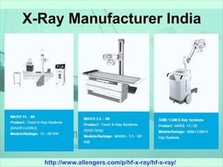
- 2. http://www.allengers.com/p/hf-x-ray/hf-x-ray/ Types Of X-Ray Systems ➢ High Frequency Mobile X-Ray Machines ➢ Fixed X-Ray Machines ➢ Medical X-Ray Machines ➢ Mobile X-Ray Machines ➢ X-Rays Machines
- 3. http://www.allengers.com/p/hf-x-ray/hf-x-ray/ High Frequency Mobile X-Ray Machines FEATURES: ➢ Substantially lower exposure factors, (Radiation safety to patients and operators) ➢ Elimination of Motion Artifacts due to lower exposure time, (Excellent film quality) ➢ Negligible skin dose, (Radiation safety to patient)
- 4. http://www.allengers.com/p/hf-x-ray/hf-x-ray/ Fixed X-Ray Machines FEATURES: ➢ Machine On/OFF switch ➢ Digital Display of parameters ➢ Parameter increase/decrease switches ➢ Ready and X-Ray ON switch with indicators ➢ Bucky selection switch ➢ Self diagnostic program with indicators for earth fault interlock, KV interlock, filament interlock and tubes thermal interlock ➢ A dual action hand switch with retractable cord for radiation protection of operator
- 5. http://www.allengers.com/p/hf-x-ray/hf-x-ray/ Medical X-Ray Machines FEATURES: ➢ Machine On/OFF switch ➢ Digital Display of parameters ➢ Parameter increase/decrease switches ➢ Ready and X-Ray ON switch with indicators ➢ Bucky selection switch ➢ Self diagnostic program with indicators for earth fault interlock, KV interlock, filament interlock and tubes thermal interlock ➢ A dual action hand switch with retractable cord for radiation protection of operator
- 6. http://www.allengers.com/p/hf-x-ray/hf-x-ray/ Mobile X-Ray Machines FEATURES: ➢ ➢ X-Ray Generators: 2.5 kw, 3.5 kw, 6 kw High Frequency (40 kHz) ➢ Totally feather touch micro controller based control panel ➢ Self diagnostic program with indicators to indicate earth fault, KV fault, Filament circuit fault & Thermal overload ➢ Easy maneuverability makes it most suitable for trauma cases & bed side X-rays in hospital wards ➢ Substantially lower exposure factors hence less radiation hazards, safe for the operation & operators
- 7. http://www.allengers.com/p/hf-x-ray/hf-x-ray/ X-Rays Machines FEATURES: ➢ Power Supply: 230/440 V AC, 50/60Hz. ➢ Compatible Tables: Allpose (Motorized, Multiposition, (Five Position Manual Tilt), Floatex Table (Four Way Floating), Horizontal Table) ➢ Optional: 2nd X-Ray tube, Image Memory Unit, Vertical Bucky Stand.