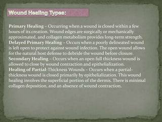
Downloaded 265 times


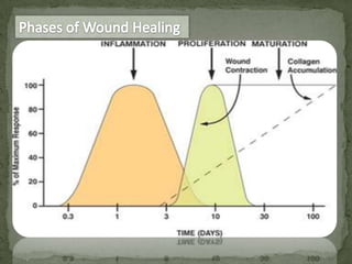

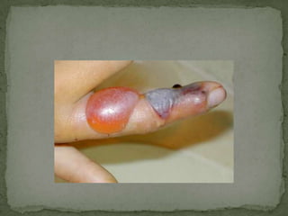
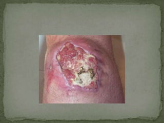
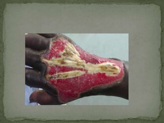
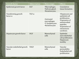


Primary intention healing occurs when a wound is closed within hours of injury through surgical or mechanical approximation of wound edges. Delayed primary healing allows time for wound debridement before closure to prevent infection. Secondary intention healing involves closure of full thickness wounds through wound contraction and epithelialization without surgical closure. Wound healing progresses through inflammatory, proliferative, and remodeling phases regardless of closure method and can move between phases based on intrinsic and extrinsic patient factors.


![Wound healing [including healing after periodontal therapy]](https://cdn.slidesharecdn.com/ss_thumbnails/woundhealingjr-150516123855-lva1-app6891-thumbnail.jpg?width=640&height=640&fit=bounds)


![CTEV [ clubfoot] DR ARUN LAL ,DR MOHAMED ASHRAF travancore medical college k...](https://cdn.slidesharecdn.com/ss_thumbnails/ctevclubfootdrarunlaldrmohamedashraftravancoremedicalcollegekollamkeralaindia-260208063247-18fc466c-thumbnail.jpg?width=640&height=640&fit=bounds)


![ONFH[AVN HIP] -TRIPLE REGIME -A NOVAL SURGICAL CONCEPT .pptx](https://cdn.slidesharecdn.com/ss_thumbnails/onfhavnhip2026koaconcalicutdrgokuldevdrmashraf-260210064517-213ec005-thumbnail.jpg?width=640&height=640&fit=bounds)


