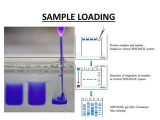
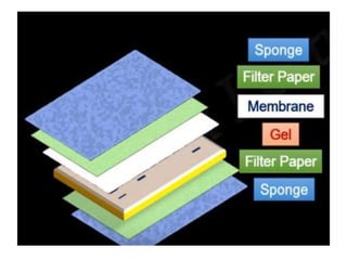
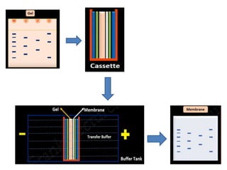
WESTERN BLOTTING
WESTERN BLOTTING
• The Western blot is an analytical technique
used to detect specific proteins in a given
sample of tissue homogenate or extract.
• Western blot for Proteins was Developed by
George Stark using antibodies to locate Proteins.
The Western blotting workflow
1. Sample preparation
2. SDS-PAGE
3. Transfer
4. Blocking
5. Antibody probing
6. Detection
7. Analysis
SAMPLE PREPARATION
• Protein from all sources can be used for western blotting
such as from single cell, whole tissues, extracellular matrices,
biological fluids.
• Protein can be extracted from the sample in three distinct
steps:
• Cell lysis: Physical method (ultra sonication, homogenization
etc.)
Chemical method (use of detergents)
Biological method (enzymatic digestion)
• Protein isolation from the rest of cellular components:
Centrifugation, DNase and other lysis buffer is used for the
removal of cell organelles, nucleic acids, polysaccharides etc.
SDS-PAGE
SDS-PAGE separates proteins based on their
molecular weight & all the separated proteins
contain a negative charge due to presence of
anionic detergent SDS.
SAMPLE LOADING
PROTEIN TRANSFER
Protein are transferred from the gel to a solid support
membrane such as nitrocellulose or polyvinylidene
difluoride (PVDF).
 The proteins transferred from the gels are immobilized
which makes it possible to detect the proteins on the
membrane using specific antibodies.
 Electro-blotting is mainly used which uses electric field
to pull proteins from the gel onto the PVDF or
nitrocellulose membrane.
 The proteins move from within the gel onto the
membrane while maintaining the organization they
had within the gel.
PROTEIN STAINING
After gel electrophoresis, it may be necessary to
confirm that all the proteins in the gel have been
completely eluted.
As proteins are not directly visible in the gel, the
gel must be stained.
Proteins are usually stained with dyes such as
coomassie blue, silver stain, or deep purple.
BLOCKING
 For meaningful results, the antibodies must bind
only to the protein of interest and not to the
membrane.
 Non-specific binding (NSB) of antibodies can be
reduced by blocking the unoccupied sites of
membrane with an inert protein or non-ionic
detergent.
 Blocking agents should possess greater affinity
towards membrane than the antibodies.
BLOCKING AGENTS
The most common blocking agents are:
a) Bovine serum albumin(BSA)
b) Non-fat milk
c) Casein
d) Gelatin
e) Dilute solution of Tween 20.
ANTIBODY PROBING
 Western blotting protocols usually uses a non-labeled
primary antibody directed against the target protein
and labeled secondary antibody directed against the
constant region of the primary Antibody.
 The secondary antibody serves not only as a carrier of
the label but is also a mechanism to amplify the
emitted signals because secondary antibodies are most
often polyclonal and can bind primary antibody at
different epitopes simultaneously.
WASHING
Unbound antibodies can cause high
background and poor detection.Hence
Washing the blot removes unbound
antibodies from the membrane.
A dilute solution of tween-20 in TBS or PBS
buffer is commonly used for washing.
PROTEIN DETECTION
 After the unbound probes are washed away,
the western blotting is now ready for detection
of the probes that are labeled and bound to the
protein of interest.
 Enzymes such as alkaline phosphatase(AP), &
Horse-radish peroxidase(HRP) are widely used
in detection of proteins.
ANALYSIS & IMAGING
This is the last & major step of the western blotting
technique.
Detection of signals, using either X-Ray film, scanners
or a CCD, results in one or more visible protein bands
on the membrane image.
The molecular weight of the protein can be estimated
by comparison with marker proteins and the amount
ofprotein can be determined as this is related to band
intensity.
Qualitative & quantitative analysis can be done in order
to verify the absence or presence of specific proteins
ofinterest.
THANK YOU
By:-
SHANKARNARAYAN A M
BLH6039

Western blotting

  • 1.
  • 2.
    WESTERN BLOTTING • TheWestern blot is an analytical technique used to detect specific proteins in a given sample of tissue homogenate or extract. • Western blot for Proteins was Developed by George Stark using antibodies to locate Proteins.
  • 3.
    The Western blottingworkflow 1. Sample preparation 2. SDS-PAGE 3. Transfer 4. Blocking 5. Antibody probing 6. Detection 7. Analysis
  • 4.
    SAMPLE PREPARATION • Proteinfrom all sources can be used for western blotting such as from single cell, whole tissues, extracellular matrices, biological fluids. • Protein can be extracted from the sample in three distinct steps: • Cell lysis: Physical method (ultra sonication, homogenization etc.) Chemical method (use of detergents) Biological method (enzymatic digestion) • Protein isolation from the rest of cellular components: Centrifugation, DNase and other lysis buffer is used for the removal of cell organelles, nucleic acids, polysaccharides etc.
  • 5.
    SDS-PAGE SDS-PAGE separates proteinsbased on their molecular weight & all the separated proteins contain a negative charge due to presence of anionic detergent SDS.
  • 6.
  • 7.
    PROTEIN TRANSFER Protein aretransferred from the gel to a solid support membrane such as nitrocellulose or polyvinylidene difluoride (PVDF).  The proteins transferred from the gels are immobilized which makes it possible to detect the proteins on the membrane using specific antibodies.  Electro-blotting is mainly used which uses electric field to pull proteins from the gel onto the PVDF or nitrocellulose membrane.  The proteins move from within the gel onto the membrane while maintaining the organization they had within the gel.
  • 10.
    PROTEIN STAINING After gelelectrophoresis, it may be necessary to confirm that all the proteins in the gel have been completely eluted. As proteins are not directly visible in the gel, the gel must be stained. Proteins are usually stained with dyes such as coomassie blue, silver stain, or deep purple.
  • 12.
    BLOCKING  For meaningfulresults, the antibodies must bind only to the protein of interest and not to the membrane.  Non-specific binding (NSB) of antibodies can be reduced by blocking the unoccupied sites of membrane with an inert protein or non-ionic detergent.  Blocking agents should possess greater affinity towards membrane than the antibodies.
  • 13.
    BLOCKING AGENTS The mostcommon blocking agents are: a) Bovine serum albumin(BSA) b) Non-fat milk c) Casein d) Gelatin e) Dilute solution of Tween 20.
  • 14.
    ANTIBODY PROBING  Westernblotting protocols usually uses a non-labeled primary antibody directed against the target protein and labeled secondary antibody directed against the constant region of the primary Antibody.  The secondary antibody serves not only as a carrier of the label but is also a mechanism to amplify the emitted signals because secondary antibodies are most often polyclonal and can bind primary antibody at different epitopes simultaneously.
  • 15.
    WASHING Unbound antibodies cancause high background and poor detection.Hence Washing the blot removes unbound antibodies from the membrane. A dilute solution of tween-20 in TBS or PBS buffer is commonly used for washing.
  • 16.
    PROTEIN DETECTION  Afterthe unbound probes are washed away, the western blotting is now ready for detection of the probes that are labeled and bound to the protein of interest.  Enzymes such as alkaline phosphatase(AP), & Horse-radish peroxidase(HRP) are widely used in detection of proteins.
  • 17.
    ANALYSIS & IMAGING Thisis the last & major step of the western blotting technique. Detection of signals, using either X-Ray film, scanners or a CCD, results in one or more visible protein bands on the membrane image. The molecular weight of the protein can be estimated by comparison with marker proteins and the amount ofprotein can be determined as this is related to band intensity. Qualitative & quantitative analysis can be done in order to verify the absence or presence of specific proteins ofinterest.
  • 18.