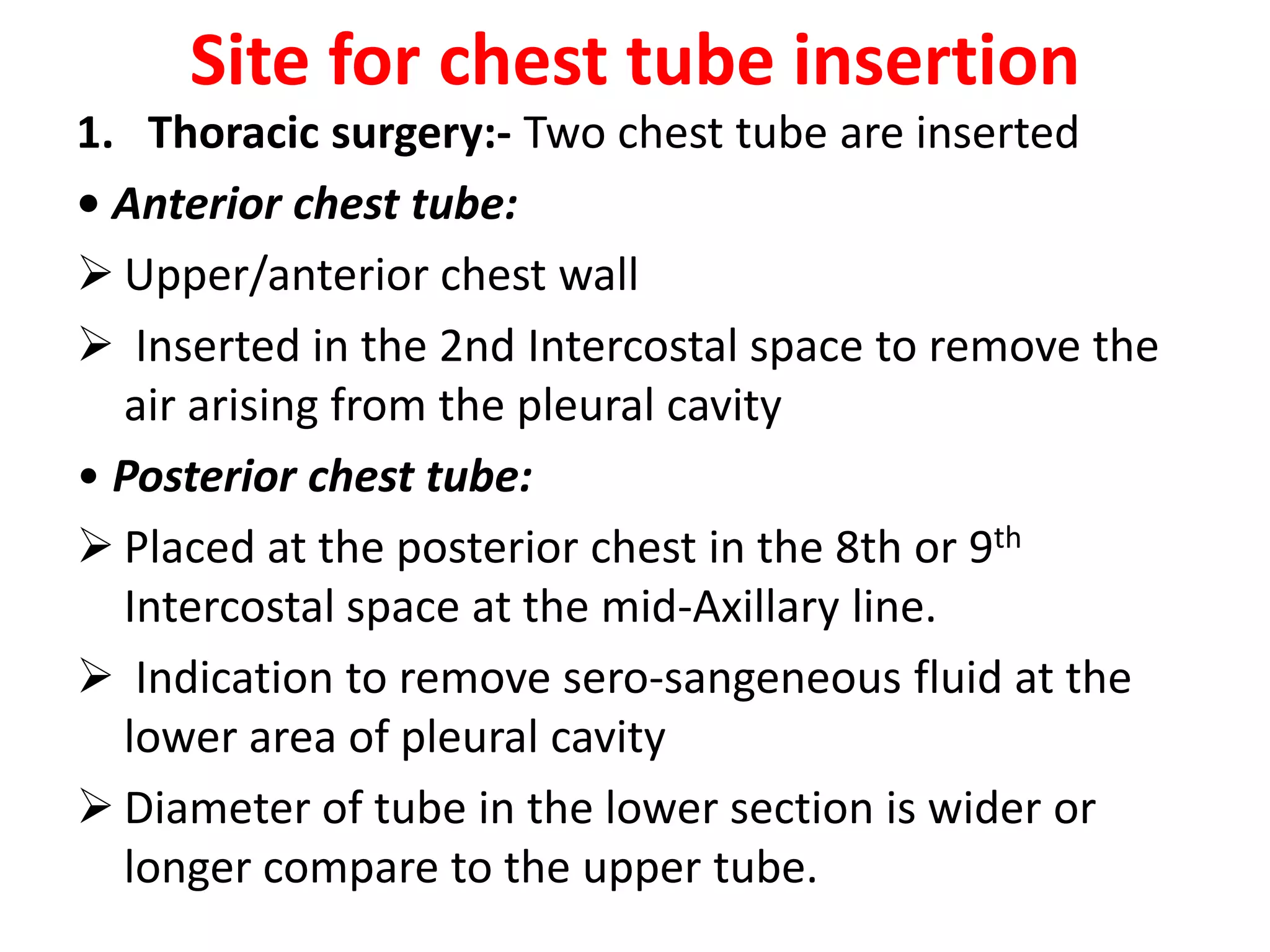
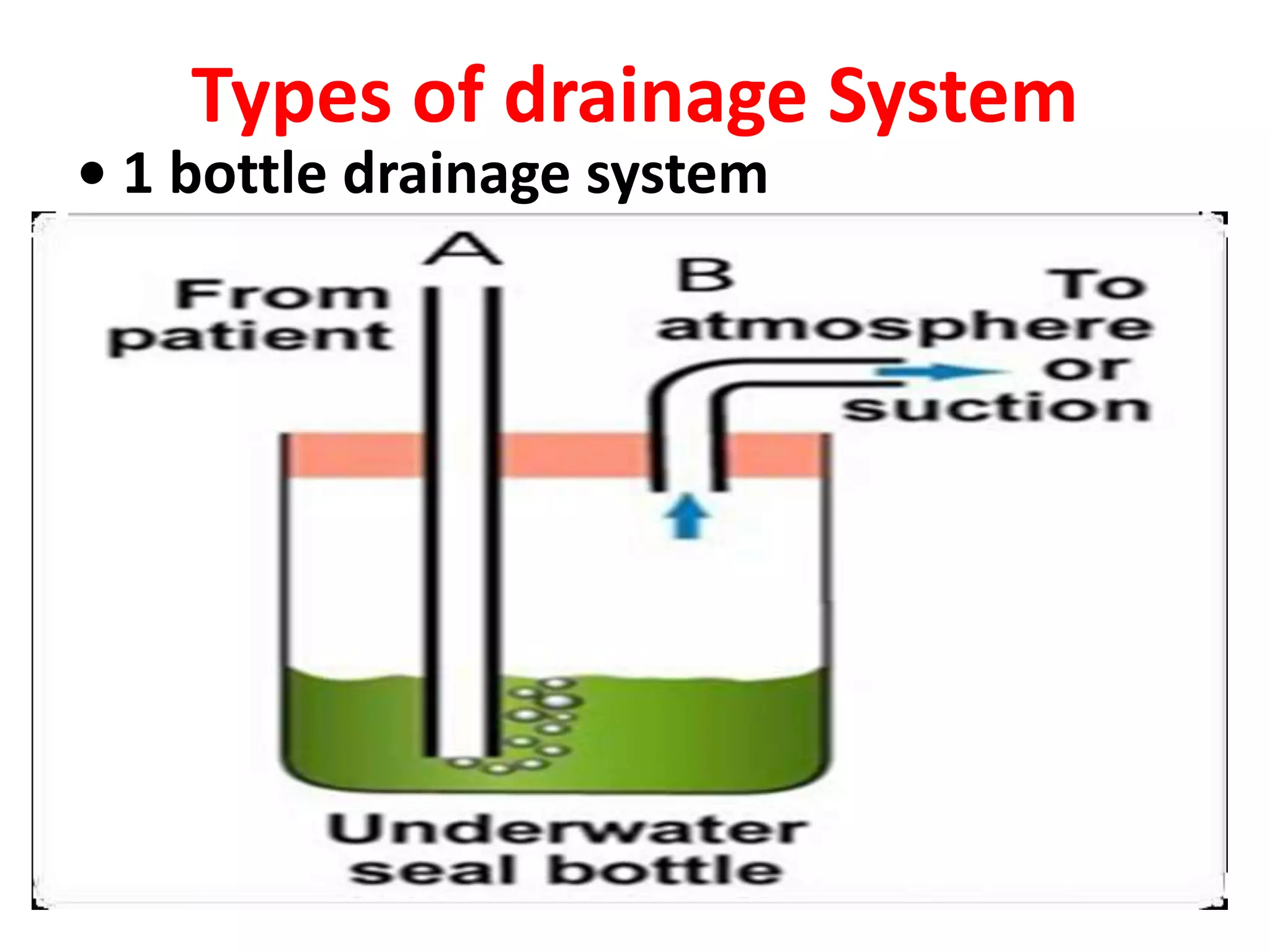
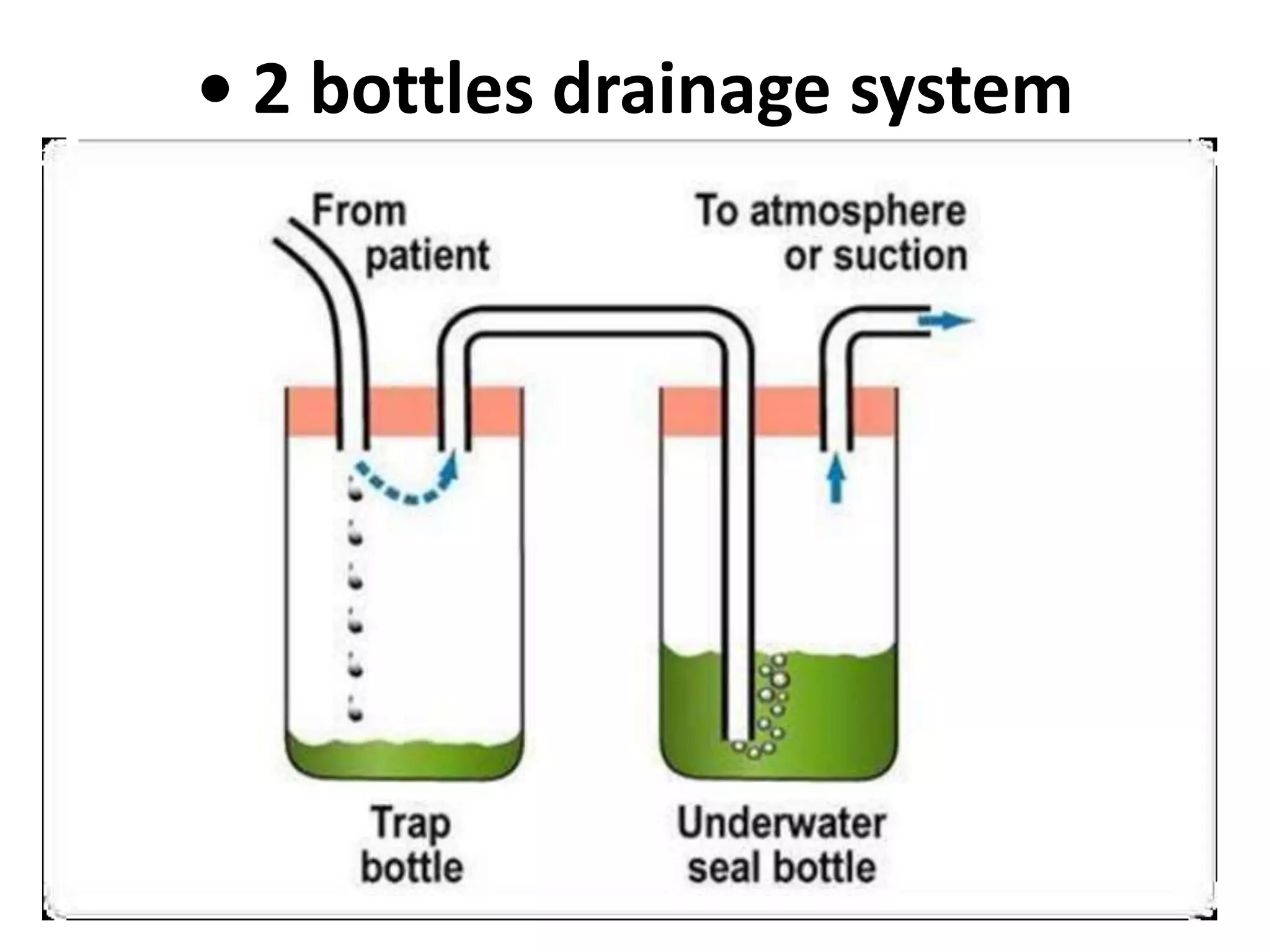
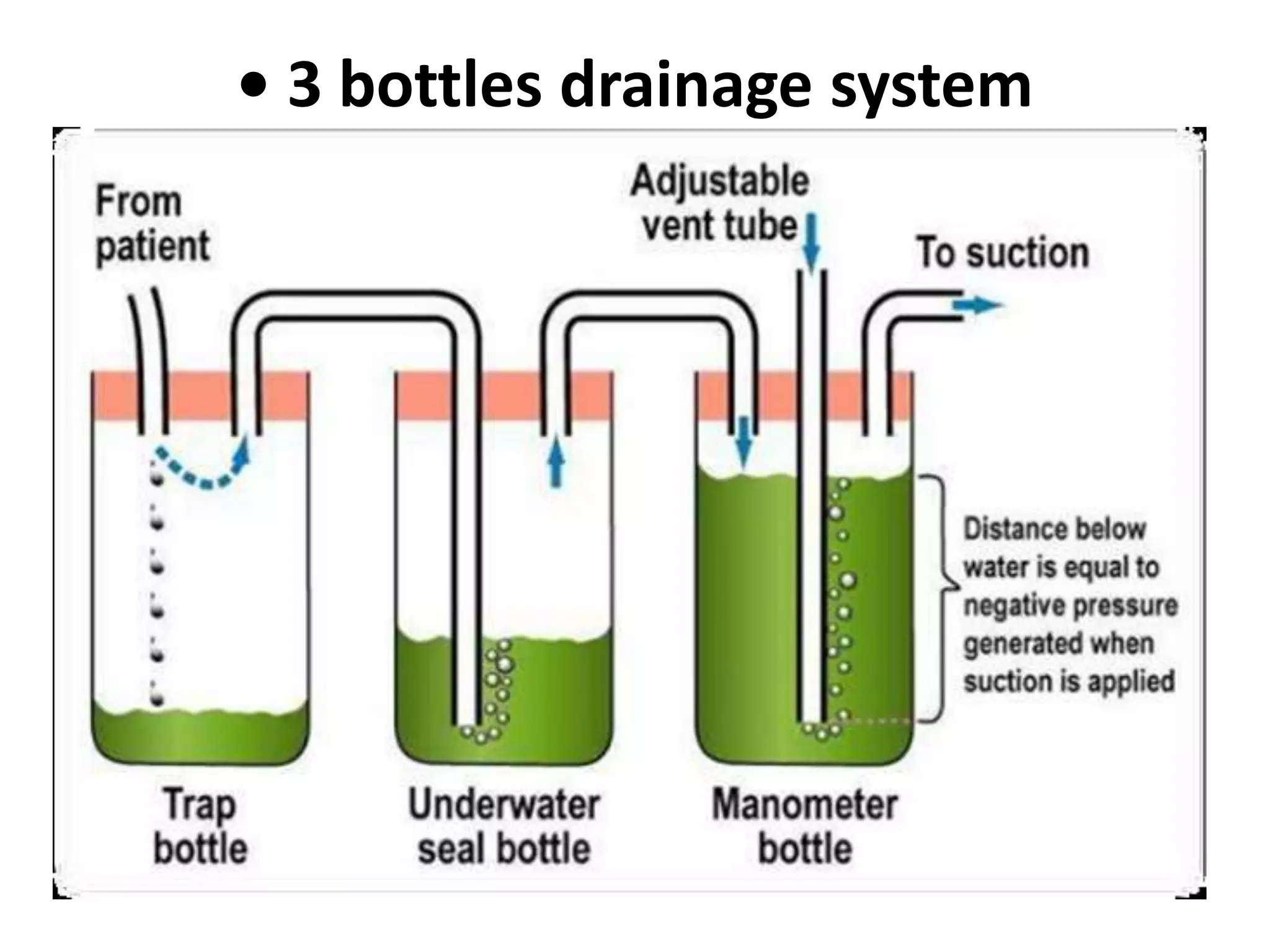
Water seal drainage is a closed chest drainage system used to drain air and fluid from the pleural space during exhalation while preventing backflow during inhalation. It is indicated for conditions causing pneumothorax or pleural effusions. The system uses gravity and a water seal to establish negative pressure and allow for lung expansion while continuously draining the pleural space. Proper placement and maintenance of the drainage system and tubes is important to ensure effective drainage and prevent complications.
Thoracentesis (thor-a-sen-tee-sis) is a procedure that is done to remove a sample of fluid from around the lung.
The lung is covered with a tissue called the pleura. The inside of the chest is also lined with pleura.
The space between these two areas is called the pleural space.
This space normally contains just a thin layer of fluid, however, some conditions such as pneumonia, some types of cancer, or congestive heart failure may cause excessive fluid to develop (pleural effusion).
Thoracentesis, also known as pleural fluid analysis, is a procedure in which a needle is inserted through the back of the chest wall into the pleural space (a space that exists between the two lungs and the anterior chest wall) to remove fluid or air.
Pleural fluid analysis is the microscopic and chemical lab analysis of the fluid obtained during thoracentesis.
IndDiagnostic: determination of pleural effusion etiology (e.g. transudative versus exudative) usually requires the removal of 50 to 100mL of pleural fluid for laboratory studies. Most new effusions require diagnostic thoracentesis, an exception being a new effusion with a clear clinical diagnosis (e.g. CHF) with no evidence for superimposed pleural space infection
Therapeutic: reduce dyspnea and respiratory compromise in patients with large pleural effusions. This is typically achieved by removing a much larger volume of fluid compared to the diagnostic thoracentesis
ications
this is the power point presentation for coughing and breathing exercises, most probably we used this for the respiratory problems, it is very helpful for the COPD patient
Thoracentesis is a procedure in which a needle is inserted into the pleural space between the lungs and the chest wall. This procedure is done to remove excess fluid, known as a pleural effusion, from the pleural space to help you breathe easier.
Sutures are used by your doctor to close wounds to your skin or other tissues. When your doctor sutures a wound, they’ll use a needle attached to a length of “thread” to stitch the wound shut.
There are a variety of available materials that can be used for suturing. Your doctor will choose a material that’s appropriate for the wound or procedure.
The different types of sutures can be classified in many ways.
First, suture material can be classified as either absorbable or nonabsorbable.
Absorbable sutures don’t require your doctor to remove them. This is because enzymes found in the tissues of your body naturally digest them.
Nonabsorbable sutures will need to be removed by your doctor at a later date or in some cases left in permanently.
There are numerous types of brain surgery. The type used is based on the area of the brain and the condition being treated.
Brain surgery is a critical and complicated process. The type of brain surgery done depends highly on the condition being treated.
Intracranial surgery refers to various medical procedures that involve repairing structural problems in the brain.
Craniotomy
A craniotomy involves making an incision in the scalp and creating a hole known as a bone flap in the skull. The hole and incision are made near the area of the brain being treated.
During open brain surgery, it is done to remove tumors, clip off an aneurysm, drain blood or fluid from an infection & remove abnormal brain tissue
Decompressive craniectomy
It is a neurosurgical procedure in which part of the skull is removed to allow a swelling brain room to expand without being squeezed. It is performed on victims of traumatic brain injury, stroke and other conditions associated with raised intracranial pressure.
How to Create Map Views in the Odoo 17 ERPCeline George
The map views are useful for providing a geographical representation of data. They allow users to visualize and analyze the data in a more intuitive manner.
This is a presentation by Dada Robert in a Your Skill Boost masterclass organised by the Excellence Foundation for South Sudan (EFSS) on Saturday, the 25th and Sunday, the 26th of May 2024.
He discussed the concept of quality improvement, emphasizing its applicability to various aspects of life, including personal, project, and program improvements. He defined quality as doing the right thing at the right time in the right way to achieve the best possible results and discussed the concept of the "gap" between what we know and what we do, and how this gap represents the areas we need to improve. He explained the scientific approach to quality improvement, which involves systematic performance analysis, testing and learning, and implementing change ideas. He also highlighted the importance of client focus and a team approach to quality improvement.
Unit 8 - Information and Communication Technology (Paper I).pdfThiyagu K
This slides describes the basic concepts of ICT, basics of Email, Emerging Technology and Digital Initiatives in Education. This presentations aligns with the UGC Paper I syllabus.
Ethnobotany and Ethnopharmacology:
Ethnobotany in herbal drug evaluation,
Impact of Ethnobotany in traditional medicine,
New development in herbals,
Bio-prospecting tools for drug discovery,
Role of Ethnopharmacology in drug evaluation,
Reverse Pharmacology.
The Indian economy is classified into different sectors to simplify the analysis and understanding of economic activities. For Class 10, it's essential to grasp the sectors of the Indian economy, understand their characteristics, and recognize their importance. This guide will provide detailed notes on the Sectors of the Indian Economy Class 10, using specific long-tail keywords to enhance comprehension.
For more information, visit-www.vavaclasses.com
Palestine last event orientationfvgnh .pptxRaedMohamed3
An EFL lesson about the current events in Palestine. It is intended to be for intermediate students who wish to increase their listening skills through a short lesson in power point.
2024.06.01 Introducing a competency framework for languag learning materials ...Sandy Millin
http://sandymillin.wordpress.com/iateflwebinar2024
Published classroom materials form the basis of syllabuses, drive teacher professional development, and have a potentially huge influence on learners, teachers and education systems. All teachers also create their own materials, whether a few sentences on a blackboard, a highly-structured fully-realised online course, or anything in between. Despite this, the knowledge and skills needed to create effective language learning materials are rarely part of teacher training, and are mostly learnt by trial and error.
Knowledge and skills frameworks, generally called competency frameworks, for ELT teachers, trainers and managers have existed for a few years now. However, until I created one for my MA dissertation, there wasn’t one drawing together what we need to know and do to be able to effectively produce language learning materials.
This webinar will introduce you to my framework, highlighting the key competencies I identified from my research. It will also show how anybody involved in language teaching (any language, not just English!), teacher training, managing schools or developing language learning materials can benefit from using the framework.
Read| The latest issue of The Challenger is here! We are thrilled to announce that our school paper has qualified for the NATIONAL SCHOOLS PRESS CONFERENCE (NSPC) 2024. Thank you for your unwavering support and trust. Dive into the stories that made us stand out!
We all have good and bad thoughts from time to time and situation to situation. We are bombarded daily with spiraling thoughts(both negative and positive) creating all-consuming feel , making us difficult to manage with associated suffering. Good thoughts are like our Mob Signal (Positive thought) amidst noise(negative thought) in the atmosphere. Negative thoughts like noise outweigh positive thoughts. These thoughts often create unwanted confusion, trouble, stress and frustration in our mind as well as chaos in our physical world. Negative thoughts are also known as “distorted thinking”.
The Art Pastor's Guide to Sabbath | Steve ThomasonSteve Thomason
What is the purpose of the Sabbath Law in the Torah. It is interesting to compare how the context of the law shifts from Exodus to Deuteronomy. Who gets to rest, and why?
2. Definition
Water seal drainage system is a closed
chest drainage system used to allow air
and fluid to escape from the plural space
with each exhalation and to prevent their
return flow with each inhalation.
4. Purposes
• To permit drainage of air and fluid from
the pleural cavity
• To establish normal negative pressure in
the pleural cavity for lung expansion
• To equalize pressure on both sides of the
thoracic cavity
• To provide continuous suction to prevent
tension pneumothorax
5. Site for chest tube insertion
1. Thoracic surgery:- Two chest tube are inserted
• Anterior chest tube:
Upper/anterior chest wall
Inserted in the 2nd Intercostal space to remove the
air arising from the pleural cavity
• Posterior chest tube:
Placed at the posterior chest in the 8th or 9th
Intercostal space at the mid-Axillary line.
Indication to remove sero-sangeneous fluid at the
lower area of pleural cavity
Diameter of tube in the lower section is wider or
longer compare to the upper tube.
6. Site for chest tube insertion
2.Pneumothorax :
Tube placed at the 2nd or 3rd Intercostal
space along mid- clavicle or anterior Axillary
line.
10. Principles of the chest tube
1. Gravity
• Enhances flow from high to low
Chest drain is placed below client’s
bed
11. 2. Under water seal
• Is a barrier to prevents backflow into
pleural space.
• Rod – depth determines the negative
pressure
• Air bubbles is released through the rod
• Air vent – to allow drained air to
escape to prevent pressure build up
12. 3. Suction
• Is a pull force
• MUST be in another bottle
• Purpose for the suction is, when :
gravity drainage is not enough.
patient’s respiration and cough are too weak
air leak is fast into the pleural space ie. - need
to speed up removal from pleural space
13. Factors affecting water seal
drainage
1. Proper placement of chest catheter
2. Proper placement of chest drainage apparatus
3. Length of drainage tubing
4. Patency of chest tubing
5. Maintenance of air tight drainage system
6. Position of the client
7. Application of mechanical suction
8. Activity of the client
14. 1. Proper placement of chest catheter
• 2 & 3rd intercostal space and 8 &9th
intercostal space
• These catheter should be connected to
the separate bottle
• When there is single tube it is usually
placed in lower intercostal space
15. 2. Proper placement of drainage
apparatus
• Drainage apparatus should be placed at
lower level then the chest
• It helps in gravity and also prevent the
back flow of air and fluid into pleural cavity
• While transferring patient apparatus
should be placed over the bed or trolley
after clamping the tubing at two places
16. 3. Length of drainage tubing
• Drainage tubing neither too short nor too long
• It should fall in straight line to the drainage
system
• There should not be any loop of drainage
tubing
• Too short drainage tubing may restrict the
movement of patient on bed or it may get
disconnected from the catheter
17. 4. Patency of chest tubing
• Patency of chest tube should be checked
frequently
• Any kink or pressure over chest tubing may
obstruct the flow from pleural space
• Ensure that patient is not lying over the tubing
• Any clot or mucus plug in pleural space may also
obstruct the flow
• Milking the tube helps to dislodge the plug
• Never clamp the tubing until it is necessary.
18. 5. Maintenance of air tight drainage
system
• Drainage system should be air tight with
stoppers and all the tubing should be taped
well
6. Position of the client
• Fowler’s position
• So that fluid can be localized in lower
pleural space and can be drained out easily
19. 7. Application of mechanical
suction
• Continuous and gentle suction is used
when :
Gravity drainage is not enough.
Patient’s respiration and cough are too
weak
Air leak is fast into the pleural space
Need to speed up removal from pleural
space
20. 8. Activity of the client
• Movement of the patient on bed helps
the fluid to drain from chest.
• Patient should be encouraged to cough
and deep breath (Breathing exercise).
• Which helps in rising the intra-pleural
and intra- pulmonary pressure
21. Nursing Responsibilities
• Criteria for good functioning of
water seal drainage apparatus
• Nursing care of the patient with
water seal drainage.
22. Criteria for good functioning of water
seal drainage apparatus
• Observe for the fluctuating movement of
fluid inside the tubing
• Observe the chest drainage
• Watch for bubbling in water seal bottle
(intermittent bubbling is normal)
Reason for mal function of suction are:
• Air leaking into the pleural space
• Air leaking into the drainage apparatus
• Mechanical problem to the pump.
23. Nursing care of the patient with
water seal drainage
• As a nurse we should have proper
knowledge of purpose, principles,
equipments used and early sign and
symptoms of complication of water seal
drainage.
• Instruct the client and bystanders how to
clamp the drainage in emergency situation
and how to handle the water seal drainage.
24. Cont….
Affix a sign board “do n. ot handle”
• • Sutured site should be well padded and
• secured with adhesive tapes
• • Clamp or hemostat clamp should be readily
• available near to the patient.
• • Never clamp the chest tubing until it is
• necessary or ordered by the physician
25. Cont….
• Apparatus of chest drainage should be kept
lower than the chest level of the patient
• During transportation clamp the tubing and
keep the apparatus top of the bed.
• Make sure that position of the client should
not exert pressure over the chest tubing.
• Chest tubing neither too short nor too long
26. Cont….
• Do not use pins to secure the tubing with bed
• Check the patency of chest tubing regularly
• Ensure the air tightening of the chest drainage
• Follow the strict aseptic technique
• Encourage the deep breathing and coughing
exercises
• Ensure that tubes are immersed in water well
to create water seal drainage
27. Instructions for replacing
1. Assemble the bottle with tube and
stopper and check their functioning
2. Clamp the chest tube near to the
chest prior to disconnecting it
3. Disconnect the bottle and connect
the new bottle but remember that
chest tubing should not be
contaminated
28. Cont…
4.Be certain that bottle/s are kept well
below the chest level
5. Unclamp the chest tubing and check the
functioning of the system
6. Observe the patient for any complication
before leaving.
29. Criteria for removal
1. Absence of air leak
2. Drained fluid is less than 75 ml/day
3. Chest X-ray should reveal the re-
expansion of lungs well contd..
30. Cont…
• Prior to removal of the water seal drainage
system chest tubing should be clamped at least
for two hours.
• After removal, wound should be covered with
sterile petroleum gauze and firm dressing
secured with wide strip of adhesive tapes
• Observe the patient for any respiratory distress
post removal the water seal drainage system
31. Reference
• Smeltzer – Brunner & Suddharth Textbook of
Medical Surgical Nursing, Wolters kluwer
publishers, 12th edition 2009.
• Black – Medical Surgical Nursing, Elsevier
publishers, 8th edition 2009.
• Nettina – Lippincott manual of Nursing
Practice, Wolters kluwer publishers, 7th edition
2014.
• Lewis – Medical Surgical Nursing, Elsevier
publishers, 10th edition, 2017