Download to read offline
![tween WC and health outcome changes
much less with increasing age than does
the relationship between BMI and health
outcome (31). However, it is not known
whether WC can provide a better assess-
ment of health risk in one sex, racial/
ethnic group, or age category than
another.
The shape of the relationship between
WC and health outcomes (e.g., linear,
monotonic, step-function, or U-shaped)
influences the WC value that can most
efficiently distinguish between “normal”
and “abnormal” and serve as a basis for
considering clinical action. Data from
most studies suggest that the shape of the
relationship between WC and health out-
come lends itself to identifying clinically
meaningful cut point values because risk
often accelerates monotonically above,
and can be relatively flat below, a specific
WC value. Optimum WC cut points will
likely vary according to the population
studied, the health outcome of interest,
and demographic factors.
Data from most clinical weight loss
and exercise training trials have shown
that reductions in WC occur concomi-
tantly with reductions in obesity-related
cardiometabolic risk factors and disease.
However, these results do not prove that
the reduction in WC was responsible for
the beneficial effect on health outcome.
Additional studies are needed to evaluate
the effect of decreasing WC on cardio-
metabolic outcomes.
QUESTION 4: Should waist
circumference be measured in
clinical practice?
The panel concluded that determining
whether waist circumference should be
measured in clinical practice depends on
the responses to the following four key
questions:
1. Can waist circumference be reli-
ably measured? Answer: Yes.
Health care personnel and even pa-
tients themselves, who are given appro-
priate training in technique, can provide
highly reproducible measurements of
WC in men and women. However, it is
not know whether measurement of one
anatomical site is a better indicator of car-
diometabolic risk than measurement at
other sites.
2. Does waist circumference provide:
a) good prediction of diabetes, CHD, and
mortality rate? Answer: Yes; b) incremen-
tal value in predicting diabetes, CHD, and
mortality rate above and beyond that pro-
vided by BMI? Answer: Yes; c) sufficient
incremental value in these predictions
above and beyond that offered by BMI
and commonly evaluated cardiometa-
bolic risk factors, such as blood glucose
concentration, lipid profile and blood
pressure? Answer: Uncertain.
Data from many large population
studies have found waist circumference to
be a strong correlate of clinical outcome,
particularly diabetes, and to be indepen-
dent of BMI. In addition, data from a lim-
ited number of studies demonstrates that
WC remains a predictor of diabetes,
CHD, and mortality rate, even after ad-
justing for BMI and several other cardio-
metabolic risk factors. Additional studies
are needed to confirm that WC remains
an independent predictor of risk.
3. Do the current definitions used to
determine a high WC identify a nontrivial
number of patients who are at increased
cardiometabolic risk, but who would not
otherwise be identified by having a BMI
Ն25 kg/m2
and an assessment of com-
monly evaluated cardiometabolic risk fac-
tors? Answer: Yes.
The recommended WC thresholds
for increased cardiometabolic risk in men
(Ͼ40 inches [102 cm]) and women (Ͼ35
inches [88 cm]) were derived from WC
values that correlated with a BMI Ն30
kg/m2
(2). The National Health and Nu-
trition Examination Survey III (NHANES
III) found that about 14% of women and
about 1% of men had a “high” WC but a
normal BMI (18.5–24.9 kg/m2
) (36). In
addition, ϳ70% of women who were
overweight (BMI 25.0–29.9 kg/m2
) had a
WC Ͼ35 inches and ϳ25% of men who
were overweight had a WC Ͼ40 inches.
An estimate based on data available from
the WHO Monica Project, conducted in
more than 32,000 men and women from
Europe, Australia, and New Zealand, sug-
gest that about 10% of participants who
had BMI values Ͻ30 kg/m2
had a WC
above the recommended cut points for in-
creased risk (36). It is not known what
portion of subjects who had a large WC
would have been identified as having in-
creased cardiometabolic risk based on
findings from a standard medical evalua-
tion. Therefore, the optimal WC criteria
needed to identify patients at increased
risk of metabolic disease, who would oth-
erwise not be identified by evaluating BMI
and/or other standard cardiometabolic
risk factors, is not known and will likely
require adjustments based on BMI, sex,
age, and race/ethnicity.
4. Would assessment of WC in pa-
tients who have a BMI Ն25 kg/m2
affect
clinical management if NHLBI obesity
treatment guidelines are followed? An-
swer: Probably not.
Measurement of WC in clinical prac-
tice is not trivial, because providing this
assessment competes for the limited time
available in a busy office practice and re-
quires specific training to ensure that re-
liable data are obtained. Therefore, waist
circumference should only be measured if
it can provide additional information that
influences patient management. Based on
NHANES III data, 99.9% of men and
98.4% of women would have received the
same treatment recommendations pro-
posed by the NHLBI Expert Panel by eval-
uating BMI and other cardiovascular risk
factors, without an assessment of WC
(37). However, it is likely that different
WC cut point values could provide more
useful clinical information. For example,
an analysis of data obtained from the
NHANES III and the Canadian Heart
Health Surveys found that BMI-specific
WC cut points provided a better indicator
of cardiometabolic risk than the recom-
mended WC thresholds (35). For nor-
mal-weight (BMI 18.5–24.9 kg/m2
),
overweight (BMI 25.0–29.9 kg/m2
), class
I obesity (BMI 30.0–34.9 kg/m2
), and
class II/class III obesity (BMI Ն35.0 kg/
m2
), the optimal WC cut points were 87,
98, 109, and 124 cm in men and 79, 92,
103, and 115 cm in women, respectively.
Therefore, it is possible that WC measure-
ment could be an effective clinical tool for
identifying “metabolically obese, lean”
patients who might benefit from lifestyle
therapy but would not have been consid-
ered for treatment because of a normal
BMI. Waist circumference could also
identify “metabolically normal, obese”
subjects who do not require aggressive
obesity therapy because they do not have
a marked increase in cardiometabolic
risk.
CONCLUSIONS
Waist circumference provides a unique
indicator of body fat distribution, which
can identify patients who are at increased
risk for obesity-related cardiometabolic
disease, above and beyond the measure-
ment of BMI. However, the current WC
cut points recommended to determine
health risk (2) were derived by regression
from an “obese” BMI and are unlikely to
affect clinical management when BMI and
other obesity-related cardiometabolic risk
factors are already being determined.
Therefore, the clinical usefulness of mea-
suring WC, when risk is based on the cur-
Consensus Statement
1650 DIABETES CARE, VOLUME 30, NUMBER 6, JUNE 2007](https://image.slidesharecdn.com/waistcircumference-131001105604-phpapp02/85/Waist-circumference-4-320.jpg)

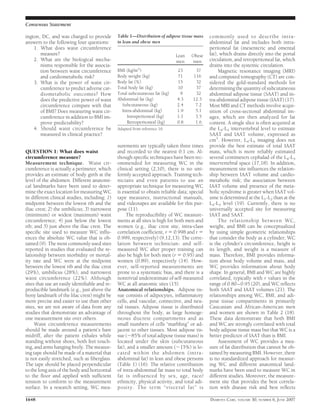
This document discusses waist circumference (WC) as a measure of abdominal fat and cardiometabolic risk. It finds that WC is a reliable surrogate for abdominal fat mass and strongly associated with health risks like diabetes and heart disease. The consensus panel addressed four questions: 1) WC measures abdominal girth and estimates abdominal fat levels. 2) Excess abdominal fat increases cardiometabolic risk through mechanisms like insulin resistance. 3) WC predicts health outcomes comparably or better than BMI and provides additional predictive value when used with BMI. 4) WC should be regularly measured in clinical practice to assess cardiometabolic risk and weight management efforts.


![Obesity by bijay [autosaved]](https://cdn.slidesharecdn.com/ss_thumbnails/obesitybybijayautosaved-200407031519-thumbnail.jpg?width=640&height=640&fit=bounds)


