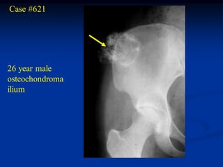
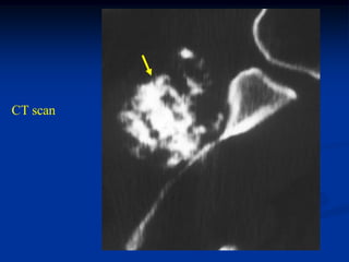
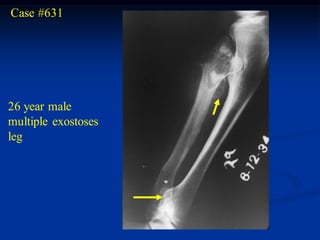
Osteochondroma is one of the most common benign bone tumors. It originates from the growth plate and continues growing during development, stopping at maturity. It is made up of a bony base with a pedunculated stalk containing fatty marrow and a cartilaginous cap. Solitary osteochondromas usually arise from large bones like the knee, femur, and humerus. Multiple hereditary osteochondromas present with multiple lesions and are inherited. Malignant conversion of a solitary osteochondroma can occur in adults but is extremely rare.
263778731218 Abortion Clinic /Pills In Harare ,sisternakatoto
263778731218 Abortion Clinic /Pills In Harare ,ABORTION WOMEN’S CLINIC +27730423979 IN women clinic we believe that every woman should be able to make choices in her pregnancy. Our job is to provide compassionate care, safety,affordable and confidential services. That’s why we have won the trust from all generations of women all over the world. we use non surgical method(Abortion pills) to terminate…Dr.LISA +27730423979women Clinic is committed to providing the highest quality of obstetrical and gynecological care to women of all ages. Our dedicated staff aim to treat each patient and her health concerns with compassion and respect.Our dedicated group ABORTION WOMEN’S CLINIC +27730423979 IN women clinic we believe that every woman should be able to make choices in her pregnancy. Our job is to provide compassionate care, safety,affordable and confidential services. That’s why we have won the trust from all generations of women all over the world. we use non surgical method(Abortion pills) to terminate…Dr.LISA +27730423979women Clinic is committed to providing the highest quality of obstetrical and gynecological care to women of all ages. Our dedicated staff aim to treat each patient and her health concerns with compassion and respect.Our dedicated group of receptionists, nurses, and physicians have worked together as a teamof receptionists, nurses, and physicians have worked together as a team wwww.lisywomensclinic.co.za/
DISSERTATION on NEW DRUG DISCOVERY AND DEVELOPMENT STAGES OF DRUG DISCOVERYNEHA GUPTA
The process of drug discovery and development is a complex and multi-step endeavor aimed at bringing new pharmaceutical drugs to market. It begins with identifying and validating a biological target, such as a protein, gene, or RNA, that is associated with a disease. This step involves understanding the target's role in the disease and confirming that modulating it can have therapeutic effects. The next stage, hit identification, employs high-throughput screening (HTS) and other methods to find compounds that interact with the target. Computational techniques may also be used to identify potential hits from large compound libraries.
Following hit identification, the hits are optimized to improve their efficacy, selectivity, and pharmacokinetic properties, resulting in lead compounds. These leads undergo further refinement to enhance their potency, reduce toxicity, and improve drug-like characteristics, creating drug candidates suitable for preclinical testing. In the preclinical development phase, drug candidates are tested in vitro (in cell cultures) and in vivo (in animal models) to evaluate their safety, efficacy, pharmacokinetics, and pharmacodynamics. Toxicology studies are conducted to assess potential risks.
Before clinical trials can begin, an Investigational New Drug (IND) application must be submitted to regulatory authorities. This application includes data from preclinical studies and plans for clinical trials. Clinical development involves human trials in three phases: Phase I tests the drug's safety and dosage in a small group of healthy volunteers, Phase II assesses the drug's efficacy and side effects in a larger group of patients with the target disease, and Phase III confirms the drug's efficacy and monitors adverse reactions in a large population, often compared to existing treatments.
After successful clinical trials, a New Drug Application (NDA) is submitted to regulatory authorities for approval, including all data from preclinical and clinical studies, as well as proposed labeling and manufacturing information. Regulatory authorities then review the NDA to ensure the drug is safe, effective, and of high quality, potentially requiring additional studies. Finally, after a drug is approved and marketed, it undergoes post-marketing surveillance, which includes continuous monitoring for long-term safety and effectiveness, pharmacovigilance, and reporting of any adverse effects.
Integrating Ayurveda into Parkinson’s Management: A Holistic ApproachAyurveda ForAll
Explore the benefits of combining Ayurveda with conventional Parkinson's treatments. Learn how a holistic approach can manage symptoms, enhance well-being, and balance body energies. Discover the steps to safely integrate Ayurvedic practices into your Parkinson’s care plan, including expert guidance on diet, herbal remedies, and lifestyle modifications.
Here is the updated list of Top Best Ayurvedic medicine for Gas and Indigestion and those are Gas-O-Go Syp for Dyspepsia | Lavizyme Syrup for Acidity | Yumzyme Hepatoprotective Capsules etc
Local Advanced Lung Cancer: Artificial Intelligence, Synergetics, Complex Sys...Oleg Kshivets
Overall life span (LS) was 1671.7±1721.6 days and cumulative 5YS reached 62.4%, 10 years – 50.4%, 20 years – 44.6%. 94 LCP lived more than 5 years without cancer (LS=2958.6±1723.6 days), 22 – more than 10 years (LS=5571±1841.8 days). 67 LCP died because of LC (LS=471.9±344 days). AT significantly improved 5YS (68% vs. 53.7%) (P=0.028 by log-rank test). Cox modeling displayed that 5YS of LCP significantly depended on: N0-N12, T3-4, blood cell circuit, cell ratio factors (ratio between cancer cells-CC and blood cells subpopulations), LC cell dynamics, recalcification time, heparin tolerance, prothrombin index, protein, AT, procedure type (P=0.000-0.031). Neural networks, genetic algorithm selection and bootstrap simulation revealed relationships between 5YS and N0-12 (rank=1), thrombocytes/CC (rank=2), segmented neutrophils/CC (3), eosinophils/CC (4), erythrocytes/CC (5), healthy cells/CC (6), lymphocytes/CC (7), stick neutrophils/CC (8), leucocytes/CC (9), monocytes/CC (10). Correct prediction of 5YS was 100% by neural networks computing (error=0.000; area under ROC curve=1.0).
micro teaching on communication m.sc nursing.pdfAnurag Sharma
Microteaching is a unique model of practice teaching. It is a viable instrument for the. desired change in the teaching behavior or the behavior potential which, in specified types of real. classroom situations, tends to facilitate the achievement of specified types of objectives.
- Video recording of this lecture in English language: https://youtu.be/kqbnxVAZs-0
- Video recording of this lecture in Arabic language: https://youtu.be/SINlygW1Mpc
- Link to download the book free: https://nephrotube.blogspot.com/p/nephrotube-nephrology-books.html
- Link to NephroTube website: www.NephroTube.com
- Link to NephroTube social media accounts: https://nephrotube.blogspot.com/p/join-nephrotube-on-social-media.html
These simplified slides by Dr. Sidra Arshad present an overview of the non-respiratory functions of the respiratory tract.
Learning objectives:
1. Enlist the non-respiratory functions of the respiratory tract
2. Briefly explain how these functions are carried out
3. Discuss the significance of dead space
4. Differentiate between minute ventilation and alveolar ventilation
5. Describe the cough and sneeze reflexes
Study Resources:
1. Chapter 39, Guyton and Hall Textbook of Medical Physiology, 14th edition
2. Chapter 34, Ganong’s Review of Medical Physiology, 26th edition
3. Chapter 17, Human Physiology by Lauralee Sherwood, 9th edition
4. Non-respiratory functions of the lungs https://academic.oup.com/bjaed/article/13/3/98/278874
Adv. biopharm. APPLICATION OF PHARMACOKINETICS : TARGETED DRUG DELIVERY SYSTEMSAkankshaAshtankar
MIP 201T & MPH 202T
ADVANCED BIOPHARMACEUTICS & PHARMACOKINETICS : UNIT 5
APPLICATION OF PHARMACOKINETICS : TARGETED DRUG DELIVERY SYSTEMS By - AKANKSHA ASHTANKAR
Muktapishti is a traditional Ayurvedic preparation made from Shoditha Mukta (Purified Pearl), is believed to help regulate thyroid function and reduce symptoms of hyperthyroidism due to its cooling and balancing properties. Clinical evidence on its efficacy remains limited, necessitating further research to validate its therapeutic benefits.
NVBDCP.pptx Nation vector borne disease control programSapna Thakur
NVBDCP was launched in 2003-2004 . Vector-Borne Disease: Disease that results from an infection transmitted to humans and other animals by blood-feeding arthropods, such as mosquitoes, ticks, and fleas. Examples of vector-borne diseases include Dengue fever, West Nile Virus, Lyme disease, and malaria.
4. Enchondroma
The enchondroma is one of the most common benign tumors seen
in the skeletal system. It is centrally located and in 50% of the cases
will be found in small tubular bones of hands and feet where it
arises as a hamartomatous process and frequently remains un-
diagnosed throughout the life of the patient until which time as
they develop a pathologic fracture thru the lesion. The lesions
have a typical geographic pattern with sharp margination and
frequently demonstrate matrix calcification, especially as the
patient ages. In the hand or foot, the lesions frequently cause
marked thinning of the surrounding cortex and dilatation of the
bone, whereas with enchondromas found in larger bones, there
is minimal evidence of cortical invasion and little if any evidence
of cortical thinning or dilatation. It is rare for an enchondroma to
convert into a chondosarcoma in the hand or foot, but in larger
bones such as the femur or pelvis a primary enchondroma can
5. convert very gradually into a low grade chondrosarcoma. The
incidence of conversion is less than 5% of cases and almost always
in adults.
In most cases, the enchondroma will be discovered as an
incidental finding, because it is asymptomatic and non-disabling.
There is no particular need to biopsy or bone graft the lesion unless
the patient develops an interest in a specific sport or occupational
activity that would put him at risk for fracture. If an acute
fracture occurs, it is best to wait until the fracture heals and then
consider the patient for a bone grafting procedure at a later date.
74. Multiple Enchondromatosis (Ollier’s)
Multiple enchondromatosis or Ollier’s disease, is a rare non-
familial cartilagenous dysplasia that is typically seen on one half
of the body and appears similar to fibrous dysplasia. Extensive
disease of the metaphyseal areas of long bone can result in bowing
and shortening. In the case of the femur a major bowing is seen
distally compared to the proximal bowing seen in fibrous dysplasia.
The cortical thinning and epiphyseal involvement seen Ollier’s
disease is rarely seen in solitary enchondromas. The chance for
malignant conversion to a low grade chondrosarcoma in adult life
is only about 25%.
75. CLASSIC
Case #568
3 year male with multiple enchondromatosis
110. Maffucci’s Syndrome
Patients with multiple enchondromatosis or Ollier’s disease will on
occasion develop soft tissue hemangiomatosis in the same areas
as the enchondromatosis is seen. This combination of both a bony
cartilaginous and a soft tissue angiomatosis is known as Maffucci’s
syndrome. The clinical appearance of the multiple enchondromatosis
is the same as Ollier’s disease but with a higher potential for a
malignant conversion to a low grade chondrosarcoma in adult life.
125. Periosteal Chondroma
The periosteal chondroma has the same basic pathology as the
enchondroma except that it occurs on the surface of a tubular
bone. It can occur as a hamartomatous process in growing patients
but as with enchondroma it is usually asymptomatic and growth
stops at bone maturity. The lesions are found in large bones, such
as the humerus and femur, and in the small bones of the hand and
foot. Radiographically one sees slight erosion into the adjacent
cortex but the lesion will not penetrate into the medullary canal.
There will be a sclerotic response at the base that extends around
the periphery of the lesion taking on the appearance of a blister
on the surface of the bone with matrix calcification seen mainly
at the periphery but found also in the central area. The lesions
usually stop growing before they reach the upper limit of 3-4 cm
in diameter. If growth continues beyond this, one must strongly
consider the possibility of a peripheral surface type chondrosarcoma
126. that would continue to grow after bone maturity.
Periosteal sarcoma is a similar chondroid lesion found typically
on large bones. It demonstrates a more aggressive pattern,
permeation into surrounding cortical structures and a more
aggressive histology with some osteoid formation. Osteo-
chondroma also has a similar appearance but would demonstrate
a more mature bony base on which a cap of cartilage is typically
seen. In the case of the periosteal chondroma, the cartilage
extends down into the depths of the tumor.
Treatment consists of a simple resection without disturbing
the subadjacent cortex so that bone grafting or internal fixation
devices are not necessary. The recurrence rate is very low after
maturity.
188. Osteochondroma
The solitaty osteochondroma or exostosis is one of the most
common benign bone tumors seen in the skeleton. As in the
enchondroma, this condition is developmental, or hamartomatous
in nature arising from the outer edge of the growth plate and
growing down the metaphyseal side where it tends to point away
from the adjacent joint. Because it originates from the growth plate,
it continues to grow during the growing years of the patient and
then stops at maturity. It is made up of a bony base with a pedunc-
ulated stock with fatty marrow extending up inside the stock that
has a cartilagenous cap giving it the appearance of a cauliflower.
Histologically, the cap has features of a normal growth plate if a
biopsy is performed during the growing years. Most of these lesions
arise from large bones, especially about the knee joint, proximal
femur, and proximal humerus. In the proximal humerus, the
osteochondroma is usually sessile-based without a typical pedunc-
189. ulated stock and is frequently misdiagnosed for that reason.
Multiple hereditary exostosis presents with multiple lesions through-
out the skeletal system and is considered to be an autosomal
dominant disorder, being one-tenth as common as the solitary
osteochondroma which is not inherited.
The conversion of a solitary osteochondoma to a chondro-
sarcoma can only occur in the adult. It is an extremely rare event
compared to the 1% chance of a malignant conversion in multiple
hereditary exostosis. It is the cartilaginous cap that converts into a
low grade secondary type of peripheral chondrosarcoma with an
excellent prognosis for survival compared to a central chondro-
sarcoma. These malignant conversions usually arise from large
osteochondromas seen in more proximal locations, such as around
the hip or pelvis. Because osteochondromata are usually without
symptoms, surgical treatment is frequently unnecessary unless the
lesions create mechanical problems such as around the knee joint
190. where larger lesions can interfere with normal ambulation.
Sometimes, a large proximal lesion with a cartilage cap exceeding
2 cm should be prophylactically resected in order to avoid a
possible chondrosarcoma. When removing an osteochondroma, it
is necessary only to remove the complete cartilaginous cap,
leaving most of the bony base intact to avoid a facture.
264. Chondroblastoma
The chondroblastoma is a benign, cartilage-forming tumor seen
in the metaepiphyseal area of bone in children and young adults.
The histologic appearance is typical of a giant cell tumor in young
adults. The chondroblastoma is one fifth as common as the giant
cell tumor and the majority of cases will occur during the second
decade of life. It is rare to find the giant cell tumor in patients
under the age of 13 years. The most common location for the
chondroblastoma is in the epiphysis of the distal femur, proximal
tibia, and proximal humerus. These lesions are usually located
near a joint and are quite painful because of a secondary inflamm-
atory synovitis of the adjacent joint that can masquerade as a
pyarthrosis. It is not unusual to find an aneurysmal bone cyst
associated with the chondroblastoma, similar to the situation seen
with giant cell tumors. Histologically, one sees round polyhedral-
shaped stromal cells located in clear cytoplasmic halos that gives
265. the appearance of chicken wire under the microscope. Giant cells
are frequently associated with the chondroid tissue that gives the
appearance of a giant cell tumor. It is very rare for a chondro-
blastoma to convert to a malignant tumor, however, as in the case
of giant cell tumor, they can metastasize to the lung and still
carry an excellent prognosis for survival.
Treatment consists of a simple curettement of the lesion
followed by packing the defect with either bone graft or bone
cement. There is a relatively low (10%) recurrence rate.
266. CLASSIC Case #123
12 year female with chondroblastoma proximal tibia
329. Chondromyxoid Fibroma
Chondromyxoid fibroma is a very rare solitary benign tumor seen
in bone. It occurs typically in the second or third decade of life and
affects men more than women. By far the more common location
for this lesion is in the proximal tibial metaphysis, followed
second by the distal femur and the first ray of the foot. This lesion
is slow growing and is associated with mild symptoms of pain.
Radiographically, there is a lytic lesion of bone with a soap-
bubbly appearance secondary to the thin sclerotic peripheral
margin giving it a pseudoloculated appearance similar to that of
a solitary bone cyst. The adjacent cortex is frequently thinned out
and slightly dilated, similar to the pattern that is seen in fibrous
dysplasia. Histologically, there will be evidence of benign fibrous
tissue with large areas of myxomatous degeneration with reactive
macrophage activity seen at the periphery of the lesion that could
suggest the diagnosis of a chondrosarcoma.
330. Treatment for this lesion consists of a simple curettement and bone
grafting. The fairly high recurrence rate of 25% can be reduced by
a more aggressive, marginal resection of the tumor. Occasionally,
this lesion can convert into a chondrosarcoma.