Download to read offline

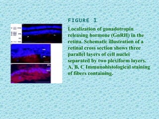


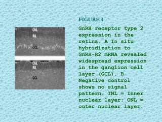


The document discusses research on the localization and expression patterns of gonadotropin-releasing hormone (GnRH) and its receptors in the retina. It includes 5 figures showing immunohistological staining and in situ hybridization results revealing: 1) GnRH fibers in the retina, 2) expression of GnRH receptor type 1 in some amacrine cells, 3) tyrosine hydroxylase-positive cells in the amacrine layer, 4) widespread expression of GnRH receptor type 2 in ganglion cells, and 5) a proposed model of how GnRH may affect retinal circuitry through these receptor types.


![melissa Poster SGM mel[1]](https://cdn.slidesharecdn.com/ss_thumbnails/cbc72576-c985-4bc6-9f04-631b12a7f357-160813003809-thumbnail.jpg?width=640&height=640&fit=bounds)


