Circulatory system
• Thecirculatory system of vertebrates is
basically a set of connecting tubes and
pumps that move fluid.
• The ability of the organism to adjust to
immediate physiological changes in physical
and metabolic activity depends on the
rapid response of this system.
• The circulatory system includes the blood
and lymph vascular systems.
• Lymphatic vessels and lymph, the fluid
they circulate, collectively constitute
the
lymphatic system.
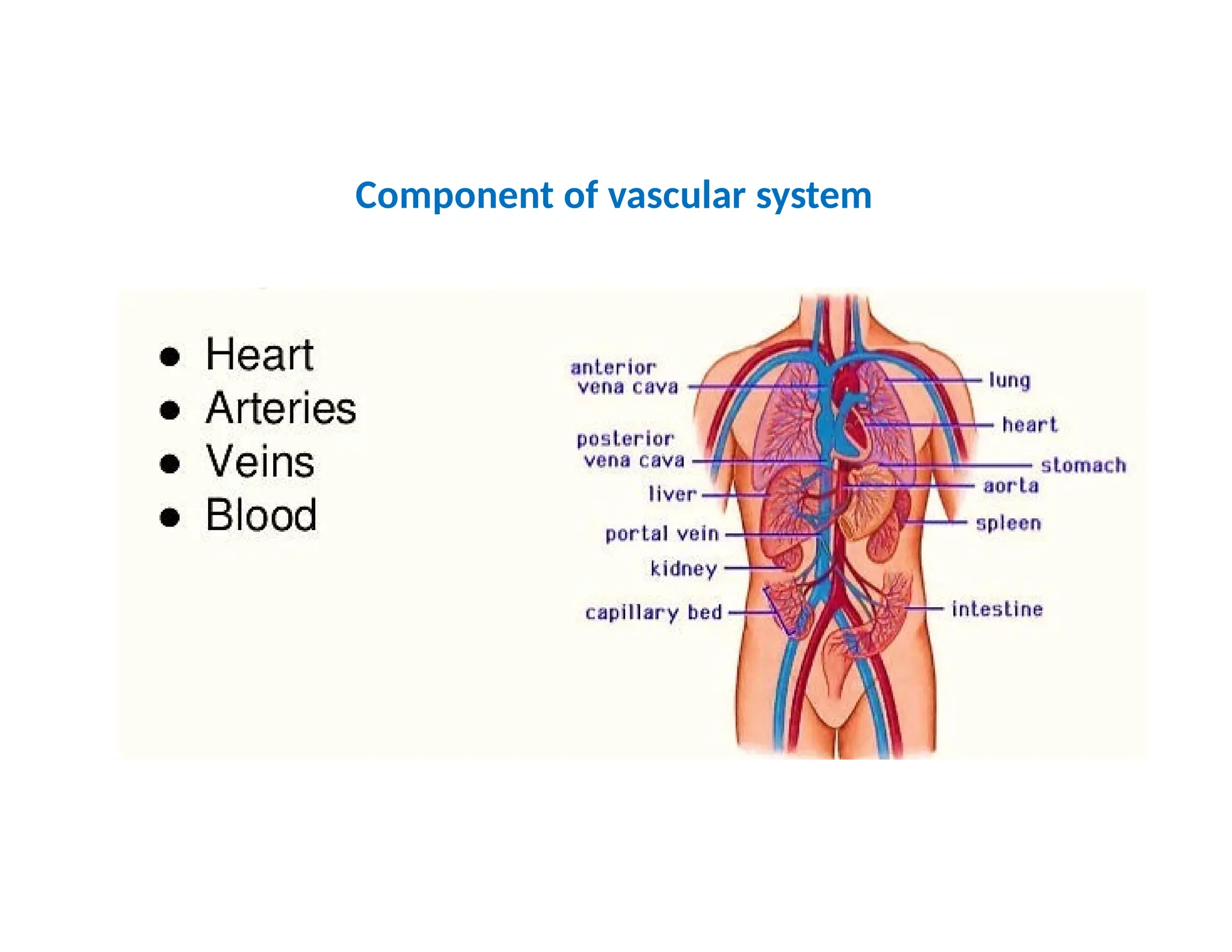
• The vascular system includes the blood
vessels that carry blood pumped by the
heart. Together blood, vessels, and heart
constitute the cardiovascular system.
Blood
• Cells producedby hemopoietic tissues usually enter the circulation to become the
peripheral or circulating blood.
• Circulating blood is comprised of plasma and formed elements.
• The plasma is the fluid component and can be thought of as the ground substance of
blood, a special connective tissue.
• The formed elements are the cellular components of blood.
• Red blood cells, or erythrocytes, are one cell type of the formed elements. All
erythrocytes have nuclei, except those in mammals. Mature red blood cells in mammals
lack nuclei.
• Hemoglobin, the major oxygen transport molecule.
• White blood cells, or leucocytes, are a second major cellular constituent of the formed
elements. Leucocytes defend the body from infection and disease.
• The platelets are a third formed element in the blood. They release factors that produce
a cascade of chemical events leading to the formation of a clot, or thrombus, at sites of
tissue damage.
• In addition to functioning in respiration and disease protection, blood also plays a part
in nutrition (carries carbohydrates, fats, proteins), excretion (carries spent metabolites),
regulation of body temperature (carries and distributes heat), maintenance of water
balance, and transport of hormones.
5.
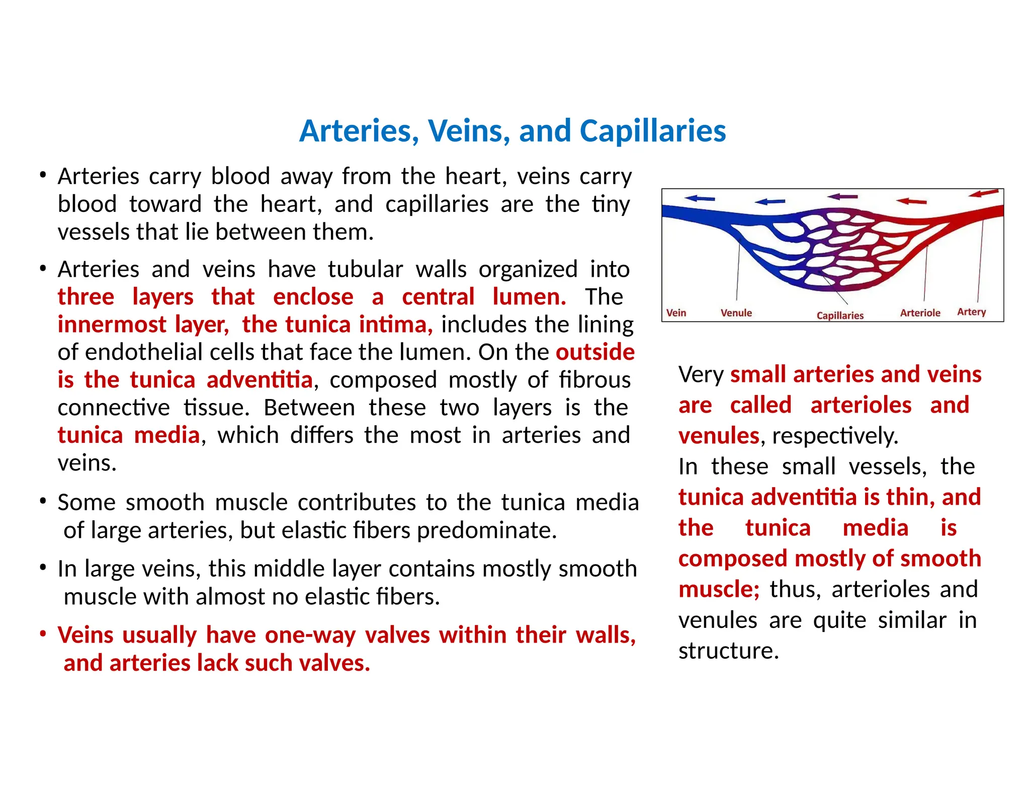
• Arteries carryblood away from the heart, veins carry
blood toward the heart, and capillaries are the tiny
vessels that lie between them.
• Arteries and veins have tubular walls organized into
three layers that enclose a central lumen. The
innermost layer, the tunica intima, includes the lining
of endothelial cells that face the lumen. On the outside
is the tunica adventitia, composed mostly of fibrous
connective tissue. Between these two layers is the
tunica media, which differs the most in arteries and
veins.
• Some smooth muscle contributes to the tunica media
of large arteries, but elastic fibers predominate.
• In large veins, this middle layer contains mostly smooth
muscle with almost no elastic fibers.
• Veins usually have one-way valves within their walls,
and arteries lack such valves.
Arteries, Veins, and Capillaries
Very small arteries and veins
are called arterioles and
venules, respectively.
In these small vessels, the
tunica adventitia is thin, and
the tunica media is
composed mostly of smooth
muscle; thus, arterioles and
venules are quite similar in
structure.
6.
The three layersof blood vessel walls change in relative thickness and size from large
arteries to small arterioles, capillaries, venules, and veins
Microcirculation
• The specificcomponent of the cardiovascular system
that
regulates and supports cell metabolism intimately is the
microcirculation.
• Capillary beds plus the arterioles that supply them and the
venules that drain them form the microcirculation.
• Blood flow to the capillary beds is controlled by smooth muscles.
• The precapillary sphincters are little rings of smooth muscle
restricting the entrance to the capillary beds.
• The walls of both arterioles and venules include thin sheets of
smooth muscles.
• Global nervous and hormonal control of these smooth muscles
regulates the flow of blood to the capillaries, as do local events in
the supplied tissues themselves.
• Whether by general events of the body (nervous, hormonal) or
local activity (autoregulation), capillary beds adjust blood flow to
match cell activity.
• Blood can be diverted through shunts that bypass some regions
entirely.
Basic structure ofHeart
• The heart probably began as a contractile vessel, much like those found within the circulatory system of
amphioxus.
• In most fishes, the heart is part of a single circulation.
• The embryonic fish heart consists of four chambers, which are also in series, so that blood flows in sequence from
the sinus venosus, to the atrium, to the ventricle, and finally to the fourth and most anterior heart chamber, the
bulbus cordis (conus arteriosus in adult chondrichthyans, holosteans and dipnoans, its contractile walls possess
cardiac muscle or bulbus arteriosus in adult teleost, elastic wall lacks cardiac muscle).
• In tetrapods, the ventral aorta often becomes reduced, sometimes persisting only as a small section of vessel at
the base of major departing aortic arches. In these cases, the term truncus arteriosus is most apt.
• The inner wall of the myocardium, especially of the ventricle, often forms projecting cones of muscle termed
trabeculae that are set off by deep recesses.
• Coronary vessels are especially well developed in elasmobranchs, crocodiles, birds, and mammals, in which they
supply most of the myocardium.
• In addition to the conal valves, the endocardium develops sets of valves between its chambers: The sinoatrial (SA)
valves form between the sinus venosus and the atrium, and the atrioventricular (AV) valves form between the
atrium and ventricle.
• Contraction of the entire heart usually begins within a restricted region in the sinus venosus called the pacemaker,
or sinoatrial (SA) node, and then spreads through a conducting system of fibers into the ventricle and other
contracting regions of the heart. In mammals, the conducting system includes, in addition to the SA node, a
second node, the atrioventricular (AV) node in the wall of the heart. The AV node consists of Purkinje fibers,
neuronlike fibers that are modified cardiac muscle cells.
• Birds and mammals have four-chambered hearts, but of the original four fish chambers, only two persist as major
receiving compartments, the atrium and the ventricle, both of which are divided into left and right
compartments to produce four anatomically separate chambers.
14.
Hagfish
• The hagfishheart is called a branchial heart to distinguish it from unique accessory blood pumps
elsewhere in its circulation.
• These supplementary circulatory pumps are sometimes called accessory “hearts,” in quotation marks
because they contract but usually lack the cardiac muscle of true branchial hearts.
• The cardinal hearts lying within the anterior cardinal veins are like sacs whose pumping action is
initiated by skeletal muscles around their outer walls.
• The paired caudal hearts, which are located in the tail, represent a unique blood pumping mechanism
among vertebrates.
• The portal heart is a single, expanded vascular sac that receives venous blood from one anterior and
one posterior cardinal vein, and then it contracts to drive the blood through the liver.
15.
The lamprey heart(branchial heart)
• It includes three compartments through
which blood flows sequentially— sinus
venosus, atrium, and ventricle but in
contrast to the hagfish heart, it is
innervated and, further, the ventricle
empties into the bulbus arteriosus,
whose walls lack cardiac muscle but
contain smooth muscle cells arranged
longitudinally and circumferentially.
• One-way valves separate compartments.
• The sinoatrial and atrioventricular valves
prevent retrograde blood flow.
• The luminal walls of the bulbus
arteriosus are thrown into leaflets,
collectively forming the semilunar
valves, which prevent reverse blood flow
and possibly aid in distributing blood to
the aortic arches.
• From the arches, blood flows to the
delicate gill capillaries next in line in the
circulation.
16.
Chondrichthyans fish
• Thehearts of chondrichthyans and bony
fishes consist of four basic chambers—
sinus venosus, atrium, ventricle, and
conus arteriosus (or bulbus arteriosus)—
with one-way valves stationed between
compartments.
• Like the other chambers, the muscular
conus arteriosus contracts, acting as an
auxiliary pump to help maintain blood
flow into the ventral aorta after the
onset of ventricular relaxation.
• Its contraction also brings together the
conal valves located on its opposing
walls. When these valves meet, they
prevent the backflow of departing
blood.
17.
Teleost fish
• Inteleosts, the conus arteriosus may
regress, leaving only remnants of a
myocardial conus, or be replaced
entirely by an elastic, noncontractile
bulbus arteriosus,
lacking muscle but
invested with
cardiac
smooth
muscle, collagen, and elastic fibers.
• A single pair of bulbar valves at the
juncture of the bulbus arteriosus and
the ventricle prevents retrograde flow.
• The S-shaped arrangement of chambers
in the fish heart places the thin-walled
sinus venosus and atrium dorsal to the
ventricle, so that atrial contraction
assists ventricular filling. Blood flows
from posterior chambers to anterior
chambers in the following sequence.
18.
Amphibians
• The heartincludes a sinus venosus, right and left atria divided by
an anatomically complete interatrial septum, a ventricle lacking
any internal subdivision, and a conus arteriosus with a spiral valve
(except for salamanders of the genus Siren, which have a partial
interventricular septum).
• The conus arteriosus of the frog heart arises from a single
trabeculate ventricle.
• Semilunar valves lie at the base of the conus and prevent
retrograde flow of blood back into the ventricle.
• Internally, a spiral valve twisting through nearly a complete
rotation establishes two channels within the conus, each of which
guides blood to specific sets of systemic and pulmocutaneous
arches.
• The systemic and pulmocutaneous arches both arise from the
truncus arteriosus, a remnant of the ventral aorta, but the two sets
of arches receive blood from different sides of the spiral valve.
• Unlike frogs, in which the pulmocutaneous artery branches give
rise to the cutaneous artery, salamanders lack a cutaneous artery.
Instead, branches from vessels supplying the systemic circulation
carry blood to the salamander skin. The pulmonary artery and the
systemic arches in salamanders arise from the truncus arteriosus
19.
Reptiles
• The sinusvenosus is reduced in comparison to amphibians but it
retains the same functions. It is still the first chamber to receive
venous blood and contains the pacemaker.
• The atrium is completely divided into
Prominent atrioventricular valves guard
ventricles.
• The conus arteriosus (or bulbus cordis)
right and left
atria.
the entrance to the
appears during early
embryonic development but becomes divided in the adult to form
the bases (trunks) of three large arteries leaving the ventricle: the
pulmonary trunk and the right and left systemic trunks.
• In snakes, a valved interaortic foramen connects the bases of
adjacent aortae. But the shunting of blood made possible by this
foramen has not been explored. Usually, the brachiocephalic artery,
delivering blood to the subclavians and carotids, emanates directly
from the right aortic arch, but in some turtles, it arises directly from
the ventricle, crowded in with the trunks of the three aortic arches.
• The ventricle is a single chamber functioning as a single fluid pump
to drive blood into the major arteries leaving the heart. Internally,
however, it has three interconnected compartments: the cavum
venosum and the cavum pulmonale separated from each other by
a muscular ridge, and the cavum arteriosum connected to the
cavum venosum via an interventricular canal.
20.
Crocodilian Hearts
• Theconus arteriosus (bulbus cordis) produces the bases of the trunks of the
three departing arteries—pulmonary and left and right aortic trunks.
• One-way lunar valves at the bases of each trunk permit blood to enter the
conus but halt reverse backflow into the ventricle.
• The sinus venosus is reduced but still functions as the receiving chamber for
returning systemic blood.
• The atrium is completely subdivided into two distinct left and
right
chambers, and the sinus venosus empties into the right atrium.
• The pulmonary vein enters the left atrium in adults, but it does not open into
the left atrium during embryonic development.
• The ventricle is divided by an anatomically complete interventricular septum
into distinct left and right chambers.
• The pulmonary trunk and left aortic arch open off the thick-walled right
ventricle.
• The right aortic arch opens off the left ventricle. A narrow channel called the
foramen of Panizza connects the left and right aortic arches shortly after they
depart from the ventricle.
21.
• When theventricles contract, blood pressure is
greatest in the left ventricle.
• The oxygenated blood it holds enters the base of
the right aortic arch, but because of its high
pressure, it also enters the left aortic arch via the
foramen of Panizza.
• High pressure in the left aortic arch keeps the
lunar valves at its base closed, leaving only the
pulmonary route of exit for blood in the right
ventricle.
• As a result, both aortic arches carry oxygenated
blood to systemic tissues, and the pulmonary
artery carries deoxygenated blood to the lungs.
Left Ventricle
Right Ventricle
right aortic arch
Left aortic arch
pulmonary artery
Air breathing :deoxygenated blood to the lungs
foramen of Panizza.
22.
Left Ventricle
Right Ventricle
foramenof Panizza.
right aortic arch
Left aortic arch
pulmonary artery
• When a crocodile dives, this pattern of cardiac blood
flow changes because of a cardiac shunt.
• Resistance to pulmonary flow increases due to
vasoconstriction of the vascular supply to the lungs and
partial constriction of a sphincter at the base of the
pulmonary artery (Lung is inactive during diving).
• As a result, systolic pressure within the right but not the
left ventricle rises substantially.
• Blood in the right ventricle now tends to exit through
the left aortic arch rather than through the pulmonary
circuit, which presents high resistance to blood flow.
• Diversion of blood in the right ventricle to the systemic
circulation represents a right-to left cardiac shunt.
• Blood in the right ventricle travels through the left aortic
arch, joining the systemic circulation and bypassing the
lungs.
Diving: deoxygenated blood to the systemic circulation
Birds and Mammals
•The hearts of birds and mammals have four chambers.
• In birds, the sinus venosus is reduced to a small but still anatomically discrete
area.
• The conus arteriosus (bulbus cordis) is only a transient embryonic chamber that
gives rise to the pulmonary trunk and a single aortic trunk in the adult.
• In mammals, the sinus venosus is reduced to a patch of Purkinje fibers, or
sinoatrial node, in the wall of the right atrium. The sinoatrial node functions as
a pacemaker, initiating the wave of contraction that spreads across the heart as
in all other vertebrates.
• Birds and mammals, both consist of parallel pumps with double circulation
circuits.
• The right side of the heart gathers deoxygenated blood from systemic tissues
and pumps it into the pulmonary circuit. The left side of the heart pumps
oxygenated blood from the lungs through the systemic circuit.
• The hearts of birds and mammals are anatomically divided into left and right
compartments; thus, there is no cardiac shunting with changing ventilation
rates.