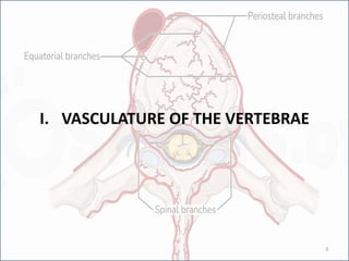
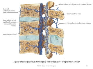
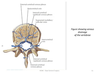
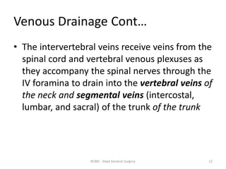
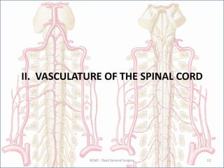
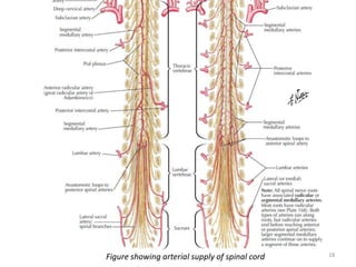
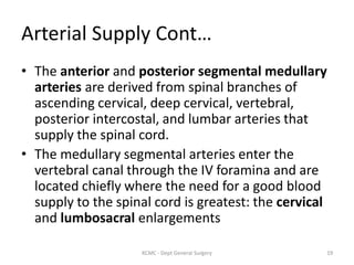
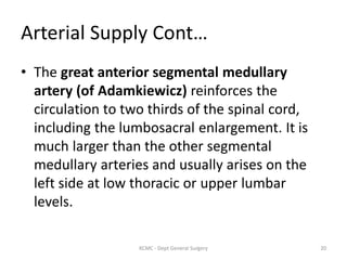
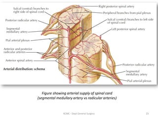
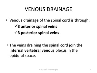
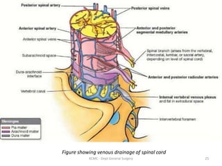
VERTEBRAL COLUMN
&
SPINAL CORD
Dr. Joshua J. Tadayo
General Surgery Resident
CONTENT
I. VASCULATURE OF THE VERTEBRAE
• Arterial Supply
• Venous Drainage
II. VASCULATURE OF THE SPINAL CORD
• Arterial Supply
• Venous Drainage
III. APPLIED ANATOMY
• Spinal Cord Ischemia
2
KCMC - Dept General Surgery
I. VASCULATURE OF THE VERTEBRAE
3
ARTERIAL SUPPLY
• Vertebrae are supplied by periosteal and
equatorial branches of the major cervical and
segmental arteries (intercostal, lumbar, and
sacral) and their spinal branches.
4
KCMC - Dept General Surgery
Arterial Supply Cont…
Spinal branches supplying the vertebrae are
branches of the:
• Vertebral and ascending cervical arteries in the
neck.
• Posterior intercostal arteries in the thoracic
region.
• Subcostal and lumbar arteries in the abdomen.
• Iliolumbar and lateral and medial sacral arteries
in the pelvis
5
KCMC - Dept General Surgery
Figure showing arterial supply of
vertebrae
6
KCMC - Dept General Surgery
Arterial Supply Cont…
• Periosteal and equatorial branches arise from
these arteries as they cross the external
surfaces of the vertebrae.
• Spinal branches enter the IV foramina and
divide into anterior and posterior vertebral
canal branches that pass to the vertebral body
and vertebral arch, respectively.
7
KCMC - Dept General Surgery
Arterial Supply Cont…
• Anterior vertebral canal branches send
nutrient arteries into the vertebral bodies.
• The spinal branches continue as terminal
radicular arteries distributed to the posterior
and anterior roots of the spinal nerves and
their coverings or as segmental medullary
arteries that continue to the spinal cord
8
KCMC - Dept General Surgery
VENOUS DRAINAGE
• The vertebral column is drained by:
Internal venous plexuses (inside vertebral canal)
External venous plexuses (outside vertebral canal)
Basivertebral veins (within the vertebral bodies)
• They communicate through the intervertebral
foramina and the intervertebral veins.
9
KCMC - Dept General Surgery
Figure showing venous drainage of the vertebrae – longitudinal section
10
KCMC - Dept General Surgery
Figure showing venous
drainage
of the vertebrae
11
KCMC - Dept General Surgery
Venous Drainage Cont…
• The intervertebral veins receive veins from the
spinal cord and vertebral venous plexuses as
they accompany the spinal nerves through the
IV foramina to drain into the vertebral veins of
the neck and segmental veins (intercostal,
lumbar, and sacral) of the trunk of the trunk
12
KCMC - Dept General Surgery
II. VASCULATURE OF THE SPINAL CORD
13
KCMC - Dept General Surgery
ARTERIAL SUPPLY
• The arteries suppling the spinal cord:
Anterior spinal artery (branch from
vertebral arteries)
2 Posterior spinal arteries (branch of
either the vertebral artery or the
posterior inferior cerebellar artery)
Segmental medullary (Ant & Post br)
Radicular arteries
14
KCMC - Dept General Surgery
Figure showing arterial supply of spinal cord
15
KCMC - Dept General Surgery
Arterial Supply Cont…
• The spinal arteries run longitudinally from the
medulla of the brainstem to the conus
medullaris of the spinal cord.
• By themselves, the anterior and posterior
spinal arteries supply only the short superior
part of the spinal cord.
16
KCMC - Dept General Surgery
Arterial Supply Cont…
• The circulation to much of the spinal cord
depends on segmental medullary and
radicular arteries running along the spinal
nerve roots.
17
KCMC - Dept General Surgery
Figure showing arterial supply of spinal cord 18
Arterial Supply Cont…
• The anterior and posterior segmental medullary
arteries are derived from spinal branches of
ascending cervical, deep cervical, vertebral,
posterior intercostal, and lumbar arteries that
supply the spinal cord.
• The medullary segmental arteries enter the
vertebral canal through the IV foramina and are
located chiefly where the need for a good blood
supply to the spinal cord is greatest: the cervical
and lumbosacral enlargements
19
KCMC - Dept General Surgery
Arterial Supply Cont…
• The great anterior segmental medullary
artery (of Adamkiewicz) reinforces the
circulation to two thirds of the spinal cord,
including the lumbosacral enlargement. It is
much larger than the other segmental
medullary arteries and usually arises on the
left side at low thoracic or upper lumbar
levels.
20
KCMC - Dept General Surgery
Arterial Supply Cont…
• NOTE:
The Artery of Adamkiewicz is sometimes called
"great radicular artery of Adamkiewicz",however
it is in fact a segmental medullary artery
21
KCMC - Dept General Surgery
Arterial Supply Cont…
• Posterior and anterior roots of the spinal
nerves and their coverings are supplied by
posterior and anterior radicular arteries,
which run along the nerve roots.
• These vessels do not reach the posterior or
anterior spinal arteries. Segmental medullary
arteries are larger vessels that make it to the
spinal arteries.
22
KCMC - Dept General Surgery
Figure showing arterial supply of spinal cord
(segmental medullary artery vs radicular arteries)
23
KCMC - Dept General Surgery
VENOUS DRAINAGE
• Venous drainage of the spinal cord is through:
3 anterior spinal veins
3 posterior spinal veins
• The veins draining the spinal cord join the
internal vertebral venous plexus in the
epidural space.
24
KCMC - Dept General Surgery
Figure showing venous drainage of spinal cord
25
KCMC - Dept General Surgery
III. APPLIED ANATOMY
26
ISCHEMIA OF SPINAL CORD
• Relatively rare i.e. : 12 in 100,000
• Median age of 52 years.
• Variable neurological deficit from minor
weakness to paraplegia or quadriplegia.
• Mortality rate in the vicinity of 20-25% for
patients admitted to hospital with spinal cord
infarction.
27
KCMC - Dept General Surgery
Etiology
• Iatrogenic during surgery: dissecting aortic
aneurysm; with aortic cross-clamping above
the renal artery (below that level anastomotic
flow via the artery of Adamkiewicz usually
provides protective circulation).
• Cardiogenic thromboembolism i.e. : atrial
fibrillation
• Compressive lesions eg: disc prolapse
• Hypo perfusion states i.e.: shock
28
KCMC - Dept General Surgery
Clinical Presentation
• Sudden and severe spinal (back) pain, which
may radiate caudad
• Bilateral weakness , paresthesias and sensory
loss
• In the acute stage: flaccid muscle tone
including absent Babinski reflexes
• Loss of sphincter control
29
KCMC - Dept General Surgery
Management
Correction of underlying pathogenic factors
Embolic, the use of anticoagulant or
antiplatelet drugs should be considered
Corticosteroids such as methyprednisolone
in cases of vasculitis
Maintaining adequate BP in cases of
hypoperfusion
30
KCMC - Dept General Surgery
Prognosis
• The prognosis of spinal cord infarction is
variable and depends on the extent of
parenchymal damage and the cause.
• Overall, about 24% of patients who suffer cord
ischemia experience no improvement; the rest
show some functional improvement, but only
20% have a good recovery with minimal
disability.
31
KCMC - Dept General Surgery
REFERENCES
• Essential Clinical Anatomy 4th Edition
Keith L. Moore
Anne M. Agur
Arthur F. Dalley
• Atlas of Human Anatomy
Frank H. Netter
• Spinal Cord Infarction Clinical Presentation
Medscape: https://emedicine.medscape.com/article/1164217-clinical#b1
32
KCMC - Dept General Surgery

VERTEBRAL COLUMN + SPINAL CORD.pdf

  • 1.
    VERTEBRAL COLUMN & SPINAL CORD Dr.Joshua J. Tadayo General Surgery Resident
  • 2.
    CONTENT I. VASCULATURE OFTHE VERTEBRAE • Arterial Supply • Venous Drainage II. VASCULATURE OF THE SPINAL CORD • Arterial Supply • Venous Drainage III. APPLIED ANATOMY • Spinal Cord Ischemia 2 KCMC - Dept General Surgery
  • 3.
    I. VASCULATURE OFTHE VERTEBRAE 3
  • 4.
    ARTERIAL SUPPLY • Vertebraeare supplied by periosteal and equatorial branches of the major cervical and segmental arteries (intercostal, lumbar, and sacral) and their spinal branches. 4 KCMC - Dept General Surgery
  • 5.
    Arterial Supply Cont… Spinalbranches supplying the vertebrae are branches of the: • Vertebral and ascending cervical arteries in the neck. • Posterior intercostal arteries in the thoracic region. • Subcostal and lumbar arteries in the abdomen. • Iliolumbar and lateral and medial sacral arteries in the pelvis 5 KCMC - Dept General Surgery
  • 6.
    Figure showing arterialsupply of vertebrae 6 KCMC - Dept General Surgery
  • 7.
    Arterial Supply Cont… •Periosteal and equatorial branches arise from these arteries as they cross the external surfaces of the vertebrae. • Spinal branches enter the IV foramina and divide into anterior and posterior vertebral canal branches that pass to the vertebral body and vertebral arch, respectively. 7 KCMC - Dept General Surgery
  • 8.
    Arterial Supply Cont… •Anterior vertebral canal branches send nutrient arteries into the vertebral bodies. • The spinal branches continue as terminal radicular arteries distributed to the posterior and anterior roots of the spinal nerves and their coverings or as segmental medullary arteries that continue to the spinal cord 8 KCMC - Dept General Surgery
  • 9.
    VENOUS DRAINAGE • Thevertebral column is drained by: Internal venous plexuses (inside vertebral canal) External venous plexuses (outside vertebral canal) Basivertebral veins (within the vertebral bodies) • They communicate through the intervertebral foramina and the intervertebral veins. 9 KCMC - Dept General Surgery
  • 10.
    Figure showing venousdrainage of the vertebrae – longitudinal section 10 KCMC - Dept General Surgery
  • 11.
    Figure showing venous drainage ofthe vertebrae 11 KCMC - Dept General Surgery
  • 12.
    Venous Drainage Cont… •The intervertebral veins receive veins from the spinal cord and vertebral venous plexuses as they accompany the spinal nerves through the IV foramina to drain into the vertebral veins of the neck and segmental veins (intercostal, lumbar, and sacral) of the trunk of the trunk 12 KCMC - Dept General Surgery
  • 13.
    II. VASCULATURE OFTHE SPINAL CORD 13 KCMC - Dept General Surgery
  • 14.
    ARTERIAL SUPPLY • Thearteries suppling the spinal cord: Anterior spinal artery (branch from vertebral arteries) 2 Posterior spinal arteries (branch of either the vertebral artery or the posterior inferior cerebellar artery) Segmental medullary (Ant & Post br) Radicular arteries 14 KCMC - Dept General Surgery
  • 15.
    Figure showing arterialsupply of spinal cord 15 KCMC - Dept General Surgery
  • 16.
    Arterial Supply Cont… •The spinal arteries run longitudinally from the medulla of the brainstem to the conus medullaris of the spinal cord. • By themselves, the anterior and posterior spinal arteries supply only the short superior part of the spinal cord. 16 KCMC - Dept General Surgery
  • 17.
    Arterial Supply Cont… •The circulation to much of the spinal cord depends on segmental medullary and radicular arteries running along the spinal nerve roots. 17 KCMC - Dept General Surgery
  • 18.
    Figure showing arterialsupply of spinal cord 18
  • 19.
    Arterial Supply Cont… •The anterior and posterior segmental medullary arteries are derived from spinal branches of ascending cervical, deep cervical, vertebral, posterior intercostal, and lumbar arteries that supply the spinal cord. • The medullary segmental arteries enter the vertebral canal through the IV foramina and are located chiefly where the need for a good blood supply to the spinal cord is greatest: the cervical and lumbosacral enlargements 19 KCMC - Dept General Surgery
  • 20.
    Arterial Supply Cont… •The great anterior segmental medullary artery (of Adamkiewicz) reinforces the circulation to two thirds of the spinal cord, including the lumbosacral enlargement. It is much larger than the other segmental medullary arteries and usually arises on the left side at low thoracic or upper lumbar levels. 20 KCMC - Dept General Surgery
  • 21.
    Arterial Supply Cont… •NOTE: The Artery of Adamkiewicz is sometimes called "great radicular artery of Adamkiewicz",however it is in fact a segmental medullary artery 21 KCMC - Dept General Surgery
  • 22.
    Arterial Supply Cont… •Posterior and anterior roots of the spinal nerves and their coverings are supplied by posterior and anterior radicular arteries, which run along the nerve roots. • These vessels do not reach the posterior or anterior spinal arteries. Segmental medullary arteries are larger vessels that make it to the spinal arteries. 22 KCMC - Dept General Surgery
  • 23.
    Figure showing arterialsupply of spinal cord (segmental medullary artery vs radicular arteries) 23 KCMC - Dept General Surgery
  • 24.
    VENOUS DRAINAGE • Venousdrainage of the spinal cord is through: 3 anterior spinal veins 3 posterior spinal veins • The veins draining the spinal cord join the internal vertebral venous plexus in the epidural space. 24 KCMC - Dept General Surgery
  • 25.
    Figure showing venousdrainage of spinal cord 25 KCMC - Dept General Surgery
  • 26.
  • 27.
    ISCHEMIA OF SPINALCORD • Relatively rare i.e. : 12 in 100,000 • Median age of 52 years. • Variable neurological deficit from minor weakness to paraplegia or quadriplegia. • Mortality rate in the vicinity of 20-25% for patients admitted to hospital with spinal cord infarction. 27 KCMC - Dept General Surgery
  • 28.
    Etiology • Iatrogenic duringsurgery: dissecting aortic aneurysm; with aortic cross-clamping above the renal artery (below that level anastomotic flow via the artery of Adamkiewicz usually provides protective circulation). • Cardiogenic thromboembolism i.e. : atrial fibrillation • Compressive lesions eg: disc prolapse • Hypo perfusion states i.e.: shock 28 KCMC - Dept General Surgery
  • 29.
    Clinical Presentation • Suddenand severe spinal (back) pain, which may radiate caudad • Bilateral weakness , paresthesias and sensory loss • In the acute stage: flaccid muscle tone including absent Babinski reflexes • Loss of sphincter control 29 KCMC - Dept General Surgery
  • 30.
    Management Correction of underlyingpathogenic factors Embolic, the use of anticoagulant or antiplatelet drugs should be considered Corticosteroids such as methyprednisolone in cases of vasculitis Maintaining adequate BP in cases of hypoperfusion 30 KCMC - Dept General Surgery
  • 31.
    Prognosis • The prognosisof spinal cord infarction is variable and depends on the extent of parenchymal damage and the cause. • Overall, about 24% of patients who suffer cord ischemia experience no improvement; the rest show some functional improvement, but only 20% have a good recovery with minimal disability. 31 KCMC - Dept General Surgery
  • 32.
    REFERENCES • Essential ClinicalAnatomy 4th Edition Keith L. Moore Anne M. Agur Arthur F. Dalley • Atlas of Human Anatomy Frank H. Netter • Spinal Cord Infarction Clinical Presentation Medscape: https://emedicine.medscape.com/article/1164217-clinical#b1 32 KCMC - Dept General Surgery