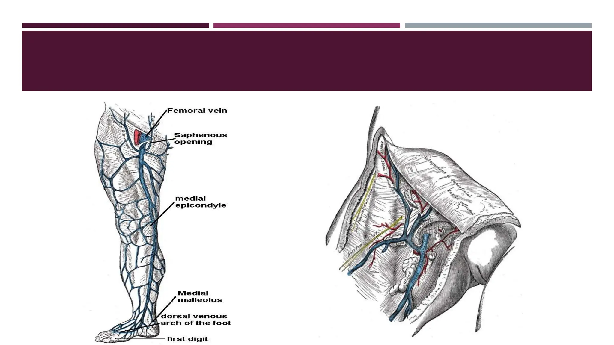
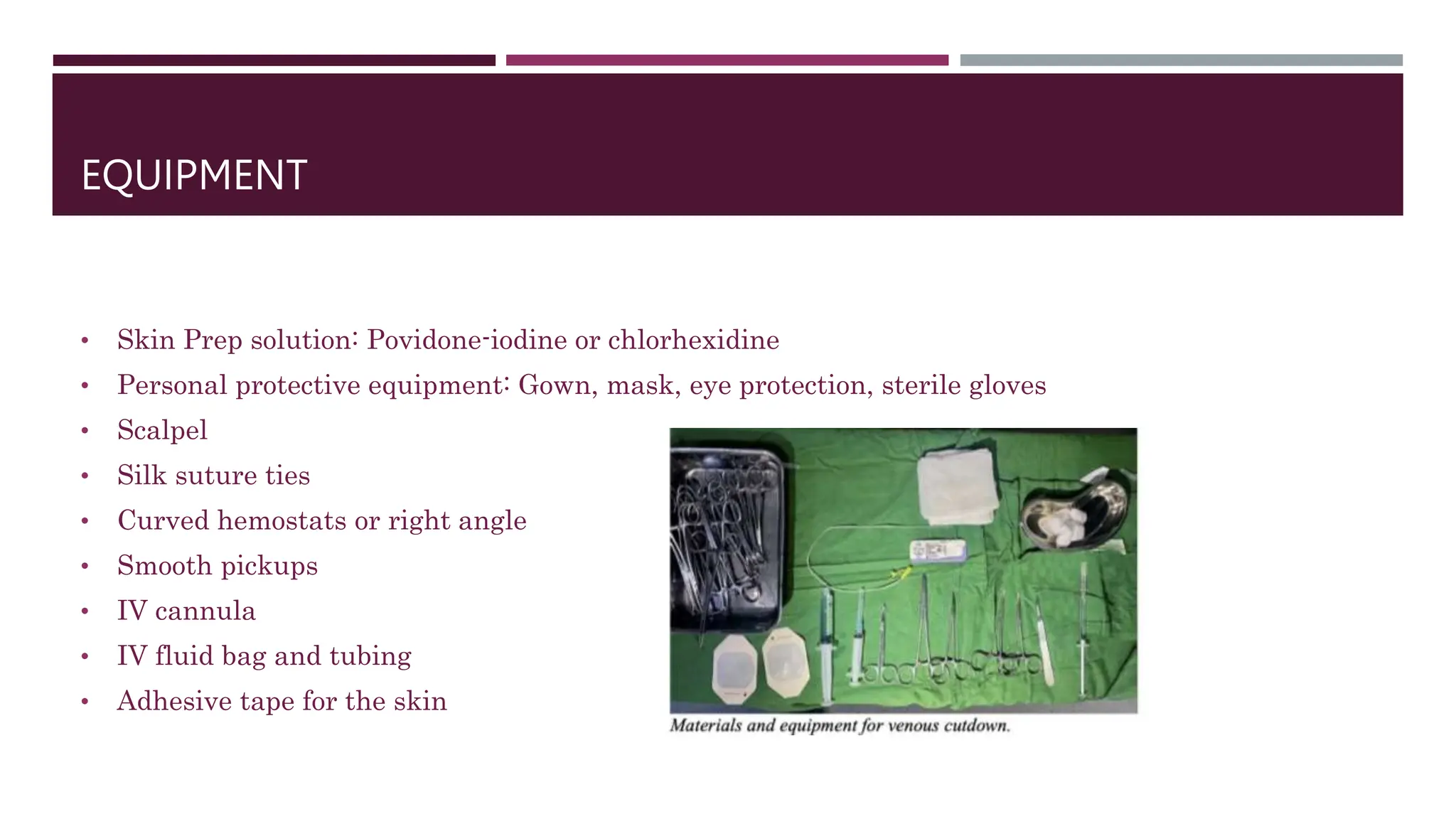
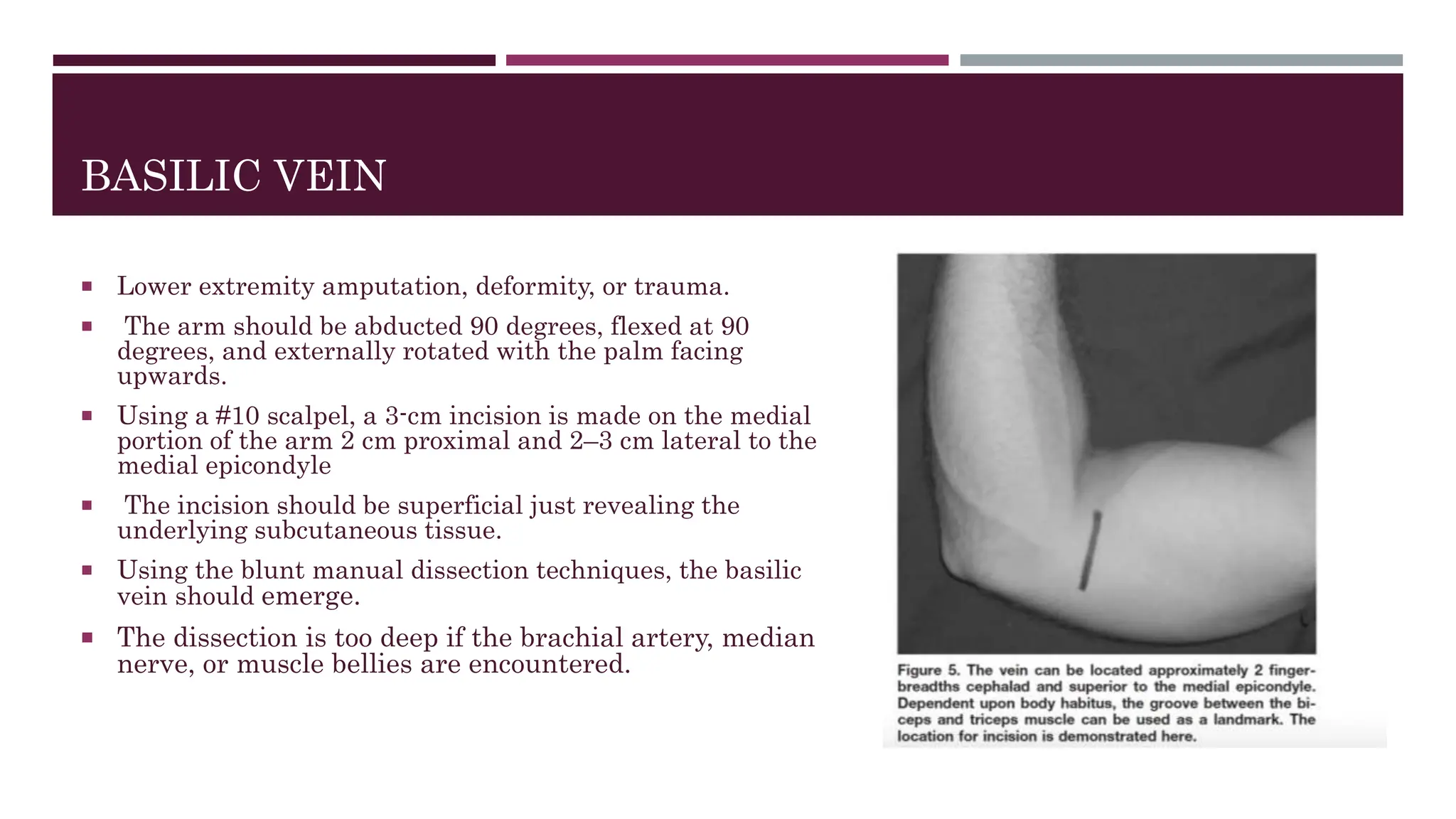
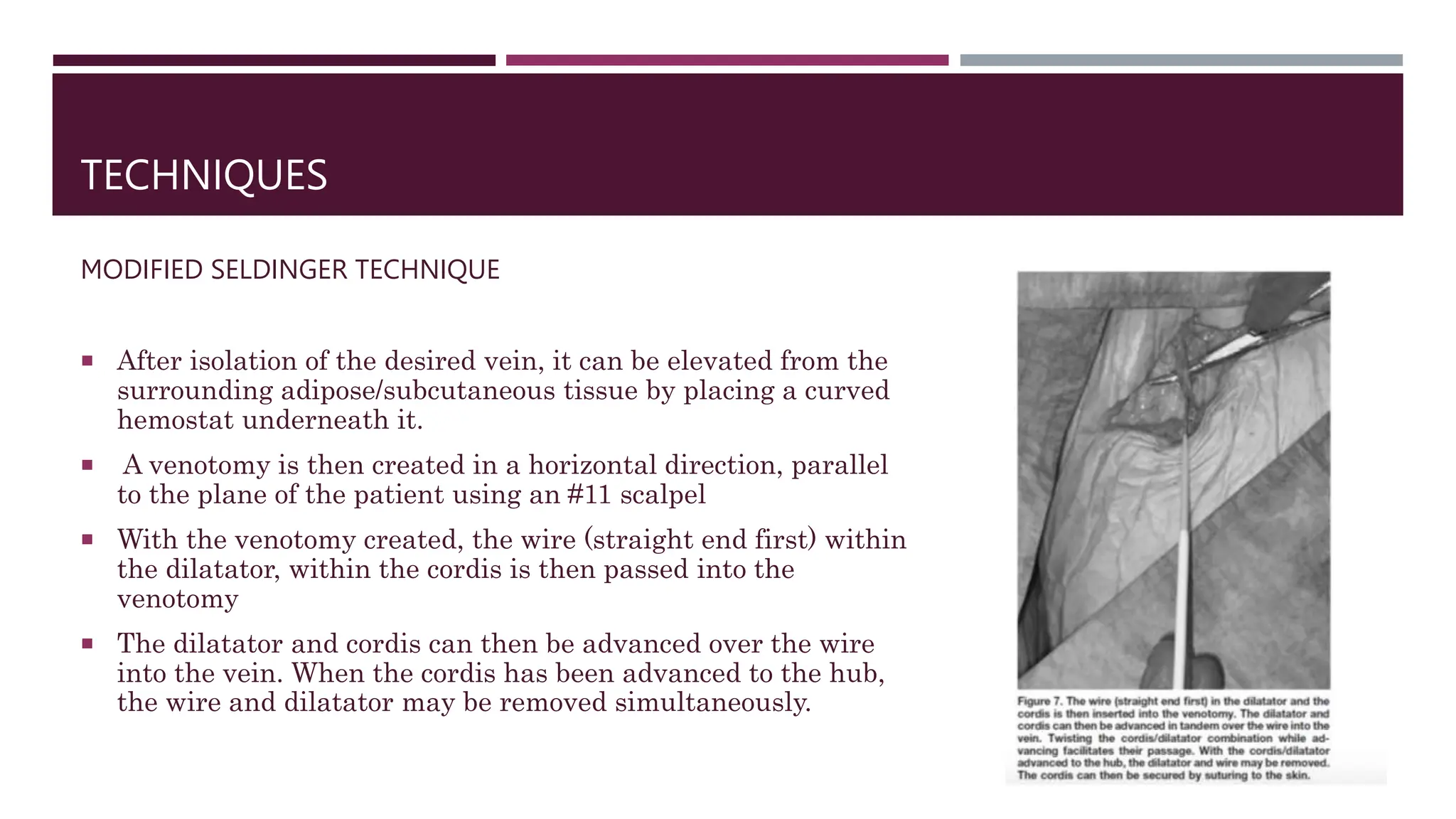
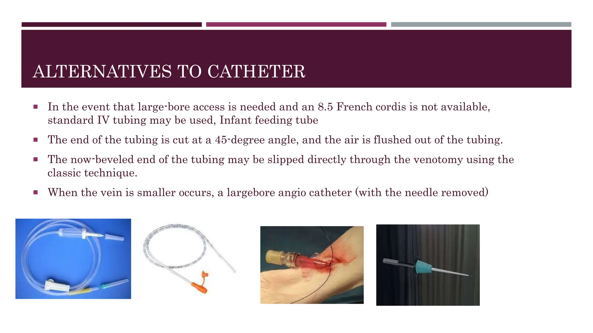
VENOUS CUT DOWN
DR ANKITA GURAV
CONTENTS
 INTRODUCTION
 ANATOMY
 INDICATIONS
 CONTRAINDICATIONS
 COMMON SITES
 EQUIPMENTS AND PREPARATION
 CUTDOWN AT ANKLE
 CUTDOWN AT THE GROIN
 CUTDOWN AT THE BASILIC VEIN
 ALTERNATE TECHNIQUES
 COMPLICATIONS
 REFERENCES
INTRODUCTION
 Venous access in the critically ill patient is of the utmost importance.
 The literature regarding peripheral venous cutdowns extends back to 1940 when Keeley introduced this
technique as an alternative
 It is essential to the treatment and resuscitation of both the medically and traumatically ill patient.
 It allows the delivery of fluids, blood products, medications, and repeated blood sampling.
 The venous cutdown technique is a surgical procedure designed to gain venous access when relatively
less invasive percutaneous procedures such as the Central lines , ultrasound-guided venous access, and
intraosseous vascular access have failed.
INTRODUCTION
 Patients with profound haemorrhagic shock, asystole, or pulseless electrical activity will lack
palpable femoral arterial pulses, complicating femoral venous access.
 The intravenous drug user or extensively injured patient without identifiable peripheral veins or
scarred, anatomically altered central venous access sites may be equally challenging.
 It is among these patients that proficiency in peripheral venous cutdown techniques may prove
invaluable and why it should remain within the emergency physician’s procedural armamentarium
COMMON SITES
There are three critical areas for venous cutdowns
For adults
 Saphenous vein at the ankle,
 The saphenous vein at the groin
 The basilic vein at the elbow.
For infants and younger children:
 Internal jugular vein
 External jugular vein
 Femoral vein
WHY IS GSV IDEAL ?
 The greater saphenous vein is the longest vein in the body
 Its superficial location and anatomic regularity make it ideal for cutdown.
 The GSV arises from the dorsal venous arch of the foot. It ascends cephalad approximately 2 cm anterior
to the medial malleolus.
 2cms cephalad to the medial malleolus, it lies just anterior to the tibial periosteum. Along with the
saphenous nerve it continues to ascend within the superficial fascia along the medial aspect of the leg.
 It eventually joins the femoral vein approximately 4 cm below and 3 cm lateral
ANATOMY OF THE SAPHENOUS VEIN
 It is part of the superficial venous collecting system of the lower extremity originating in the foot
and traveling up the medial leg and upper thigh where it enters the femoral vein within the
femoral triangle.
 Specifically, at the foot, it originates at the confluence of the dorsal vein of the first digit and the
dorsal venous arch of the foot.
 It travels up the medial leg alongside the saphenous nerve which must be preserved during vein
harvesting.
 At the knee, it runs over the posterior border of the medial condyle of the femur bone.
 It takes a slightly lateral course along the anteromedial thigh before entering an opening in the
fascia lata called the saphenous opening at the saphenofemoral junction.
INDICATIONS
• Infants, children, and adults in shock
• Burned or scarred patients
• Distorted anatomy
• Cardiac arrest without a palpable femoral pulse
• Individuals in which IV lines or alternative techniques for obtaining central access cannot be
quickly obtained
• Intravenous (IV) drug abusers where all the peripheral veins are thrombosed
• Vascular lower extremity bypass operations (femoral-popliteal or femoral-distal bypass) requiring a
vein conduit
• Vascular operations requiring a vein patch
CONTRAINDICATIONS
 Trauma to the targeted site or in patients with a massive pelvic injury with suspected avulsion of
the iliac veins.
 An active infection over the site of interest, when coagulopathies are present, or when less-invasive
methods of venous access are possible.
 In the event of bilateral lower extremity trauma compromising the integrity of the GSV, the basilic
vein in the upper extremity may be an alternative access site.
EQUIPMENT
• Skin Prep solution: Povidone-iodine or chlorhexidine
• Personal protective equipment: Gown, mask, eye protection, sterile gloves
• Scalpel
• Silk suture ties
• Curved hemostats or right angle
• Smooth pickups
• IV cannula
• IV fluid bag and tubing
• Adhesive tape for the skin
PREPARATION
 A local anesthetic (lidocaine 1% or 2% with or without
epinephrine) may be administered before skin incision
depending on the acuity of the situation and status of the
patient.
 Try to palpate the saphenous vein anterior to the medial
malleolus (harder to locate in patients with
hypovolemic shock). Have all equipment open and
available before making the skin incision
VENOUS CUT DOWN FOR GSV AT ANKLE
 The cutdown procedure begins with sterile preparation and draping of the anteromedial ankle with
the medial malleolus visible as a bony landmark.
 Traveling 1 cm anterior and 1 cm superior to the medial malleolus approximates the location of the
GSV where a 3 cm transverse skin incision is made with a scalpel.
 Curved hemostats are then used to longitudinally dissect through the subcutaneous tissue while
being cognizant of the rather superficial location of the vessel.
 Dissection continues circumferentially around the vessel and extends a length of about 2 cm to
allow passage of permanent suture material such as 3-0 silk around the proximal and distal ends of
the exposed vessel.
VENOUS CUT DOWN FOR GSV AT ANKLE
 Care must be taken to dissect off and avoid damage to the saphenous nerve which runs parallel with the vein.
 These sutures will aid in proximal and distal control as well as act as a sling to gently lift the vessel into the surgical field.
 A hemostat may be used to apply distal traction in preparation for venous entry.
 Venous access is gained by either a formal venotomy with a No. 15 scalpel to accommodate a larger IV cannula or by an introducer
needle using Seldinger technique.
 If using a scalpel, place a hemostat behind the mobilized vein to aid in dividing 40% of the transverse diameter.
 Importantly, this venous access is secured by tying down the permanent suture encircling both the vein and cannula hub tightly.
 The distal vein is also ligated with a silk suture. The cannula or IV tubing should also be secured to the skin with suture or tape to
prevent it from being dislodged.
VENOUS CUT DOWN AT GROIN LEVEL
 With a #10 scalpel, a transverse incision is made
from just distal to where the labial/scrotal fold
meets the thigh.
 The incision is extended laterally for 5–6 cm,
entering the subcutaneous tissues
 Dissection through the subcutaneous tissues may
be performed with hemostats.
 If the thigh muscles or deep investing fascia are
encountered, the dissection is too deep.
 Landmarks should be relocated and the skin
incision/dissection adjusted as needed.
 Another technique, described by Rogers, involves similar blunt manual dissection but through an
incision made parallel to the vessel
 He described making a 10-cm incision from 2 fingerbreadths lateral and inferior to the pubic
tubercle extended in the direction of the medial epicondyle of the femur.
 The dissection is accomplished by grasping the skin edges and forcibly pulling them apart,
readily revealing the vein’s course along the ante-
BASILIC VEIN
 Lower extremity amputation, deformity, or trauma.
 The arm should be abducted 90 degrees, flexed at 90
degrees, and externally rotated with the palm facing
upwards.
 Using a #10 scalpel, a 3-cm incision is made on the medial
portion of the arm 2 cm proximal and 2–3 cm lateral to the
medial epicondyle
 The incision should be superficial just revealing the
underlying subcutaneous tissue.
 Using the blunt manual dissection techniques, the basilic
vein should emerge.
 The dissection is too deep if the brachial artery, median
nerve, or muscle bellies are encountered.
TECHNIQUES
MODIFIED SELDINGER TECHNIQUE
 After isolation of the desired vein, it can be elevated from the
surrounding adipose/subcutaneous tissue by placing a curved
hemostat underneath it.
 A venotomy is then created in a horizontal direction, parallel
to the plane of the patient using an #11 scalpel
 With the venotomy created, the wire (straight end first) within
the dilatator, within the cordis is then passed into the
venotomy
 The dilatator and cordis can then be advanced over the wire
into the vein. When the cordis has been advanced to the hub,
the wire and dilatator may be removed simultaneously.
ALTERNATIVES TO CATHETER
 In the event that large-bore access is needed and an 8.5 French cordis is not available,
standard IV tubing may be used, Infant feeding tube
 The end of the tubing is cut at a 45-degree angle, and the air is flushed out of the tubing.
 The now-beveled end of the tubing may be slipped directly through the venotomy using the
classic technique.
 When the vein is smaller occurs, a largebore angio catheter (with the needle removed)
COMPLICATIONS
 Potential complications of the venous cutdown technique include infection, failed cannulation,
heamorrhage, nerve injury, and air embolism.
 Removal of the catheter within 12 to 24 hours is recommended to reduce the incidence of
complications.
SUMMARY
 In the trauma or medical patient in extremis, vascular access is essential during the resuscitative
efforts. Oftentimes, body habitus, past medical history, or clinical circumstances make peripheral or
central percutaneous intravenous access challenging.
 Greater saphenous vein cutdown technique remains an effective intervention for rapid venous
access in the case of failed percutaneous methods.
 Its remote location from the head and torso make GSV cutdown at the ankle feasible during major
resuscitation efforts, especially in the acute traumatic setting.
REFERENCES
 PERIPHERAL VENOUS CUTDOWN Stephen Chappell, MD,* Gary M. Vilke, MD,† Theodore C.
Chan, MD,† Richard A. Harrigan, MD,* and Jacob W. Ufberg, MD**Department of Emergency
Medicine, Temple University School of Medicine, Philadelphia, Pennsylvania, and †Department
ofEmergency Medicine, University of California, San Diego Medical Center, San Diego, California
 Caggiati, Alberto; Bergan, John J. (2002). "The saphenous vein: derivation of its name and its
relevant anatomy". Journal of Vascular Surgery. 35 (1): 172–5. doi:10.1067/mva.2002.118826

VENOUS CUT DOWN Procedure ppt presntation

  • 1.
    VENOUS CUT DOWN DRANKITA GURAV
  • 2.
    CONTENTS  INTRODUCTION  ANATOMY INDICATIONS  CONTRAINDICATIONS  COMMON SITES  EQUIPMENTS AND PREPARATION  CUTDOWN AT ANKLE  CUTDOWN AT THE GROIN  CUTDOWN AT THE BASILIC VEIN  ALTERNATE TECHNIQUES  COMPLICATIONS  REFERENCES
  • 3.
    INTRODUCTION  Venous accessin the critically ill patient is of the utmost importance.  The literature regarding peripheral venous cutdowns extends back to 1940 when Keeley introduced this technique as an alternative  It is essential to the treatment and resuscitation of both the medically and traumatically ill patient.  It allows the delivery of fluids, blood products, medications, and repeated blood sampling.  The venous cutdown technique is a surgical procedure designed to gain venous access when relatively less invasive percutaneous procedures such as the Central lines , ultrasound-guided venous access, and intraosseous vascular access have failed.
  • 4.
    INTRODUCTION  Patients withprofound haemorrhagic shock, asystole, or pulseless electrical activity will lack palpable femoral arterial pulses, complicating femoral venous access.  The intravenous drug user or extensively injured patient without identifiable peripheral veins or scarred, anatomically altered central venous access sites may be equally challenging.  It is among these patients that proficiency in peripheral venous cutdown techniques may prove invaluable and why it should remain within the emergency physician’s procedural armamentarium
  • 5.
    COMMON SITES There arethree critical areas for venous cutdowns For adults  Saphenous vein at the ankle,  The saphenous vein at the groin  The basilic vein at the elbow. For infants and younger children:  Internal jugular vein  External jugular vein  Femoral vein
  • 6.
    WHY IS GSVIDEAL ?  The greater saphenous vein is the longest vein in the body  Its superficial location and anatomic regularity make it ideal for cutdown.  The GSV arises from the dorsal venous arch of the foot. It ascends cephalad approximately 2 cm anterior to the medial malleolus.  2cms cephalad to the medial malleolus, it lies just anterior to the tibial periosteum. Along with the saphenous nerve it continues to ascend within the superficial fascia along the medial aspect of the leg.  It eventually joins the femoral vein approximately 4 cm below and 3 cm lateral
  • 7.
    ANATOMY OF THESAPHENOUS VEIN  It is part of the superficial venous collecting system of the lower extremity originating in the foot and traveling up the medial leg and upper thigh where it enters the femoral vein within the femoral triangle.  Specifically, at the foot, it originates at the confluence of the dorsal vein of the first digit and the dorsal venous arch of the foot.  It travels up the medial leg alongside the saphenous nerve which must be preserved during vein harvesting.  At the knee, it runs over the posterior border of the medial condyle of the femur bone.  It takes a slightly lateral course along the anteromedial thigh before entering an opening in the fascia lata called the saphenous opening at the saphenofemoral junction.
  • 9.
    INDICATIONS • Infants, children,and adults in shock • Burned or scarred patients • Distorted anatomy • Cardiac arrest without a palpable femoral pulse • Individuals in which IV lines or alternative techniques for obtaining central access cannot be quickly obtained • Intravenous (IV) drug abusers where all the peripheral veins are thrombosed • Vascular lower extremity bypass operations (femoral-popliteal or femoral-distal bypass) requiring a vein conduit • Vascular operations requiring a vein patch
  • 10.
    CONTRAINDICATIONS  Trauma tothe targeted site or in patients with a massive pelvic injury with suspected avulsion of the iliac veins.  An active infection over the site of interest, when coagulopathies are present, or when less-invasive methods of venous access are possible.  In the event of bilateral lower extremity trauma compromising the integrity of the GSV, the basilic vein in the upper extremity may be an alternative access site.
  • 11.
    EQUIPMENT • Skin Prepsolution: Povidone-iodine or chlorhexidine • Personal protective equipment: Gown, mask, eye protection, sterile gloves • Scalpel • Silk suture ties • Curved hemostats or right angle • Smooth pickups • IV cannula • IV fluid bag and tubing • Adhesive tape for the skin
  • 12.
    PREPARATION  A localanesthetic (lidocaine 1% or 2% with or without epinephrine) may be administered before skin incision depending on the acuity of the situation and status of the patient.  Try to palpate the saphenous vein anterior to the medial malleolus (harder to locate in patients with hypovolemic shock). Have all equipment open and available before making the skin incision
  • 13.
    VENOUS CUT DOWNFOR GSV AT ANKLE  The cutdown procedure begins with sterile preparation and draping of the anteromedial ankle with the medial malleolus visible as a bony landmark.  Traveling 1 cm anterior and 1 cm superior to the medial malleolus approximates the location of the GSV where a 3 cm transverse skin incision is made with a scalpel.  Curved hemostats are then used to longitudinally dissect through the subcutaneous tissue while being cognizant of the rather superficial location of the vessel.  Dissection continues circumferentially around the vessel and extends a length of about 2 cm to allow passage of permanent suture material such as 3-0 silk around the proximal and distal ends of the exposed vessel.
  • 14.
    VENOUS CUT DOWNFOR GSV AT ANKLE  Care must be taken to dissect off and avoid damage to the saphenous nerve which runs parallel with the vein.  These sutures will aid in proximal and distal control as well as act as a sling to gently lift the vessel into the surgical field.  A hemostat may be used to apply distal traction in preparation for venous entry.  Venous access is gained by either a formal venotomy with a No. 15 scalpel to accommodate a larger IV cannula or by an introducer needle using Seldinger technique.  If using a scalpel, place a hemostat behind the mobilized vein to aid in dividing 40% of the transverse diameter.  Importantly, this venous access is secured by tying down the permanent suture encircling both the vein and cannula hub tightly.  The distal vein is also ligated with a silk suture. The cannula or IV tubing should also be secured to the skin with suture or tape to prevent it from being dislodged.
  • 15.
    VENOUS CUT DOWNAT GROIN LEVEL  With a #10 scalpel, a transverse incision is made from just distal to where the labial/scrotal fold meets the thigh.  The incision is extended laterally for 5–6 cm, entering the subcutaneous tissues  Dissection through the subcutaneous tissues may be performed with hemostats.  If the thigh muscles or deep investing fascia are encountered, the dissection is too deep.  Landmarks should be relocated and the skin incision/dissection adjusted as needed.
  • 17.
     Another technique,described by Rogers, involves similar blunt manual dissection but through an incision made parallel to the vessel  He described making a 10-cm incision from 2 fingerbreadths lateral and inferior to the pubic tubercle extended in the direction of the medial epicondyle of the femur.  The dissection is accomplished by grasping the skin edges and forcibly pulling them apart, readily revealing the vein’s course along the ante-
  • 18.
    BASILIC VEIN  Lowerextremity amputation, deformity, or trauma.  The arm should be abducted 90 degrees, flexed at 90 degrees, and externally rotated with the palm facing upwards.  Using a #10 scalpel, a 3-cm incision is made on the medial portion of the arm 2 cm proximal and 2–3 cm lateral to the medial epicondyle  The incision should be superficial just revealing the underlying subcutaneous tissue.  Using the blunt manual dissection techniques, the basilic vein should emerge.  The dissection is too deep if the brachial artery, median nerve, or muscle bellies are encountered.
  • 19.
    TECHNIQUES MODIFIED SELDINGER TECHNIQUE After isolation of the desired vein, it can be elevated from the surrounding adipose/subcutaneous tissue by placing a curved hemostat underneath it.  A venotomy is then created in a horizontal direction, parallel to the plane of the patient using an #11 scalpel  With the venotomy created, the wire (straight end first) within the dilatator, within the cordis is then passed into the venotomy  The dilatator and cordis can then be advanced over the wire into the vein. When the cordis has been advanced to the hub, the wire and dilatator may be removed simultaneously.
  • 20.
    ALTERNATIVES TO CATHETER In the event that large-bore access is needed and an 8.5 French cordis is not available, standard IV tubing may be used, Infant feeding tube  The end of the tubing is cut at a 45-degree angle, and the air is flushed out of the tubing.  The now-beveled end of the tubing may be slipped directly through the venotomy using the classic technique.  When the vein is smaller occurs, a largebore angio catheter (with the needle removed)
  • 21.
    COMPLICATIONS  Potential complicationsof the venous cutdown technique include infection, failed cannulation, heamorrhage, nerve injury, and air embolism.  Removal of the catheter within 12 to 24 hours is recommended to reduce the incidence of complications.
  • 22.
    SUMMARY  In thetrauma or medical patient in extremis, vascular access is essential during the resuscitative efforts. Oftentimes, body habitus, past medical history, or clinical circumstances make peripheral or central percutaneous intravenous access challenging.  Greater saphenous vein cutdown technique remains an effective intervention for rapid venous access in the case of failed percutaneous methods.  Its remote location from the head and torso make GSV cutdown at the ankle feasible during major resuscitation efforts, especially in the acute traumatic setting.
  • 23.
    REFERENCES  PERIPHERAL VENOUSCUTDOWN Stephen Chappell, MD,* Gary M. Vilke, MD,† Theodore C. Chan, MD,† Richard A. Harrigan, MD,* and Jacob W. Ufberg, MD**Department of Emergency Medicine, Temple University School of Medicine, Philadelphia, Pennsylvania, and †Department ofEmergency Medicine, University of California, San Diego Medical Center, San Diego, California  Caggiati, Alberto; Bergan, John J. (2002). "The saphenous vein: derivation of its name and its relevant anatomy". Journal of Vascular Surgery. 35 (1): 172–5. doi:10.1067/mva.2002.118826