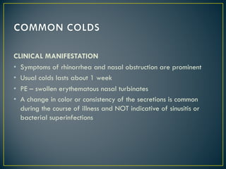
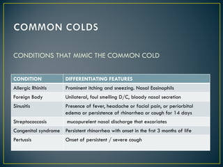
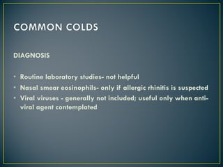
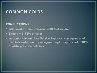
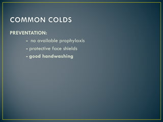
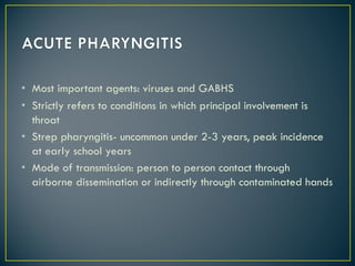
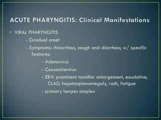
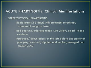
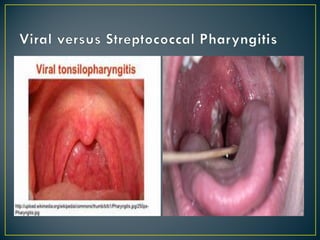
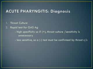
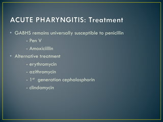
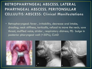
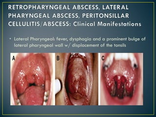
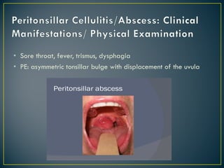
CAGAYAN STATE UNIVERSITY
College of Medicine
Department of Pediatrics
• Rhinitis, rhinosinositis – self limitedinvolvement of the sinus
mucosa
• Most common infectious condition in children
• Common pathogens
Association Pathogens Relative Frequency of
Colds Caused
Agents primarily
associated with colds
Agents primarily
associated with other
clinical syndrome that also
cause common colds
symptoms
Rhinoviruses
Coronaviruses
Respiratory Snycytial Virus
Human pneumometavirus
Influenza viruses
Parainfluenza viruses
Adenoviruses
Enteroviruses
Bocaviruses
Frequent
Occasional
Occasional
Occasional
Uncommon
Uncommon
Uncommon
Uncommon
Uncommon
EPIDEMIOLOGY:
• Average in children : 6-7 colds /year
• Period of infectivity: few hours to 1-2 days after illness
appeared
• Mode of Transmission: through inhalation or droplet nuclei;
direct inoculation/contact
CLINICAL MANIFESTATION
• Symptoms of rhinorrhea and nasal obstruction are prominent
• Usual colds lasts about 1 week
• PE – swollen erythematous nasal turbinates
• A change in color or consistency of the secretions is common
during the course of illness and NOT indicative of sinusitis or
bacterial superinfections
CONDITIONS THAT MIMIC THE COMMON COLD
CONDITION DIFFERENTIATING FEATURES
Allergic Rhinitis Prominent itching and sneezing. Nasal Eosinophils
Foreign Body Unilateral, foul smelling D/C, bloody nasal secretion
Sinusitis Presence of fever, headache or facial pain, or periorbital
edema or persistence of rhinorrhea or cough for 14 days
Streptococcosis mucopurelent nasal discharge that excoriates
Congenital syndrome Persistent rhinorrhea with onset in the frst 3 months of life
Pertussis Onset of persistent / severe cough
DIAGNOSIS
• Routine laboratory studies- not helpful
• Nasal smear eosinophils- only if allergic rhinitis is suspected
• Viral viruses - generally not included; useful only when anti-
viral agent contemplated
COMPLICATIONS
• Otitis media – most common; 5-30% of children
• Sinusitis – 5-13% of cases
• Inappropriate use of antibiotics- important consequences of
antibiotic resistance of pathogenic respiratory bacteria; 30%
of MDs- prescribe antibiotic
TREATMENT
• Primarily symptomatic
• Antibiotics of no benefits
• Antiviral – specific for rhinovirus – not available
• Ribavirin for RSV- no role
• Oseltamivir/Zanamivir- modest effect on duration of influenza;
beneficial if started w/in 2 days of onset
TREATMENT:
1. NASAL OBSTRUCTION – topical adrenergic agents
(xylometazoline, oxymetazoline, phenyleprine; not approved
for <2 years old, prolonged use should be avoided to prevent
RHINITIS MEDICAMENTOSA.
- Oral adrenergic agents – less effective, associated with
CNS stimulation, hypertension and palpitation
2. COUGH
- due to URT irritation associated with PND --> treatment
1st generation antihistamine--> helpful
- due to virus- induced reactive airway disease-->
bronchodilator treatment
- codeine
- dextromethorphan
- guiefeneisin
3. RHINORRHEA
- 1st generation antihistamine- reduce it by 25-30%; effect
due to anticholinergic property
- 2nd generation antihistamine- no-sedating; no effect on
symptom
- Ipratropium Bromide –topical anticholinergic; not
associated with sedation; most common side effects:
nasal irritation and bleeding
- Vitamin C, guaifenesin, warm humidifier air, zinc,
echinacea
PREVENTATION:
- no available prophylaxis
- protective face shields
- good handwashing
• Most important agents: viruses and GABHS
• Strictly refers to conditions in which principal involvement is
throat
• Strep pharyngitis- uncommon under 2-3 years, peak incidence
at early school years
• Mode of transmission: person to person contact through
airborne dissemination or indirectly through contaminated hands
• VIRAL PHARYNGITIS
- Gradual onset
- Symptoms: rhinorrhea, cough and diarrhea; w/ specific
features:
- Adenovirus
- Coxsackievirus
- EBV: prominent tonsillar enlargement, exudative,
CLAD, hepatosplenomegaly, rash, fatigue
- primary herpes simplex
• STREPTOCOCCAL PHARYNGITIS
- Rapid onset (2-5 days) with prominent sorethroat,
absence of cough or fever
- Red pharynx, enlarged tonsils with yellow, blood -tinged
exudates
- Petechiae/ donut lesions on the soft palate and posterior
pharynx; uvula red, stippled and swollen, enlarged and
tender CLAD
1. Throat Culture
2. Rapid test for GAS Ag
- high specificity so if (+), throat culture /sensitivity is
unnecessary
- less sensitive, so a (-) test must be confirmed by throat c/s
• Early antibiotic treatment hastens clinical recovery by 12-24
hours
• Primary benefit of treatment: prevention of Acute RF
• Antibiotics recommended even without c/s in:
- children with symptomatic pharyngitis and clinical a
positive rapid streptococcal Ag test
- clinical diagnosis of scarlet fever
- household contact with documented strep pharyngitis
- past history of Acute RF
-recent history of acute RF in family member
• GABHS remains universally susceptible to penicillin
- Pen V
- Amoxiciillin
• Alternative treatment
- erythromycin
- azithromycin
- 1st generation cephalosphorin
- clindamycin
• Neck contains deeply located lymph nodes-
retropharyngeal/lateral pharyngeal nodes that drain upper
airway and digestive tracts
• Infection- usually an extension from a localized infection of the
oropharynx
• Etiology: 67% w/ history of recent ENT infection; can result
from penetrating trauma to the oropharynx; dental infection;
vertebral osteomyelitis
• Grp A hemolytic streptococci, oral anaerobes and S aureus-
most common pathogens
• Once infected, nodes progress to 3 stages:
cellulitis --> phlegmon --> abscess
• Retropharyngeal: fever , irritability, decrease oral intake,
drooling; neck stiffness, torticollis, refusal to move the neck; sore
throat, muffled voice, stridor , respiratory distress; PE: bulge in
posterior pharyngeal wall (<50%), CLAD
• Lateral Pharyngeal: fever, dysphagia and a prominent bulge of
lateral pharyngeal wall w/ displacement of the tonsils
• I and D for culture and sensitivity for definitive treatment
• CT scan only useful but accurate only in 63%; soft tissue film –
increased width, air-fluid level in the retropharyngeal space
• Antibiotic with or w/out surgical drainage
- 3rd generation cephalosphorin + Sulbactam Ampicillin
or Clindamycin for anearobic coverage
- drainage if with respiratory distress or failure to
improve with IV antibiotics
• Significant upper airway obstruction
• Rupture --> aspiration Pneumonia
• Extension to mediastinum
• Caused by bacterial invasion through capsule of tonsils
• Typical patient – adolescent w/ recent history of ATP; group A
streptococcus and oral anaerobes- most common pathogens
• Sore throat, fever, trismus, dysphagia
• PE: asymmetric tonsillar bulge with displacement of the uvula
• Antibiotic against against GAS/anaerobe: surgical drainage
• Tonsillectomy :
- failure to improve within 24 hours of antibiotic therapy
and needle aspiration
- history of recurrent peritonsillar abscess/ recurrent
tonsillitis
- complications from peritonsillar
• Maxillary and ethmoid – present at birth; ethmoidal
pneumotized, maxillary not until 4 years of age
• Frontal sinus : begin to develop by age 7-8 years old
• Sphenoid sinus: present by age 5 year of life
• Paranasal sinus: normally sterile, maintained by mucocilliary
system
• Typically follows a viral URTI
• Nose blowing- can generate sufficient force to propel nasal
secretions into nasal cavities
üEtiology: M. catarrhalis (20%), H. influenza(20%), S pneumonia
(30%)
üMay occur at any age
üPredisposing conditions: viral URTI, allergic rhinitis, cigarette
smoke exposure
üPresence of URTI ( nasal discharge and cough) > 10- 14 days
without improvement
üSevere respiratory symptoms, including temperature of at least
39o C
üPurulent nasal discharge x 3-4 consecutive days
Clinical Manifestations
Nonspecific: nasal congestion/ purulent nasal discharge, fever,
cough
Less common: halitosis, decrease sense of smell and periorbital
edema
Rare: headache and facial pain
Additional/s: maxillary tooth discomfort, pain exacerbated by
bending forward, hyposmia
Physical Examination
- mild erythema and swelling of nasal mucosa with purulent nasal
discharge
- Sinus tenderness in adolescent
Diagnosis
- Transillumination of sinus cavity
- Sinus plain films and CT scan- can confirm presence of sinus
inflammation
- Sinus aspirate culture- the only accurate method for diagnosis
Treatment
- Amoxicillin
- Amoxicillin- clavulanic acid
- Second generation cephalosphorins if allergic with penicillin
- Treatment failure with amoxicillin after 72 hours: use
azithromycin, levofloxacin
- Decongestants, mucolytics, intranasal corticosteroids
- Saline nasal wash/ spray- liquefy secretions
Urtipediai

Urtipediai

  • 1.
    CAGAYAN STATE UNIVERSITY Collegeof Medicine Department of Pediatrics
  • 2.
    • Rhinitis, rhinosinositis– self limitedinvolvement of the sinus mucosa • Most common infectious condition in children
  • 3.
    • Common pathogens AssociationPathogens Relative Frequency of Colds Caused Agents primarily associated with colds Agents primarily associated with other clinical syndrome that also cause common colds symptoms Rhinoviruses Coronaviruses Respiratory Snycytial Virus Human pneumometavirus Influenza viruses Parainfluenza viruses Adenoviruses Enteroviruses Bocaviruses Frequent Occasional Occasional Occasional Uncommon Uncommon Uncommon Uncommon Uncommon
  • 4.
    EPIDEMIOLOGY: • Average inchildren : 6-7 colds /year • Period of infectivity: few hours to 1-2 days after illness appeared • Mode of Transmission: through inhalation or droplet nuclei; direct inoculation/contact
  • 5.
    CLINICAL MANIFESTATION • Symptomsof rhinorrhea and nasal obstruction are prominent • Usual colds lasts about 1 week • PE – swollen erythematous nasal turbinates • A change in color or consistency of the secretions is common during the course of illness and NOT indicative of sinusitis or bacterial superinfections
  • 6.
    CONDITIONS THAT MIMICTHE COMMON COLD CONDITION DIFFERENTIATING FEATURES Allergic Rhinitis Prominent itching and sneezing. Nasal Eosinophils Foreign Body Unilateral, foul smelling D/C, bloody nasal secretion Sinusitis Presence of fever, headache or facial pain, or periorbital edema or persistence of rhinorrhea or cough for 14 days Streptococcosis mucopurelent nasal discharge that excoriates Congenital syndrome Persistent rhinorrhea with onset in the frst 3 months of life Pertussis Onset of persistent / severe cough
  • 7.
    DIAGNOSIS • Routine laboratorystudies- not helpful • Nasal smear eosinophils- only if allergic rhinitis is suspected • Viral viruses - generally not included; useful only when anti- viral agent contemplated
  • 8.
    COMPLICATIONS • Otitis media– most common; 5-30% of children • Sinusitis – 5-13% of cases • Inappropriate use of antibiotics- important consequences of antibiotic resistance of pathogenic respiratory bacteria; 30% of MDs- prescribe antibiotic
  • 9.
    TREATMENT • Primarily symptomatic •Antibiotics of no benefits • Antiviral – specific for rhinovirus – not available • Ribavirin for RSV- no role • Oseltamivir/Zanamivir- modest effect on duration of influenza; beneficial if started w/in 2 days of onset
  • 10.
    TREATMENT: 1. NASAL OBSTRUCTION– topical adrenergic agents (xylometazoline, oxymetazoline, phenyleprine; not approved for <2 years old, prolonged use should be avoided to prevent RHINITIS MEDICAMENTOSA. - Oral adrenergic agents – less effective, associated with CNS stimulation, hypertension and palpitation
  • 11.
    2. COUGH - dueto URT irritation associated with PND --> treatment 1st generation antihistamine--> helpful - due to virus- induced reactive airway disease--> bronchodilator treatment - codeine - dextromethorphan - guiefeneisin
  • 12.
    3. RHINORRHEA - 1stgeneration antihistamine- reduce it by 25-30%; effect due to anticholinergic property - 2nd generation antihistamine- no-sedating; no effect on symptom - Ipratropium Bromide –topical anticholinergic; not associated with sedation; most common side effects: nasal irritation and bleeding - Vitamin C, guaifenesin, warm humidifier air, zinc, echinacea
  • 13.
    PREVENTATION: - no availableprophylaxis - protective face shields - good handwashing
  • 15.
    • Most importantagents: viruses and GABHS • Strictly refers to conditions in which principal involvement is throat • Strep pharyngitis- uncommon under 2-3 years, peak incidence at early school years • Mode of transmission: person to person contact through airborne dissemination or indirectly through contaminated hands
  • 16.
    • VIRAL PHARYNGITIS -Gradual onset - Symptoms: rhinorrhea, cough and diarrhea; w/ specific features: - Adenovirus - Coxsackievirus - EBV: prominent tonsillar enlargement, exudative, CLAD, hepatosplenomegaly, rash, fatigue - primary herpes simplex
  • 17.
    • STREPTOCOCCAL PHARYNGITIS -Rapid onset (2-5 days) with prominent sorethroat, absence of cough or fever - Red pharynx, enlarged tonsils with yellow, blood -tinged exudates - Petechiae/ donut lesions on the soft palate and posterior pharynx; uvula red, stippled and swollen, enlarged and tender CLAD
  • 19.
    1. Throat Culture 2.Rapid test for GAS Ag - high specificity so if (+), throat culture /sensitivity is unnecessary - less sensitive, so a (-) test must be confirmed by throat c/s
  • 20.
    • Early antibiotictreatment hastens clinical recovery by 12-24 hours • Primary benefit of treatment: prevention of Acute RF • Antibiotics recommended even without c/s in: - children with symptomatic pharyngitis and clinical a positive rapid streptococcal Ag test - clinical diagnosis of scarlet fever - household contact with documented strep pharyngitis - past history of Acute RF -recent history of acute RF in family member
  • 21.
    • GABHS remainsuniversally susceptible to penicillin - Pen V - Amoxiciillin • Alternative treatment - erythromycin - azithromycin - 1st generation cephalosphorin - clindamycin
  • 23.
    • Neck containsdeeply located lymph nodes- retropharyngeal/lateral pharyngeal nodes that drain upper airway and digestive tracts • Infection- usually an extension from a localized infection of the oropharynx • Etiology: 67% w/ history of recent ENT infection; can result from penetrating trauma to the oropharynx; dental infection; vertebral osteomyelitis • Grp A hemolytic streptococci, oral anaerobes and S aureus- most common pathogens
  • 24.
    • Once infected,nodes progress to 3 stages: cellulitis --> phlegmon --> abscess
  • 25.
    • Retropharyngeal: fever, irritability, decrease oral intake, drooling; neck stiffness, torticollis, refusal to move the neck; sore throat, muffled voice, stridor , respiratory distress; PE: bulge in posterior pharyngeal wall (<50%), CLAD
  • 26.
    • Lateral Pharyngeal:fever, dysphagia and a prominent bulge of lateral pharyngeal wall w/ displacement of the tonsils
  • 27.
    • I andD for culture and sensitivity for definitive treatment • CT scan only useful but accurate only in 63%; soft tissue film – increased width, air-fluid level in the retropharyngeal space
  • 28.
    • Antibiotic withor w/out surgical drainage - 3rd generation cephalosphorin + Sulbactam Ampicillin or Clindamycin for anearobic coverage - drainage if with respiratory distress or failure to improve with IV antibiotics
  • 29.
    • Significant upperairway obstruction • Rupture --> aspiration Pneumonia • Extension to mediastinum
  • 31.
    • Caused bybacterial invasion through capsule of tonsils • Typical patient – adolescent w/ recent history of ATP; group A streptococcus and oral anaerobes- most common pathogens
  • 32.
    • Sore throat,fever, trismus, dysphagia • PE: asymmetric tonsillar bulge with displacement of the uvula
  • 33.
    • Antibiotic againstagainst GAS/anaerobe: surgical drainage • Tonsillectomy : - failure to improve within 24 hours of antibiotic therapy and needle aspiration - history of recurrent peritonsillar abscess/ recurrent tonsillitis - complications from peritonsillar
  • 35.
    • Maxillary andethmoid – present at birth; ethmoidal pneumotized, maxillary not until 4 years of age • Frontal sinus : begin to develop by age 7-8 years old • Sphenoid sinus: present by age 5 year of life • Paranasal sinus: normally sterile, maintained by mucocilliary system • Typically follows a viral URTI • Nose blowing- can generate sufficient force to propel nasal secretions into nasal cavities
  • 36.
    üEtiology: M. catarrhalis(20%), H. influenza(20%), S pneumonia (30%) üMay occur at any age üPredisposing conditions: viral URTI, allergic rhinitis, cigarette smoke exposure üPresence of URTI ( nasal discharge and cough) > 10- 14 days without improvement üSevere respiratory symptoms, including temperature of at least 39o C üPurulent nasal discharge x 3-4 consecutive days
  • 37.
    Clinical Manifestations Nonspecific: nasalcongestion/ purulent nasal discharge, fever, cough Less common: halitosis, decrease sense of smell and periorbital edema Rare: headache and facial pain Additional/s: maxillary tooth discomfort, pain exacerbated by bending forward, hyposmia
  • 38.
    Physical Examination - milderythema and swelling of nasal mucosa with purulent nasal discharge - Sinus tenderness in adolescent Diagnosis - Transillumination of sinus cavity - Sinus plain films and CT scan- can confirm presence of sinus inflammation - Sinus aspirate culture- the only accurate method for diagnosis
  • 39.
    Treatment - Amoxicillin - Amoxicillin-clavulanic acid - Second generation cephalosphorins if allergic with penicillin - Treatment failure with amoxicillin after 72 hours: use azithromycin, levofloxacin - Decongestants, mucolytics, intranasal corticosteroids - Saline nasal wash/ spray- liquefy secretions