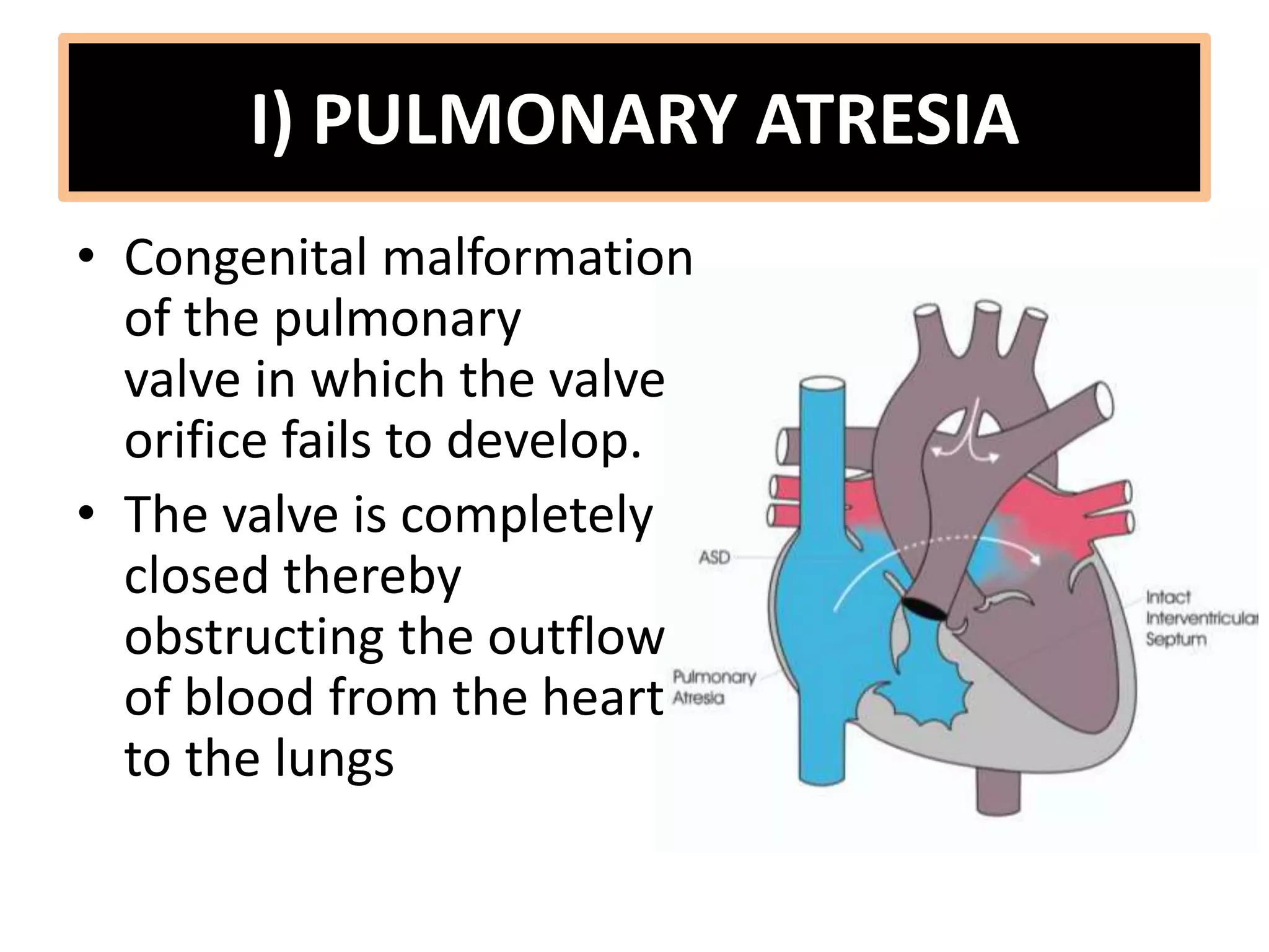
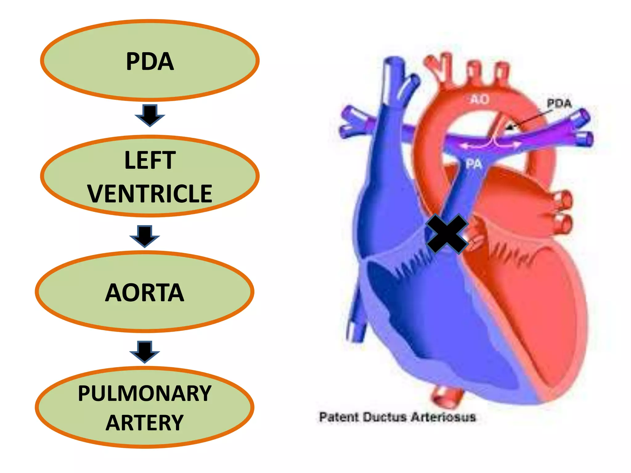
11) PATHOPHYSIOLOGY OF
DUCTAL DEPENDANT FOR
PULMONARY CIRCULATION CHD
Anatomy of Ductus Arteriosus
Connects the main
pulmonary artery to
descending aorta.
Physiology of Ductus Arteriosus
• Carries 60% of combined
vent. Output
• Diverts blood from high
resistance pulmonary
circulation to low resistance
descending aorta and
placental circulation.
• PGE1 and PGI2 formed
intramurally and in placenta
maintain ductal patency
in fetal life
Post Natal Closure of PDA
• Functional closure
In 12 hr., contraction of
medial smooth muscles
due to ↑ PO2 & ↓ PGE1.
• Anatomical closure
In 3 wk., replacement of
muscle fibres with fibrosis
creating ligamentum arteriosus.
• If ductal closure causes significant decrease in
systemic circulation, the condition is called
ductus dependent systemic blood flow
• If ductal closure causes significant decrease in
pulmonary circulation, the condition is called
ductus dependent pulmonary blood flow
BEFORE
BIRTH
AFTER
BIRTH
DUCTUS ARTERIOSUS
DEPENDENT
SYSTEMIC BLOOD
FLOW
PULMONARY
BLOOD FLOW
Lesions characterized by the
entire or part of
the systemic blood flow
depends solely on
the patency of the ductus
arteriosus.
– Coarctation of Aorta
(severe)
– Interrupted Aortic Arch
– Hypoplastic Left Heart
Hypoxic lesions characterized
by the pulmonary blood flow
depends solely on
the patency of the ductus
arteriosus.
– Pulmonary atresia
– Severe pulmonary
stenosis
– TOF with severe
pulmonary stenosis
DUCTUS ARTERIOSUS
DEPENDENT
PULMONARY BLOOD FLOW
I) PULMONARY ATRESIA
• Congenital malformation
of the pulmonary
valve in which the valve
orifice fails to develop.
• The valve is completely
closed thereby
obstructing the outflow
of blood from the heart
to the lungs
II) SEVERE PULMONARY STENOSIS
• Is a dynamic or fixed
obstruction of flow from
the right ventricle of the
heart to the pulmonary
artery.
• Usually due to isolated
valvular obstruction
(Pulmonary valve
stenosis), but may be due
to subvalvular or
supravalvular
obstruction.
III)TOF WITH SEVERE PULMONARY
STENOSIS
• Large VSD
• Overriding of the
aorta
• **Pulmonary stenosis
• RVH
***OBSTRUCTION OF
PULMONARY FLOW
RVH
RIGHT ATRIAL PRESSURE ↑
SYSTEMIC CYANOSIS
PERSISTENT OPENING
FORAMEN OVALE
Resistance Blood Flow
Fails
Shunting of unoxygenated blood from
the right atrium into the left atrium
PDA
LEFT
VENTRICLE
AORTA
PULMONARY
ARTERY

pathophysiology of ductal dependant for pulmonary

  • 1.
    11) PATHOPHYSIOLOGY OF DUCTALDEPENDANT FOR PULMONARY CIRCULATION CHD
  • 2.
    Anatomy of DuctusArteriosus Connects the main pulmonary artery to descending aorta.
  • 3.
    Physiology of DuctusArteriosus • Carries 60% of combined vent. Output • Diverts blood from high resistance pulmonary circulation to low resistance descending aorta and placental circulation. • PGE1 and PGI2 formed intramurally and in placenta maintain ductal patency in fetal life
  • 4.
    Post Natal Closureof PDA • Functional closure In 12 hr., contraction of medial smooth muscles due to ↑ PO2 & ↓ PGE1. • Anatomical closure In 3 wk., replacement of muscle fibres with fibrosis creating ligamentum arteriosus.
  • 5.
    • If ductalclosure causes significant decrease in systemic circulation, the condition is called ductus dependent systemic blood flow • If ductal closure causes significant decrease in pulmonary circulation, the condition is called ductus dependent pulmonary blood flow BEFORE BIRTH AFTER BIRTH
  • 6.
    DUCTUS ARTERIOSUS DEPENDENT SYSTEMIC BLOOD FLOW PULMONARY BLOODFLOW Lesions characterized by the entire or part of the systemic blood flow depends solely on the patency of the ductus arteriosus. – Coarctation of Aorta (severe) – Interrupted Aortic Arch – Hypoplastic Left Heart Hypoxic lesions characterized by the pulmonary blood flow depends solely on the patency of the ductus arteriosus. – Pulmonary atresia – Severe pulmonary stenosis – TOF with severe pulmonary stenosis
  • 7.
  • 8.
    I) PULMONARY ATRESIA •Congenital malformation of the pulmonary valve in which the valve orifice fails to develop. • The valve is completely closed thereby obstructing the outflow of blood from the heart to the lungs
  • 9.
    II) SEVERE PULMONARYSTENOSIS • Is a dynamic or fixed obstruction of flow from the right ventricle of the heart to the pulmonary artery. • Usually due to isolated valvular obstruction (Pulmonary valve stenosis), but may be due to subvalvular or supravalvular obstruction.
  • 10.
    III)TOF WITH SEVEREPULMONARY STENOSIS • Large VSD • Overriding of the aorta • **Pulmonary stenosis • RVH
  • 11.
    ***OBSTRUCTION OF PULMONARY FLOW RVH RIGHTATRIAL PRESSURE ↑ SYSTEMIC CYANOSIS PERSISTENT OPENING FORAMEN OVALE Resistance Blood Flow Fails Shunting of unoxygenated blood from the right atrium into the left atrium
  • 12.