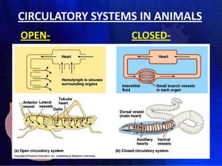
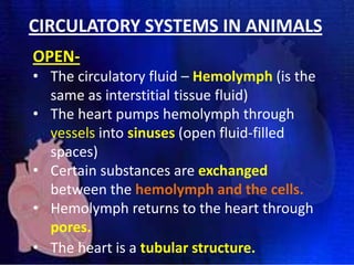
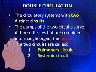
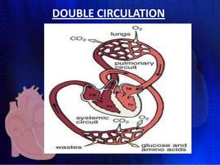
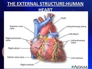
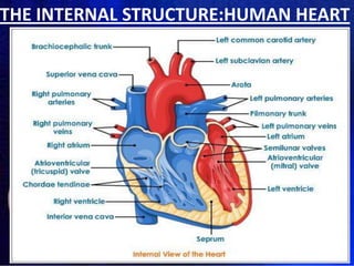
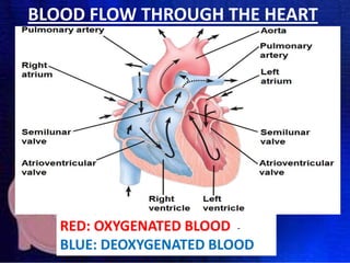
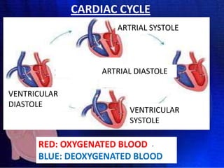
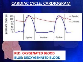
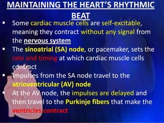
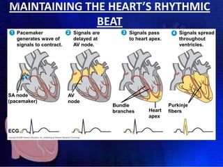
WELCOME BACK!!
• CONGRATULATIONS ON PASSING THE EXAM!
• BUT NOW KEEP UP THE GOOD WORK…IT’S
GOING TO BE A BUSY TERM!
• NEW LEARNER GUIDE – READ AND MAKE USE
OF IT!
• MAKE NOTE OF IMPORTANT DATES!
– PRAC 1 – NEXT TUESDAY, 24TH!
– CLASS TEST 1 – NEXT WEDNESDAY, 25TH!
NATIONAL SCIENCE WEEK
• Organised by the Department of Science &
Technology
• Launch hosted by UJ, Soweto Campus, 28th July
• Aims to excite youth with science at a young age!
• You are invited free of charge!
• Launch day: 07:00 – 15:00
• Register after class with ID number
• The rest of the week 14:30 – 15:30
POSTER PROJECT
•
•
•
•

GROUPS OF 5 – names on back of poster
A3 POSTER
ANY BIOLOGICAL TOPIC
INTERESTING, INFORMATIVE, PICTURES,
DIAGRAMS, COLOURFUL, FUN
• TARGET: High School Students
• DUE DATE: Next Tuesday, 24th July in class!!
TRANSPORT
SYSTEMS IN
ANIMALS
(Campbell and Reece (2010), Chapter
42: P.898-915)
CIRCULATORY SYSTEMS IN ANIMALS
 Many invertebrates do not have
a circulatory system at all.
 Their cells are close enough to
their environment for oxygen,
other gases, nutrients, and
waste products to simply diffuse
out of and into their cells.
 E.g. Hydra
CIRCULATORY SYSTEMS IN ANIMALS
• In animals with multiple layers of cells,
their cells are too far from the external
environment for simple osmosis and
diffusion.
• In higher animals, there are two
primary types of circulatory systems -open and closed.
CIRCULATORY SYSTEMS IN ANIMALS
OPEN-

CLOSED-
CIRCULATORY SYSTEMS IN ANIMALS
OPEN• The circulatory fluid – Hemolymph (is the
same as interstitial tissue fluid)
• The heart pumps hemolymph through
vessels into sinuses (open fluid-filled
spaces)
• Certain substances are exchanged
between the hemolymph and the cells.
• Hemolymph returns to the heart through
pores.
• The heart is a tubular structure.
CIRCULATORY SYSTEMS IN ANIMALS
CLOSED• Circulate blood entirely within
vessels.
• So the blood is distinct from the
interstitial fluid.
• Chemical exchange occurs between
the blood and interstitial fluid as
well as between interstitial fluid and
body cells.
DOUBLE CIRCULATION
• The circulatory systems with two
distinct circuits.
• The pumps of the two circuits serve
different tissues but are combined
into a single organ, the heart.
• The two circuits are called:
1. Pulmonary circuit
2. Systemic circuit
DOUBLE CIRCULATION
1. Pulmonary circuit
The heart pumps deoxygenated blood
to the lungs and oxygenated blood
back to the heart.
2. Systemic circuit
The heart pumps oxygenated blood to
the body cells and the deoxygenated
blood back to the heart.
DOUBLE CIRCULATION
DIFFERENT BLOOD VESSELS:
ARTERIES, VEINS AND CAPILLARIES
FUNCTIONS OF CAPILLARIES
Capillaries: Connect arteries and
veins – are structurally suited to
facilitate the exchange of
substances between the blood in the
capillaries and the interstitial tissue
fluid
FUNCTIONS OF ARTERIES
Arteries: transport oxygenated blood except
for the pulmonary artery. Main artery is called
the Aorta – pumping oxygenated blood from
heart to rest of body.
Other arteries:
Renal artery (kidney)
Hepatic artery (liver)
FUNCTIONS OF VEINS
Veins : transports deoxygenated blood except
for the pulmonary vein. Main vein is called the
vena cava – pumping deoxygenated blood
towards the heart from the rest of the body.
Other veins:
Renal vein (kidney)
Hepatic vein (liver)
THE HUMAN CIRCULATORY SYSTEM:
HEART AND ASSOCIATED VESSELS
THE HUMAN CIRCULATORY SYSTEM:
HEART AND ASSOCIATED VESSELS
 Mammals have a four-chambered heart with two
atria and two ventricles
 The left side of the heart pumps and receives only
oxygen-rich blood, while the right side receives
and pumps only oxygen-poor blood
 The mammalian cardiovascular system meets the
body’s continuous demand for O2.
 Blood begins its flow when deoxygenated blood
flow from the body into the right atrium.
 Blood then flows from the right atrium into the
right ventricle through the tricuspid valve
 The blood is then pumped into lungs, through the
semilunar valve via the pulmonary artery.
 In the lungs, the blood loads O2 and unloads CO2.
 Oxygen-rich blood from the lungs enters the heart
via the pulmonary vein at the left atrium
 Blood then flows into the left ventricle through the
bicuspid valve.
 This blood is then pumped through semilunar
valve into the aorta with takes blood to the entire
body.
 Deoxygenated blood returns to the heart through
the superior vena cava (blood from head, neck, and
forelimbs) and inferior vena cava (blood from
trunk and hind limbs)
 The superior vena cava and inferior vena cava flow
into the right atrium.
 The atrioventricular (AV) valves (tricuspid and
bicuspid valves) separate each atrium and ventricle
 The semilunar valves control blood flow to the
aorta and the pulmonary artery.
THE EXTERNAL STRUCTURE:HUMAN
HEART
THE INTERNAL STRUCTURE:HUMAN HEART
BLOOD FLOW THROUGH THE HEART

RED: OXYGENATED BLOOD BLUE: DEOXYGENATED BLOOD
CARDIAC CYCLE
 The heart contracts and relaxes in a
rhythmic cycle called the cardiac cycle
 The contraction, or pumping, phase is
called systole
 The relaxation, or filling, phase is called
diastole
 The heart rate, also called the pulse, is
the number of beats per minute
CARDIAC CYCLE
ARTRIAL SYSTOLE

ARTRIAL DIASTOLE
VENTRICULAR
DIASTOLE

VENTRICULAR
SYSTOLE

RED: OXYGENATED BLOOD BLUE: DEOXYGENATED BLOOD
CARDIAC CYCLE: CARDIOGRAM

RED: OXYGENATED BLOOD BLUE: DEOXYGENATED BLOOD
MAINTAINING THE HEART’S RHYTHMIC
BEAT

 Some cardiac muscle cells are self-excitable,
meaning they contract without any signal from
the nervous system
 The sinoatrial (SA) node, or pacemaker, sets the
rate and timing at which cardiac muscle cells
contract
 Impulses from the SA node travel to the
atrioventricular (AV) node
 At the AV node, the impulses are delayed and
then travel to the Purkinje fibers that make the
ventricles contract
MAINTAINING THE HEART’S RHYTHMIC
BEAT

Fig. 42-9-5

1 Pacemaker

generates wave of
signals to contract.

SA node
(pacemaker)

ECG

2 Signals are

delayed at
AV node.

AV
node

3 Signals pass

to heart apex.

Bundle
branches

Heart
apex

4 Signals spread

throughout
ventricles.

Purkinje
fibers
WHAT WILL INFLUENCE THE HEART’S
RHYTHMIC BEAT?
LYMPHATIC SYSTEM
 The lymphatic system returns fluid that leaks
out in the capillary beds
 This system aids in body defence
 Fluid, called lymph, re-enters the circulation
directly at the venous end of the capillary bed
and indirectly through the lymphatic system
 The lymphatic system drains into veins in the
neck.
 Lymph nodes are organs that filter lymph and
play an important role in the body’s defence
 Edema is swelling caused by disruptions in the
flow of lymph.
LYMPHATIC SYSTEM
LYMPHATIC SYSTEM VS. CIRCULATORY -
DISEASES OF THE HEART AND
CIRCULATORY SYSTEM

 Cardiovascular diseases are disorders of the
heart and the blood vessels
 They account for more than half the deaths in
the South Africa
 One type of cardiovascular disease,
atherosclerosis, is caused by the build-up of
plaque (fat) deposits within arteries.
 A heart attack is the death of cardiac muscle
tissue resulting from blockage of one or more
coronary arteries
 A stroke is the death of nervous tissue in the
brain, usually resulting from rupture or blockage
of arteries in the head.
 Hypertension, or high blood pressure, promotes
atherosclerosis and increases the risk of heart
attack and stroke.
 Hypertension can be reduced by dietary
changes, exercise, and/or medication

Unit 1 transport systems in animals

  • 1.
    WELCOME BACK!! • CONGRATULATIONSON PASSING THE EXAM! • BUT NOW KEEP UP THE GOOD WORK…IT’S GOING TO BE A BUSY TERM! • NEW LEARNER GUIDE – READ AND MAKE USE OF IT! • MAKE NOTE OF IMPORTANT DATES! – PRAC 1 – NEXT TUESDAY, 24TH! – CLASS TEST 1 – NEXT WEDNESDAY, 25TH!
  • 2.
    NATIONAL SCIENCE WEEK •Organised by the Department of Science & Technology • Launch hosted by UJ, Soweto Campus, 28th July • Aims to excite youth with science at a young age! • You are invited free of charge! • Launch day: 07:00 – 15:00 • Register after class with ID number • The rest of the week 14:30 – 15:30
  • 3.
    POSTER PROJECT • • • • GROUPS OF5 – names on back of poster A3 POSTER ANY BIOLOGICAL TOPIC INTERESTING, INFORMATIVE, PICTURES, DIAGRAMS, COLOURFUL, FUN • TARGET: High School Students • DUE DATE: Next Tuesday, 24th July in class!!
  • 4.
    TRANSPORT SYSTEMS IN ANIMALS (Campbell andReece (2010), Chapter 42: P.898-915)
  • 5.
    CIRCULATORY SYSTEMS INANIMALS  Many invertebrates do not have a circulatory system at all.  Their cells are close enough to their environment for oxygen, other gases, nutrients, and waste products to simply diffuse out of and into their cells.  E.g. Hydra
  • 6.
    CIRCULATORY SYSTEMS INANIMALS • In animals with multiple layers of cells, their cells are too far from the external environment for simple osmosis and diffusion. • In higher animals, there are two primary types of circulatory systems -open and closed.
  • 7.
    CIRCULATORY SYSTEMS INANIMALS OPEN- CLOSED-
  • 8.
    CIRCULATORY SYSTEMS INANIMALS OPEN• The circulatory fluid – Hemolymph (is the same as interstitial tissue fluid) • The heart pumps hemolymph through vessels into sinuses (open fluid-filled spaces) • Certain substances are exchanged between the hemolymph and the cells. • Hemolymph returns to the heart through pores. • The heart is a tubular structure.
  • 9.
    CIRCULATORY SYSTEMS INANIMALS CLOSED• Circulate blood entirely within vessels. • So the blood is distinct from the interstitial fluid. • Chemical exchange occurs between the blood and interstitial fluid as well as between interstitial fluid and body cells.
  • 10.
    DOUBLE CIRCULATION • Thecirculatory systems with two distinct circuits. • The pumps of the two circuits serve different tissues but are combined into a single organ, the heart. • The two circuits are called: 1. Pulmonary circuit 2. Systemic circuit
  • 11.
    DOUBLE CIRCULATION 1. Pulmonarycircuit The heart pumps deoxygenated blood to the lungs and oxygenated blood back to the heart. 2. Systemic circuit The heart pumps oxygenated blood to the body cells and the deoxygenated blood back to the heart.
  • 12.
  • 13.
  • 14.
    FUNCTIONS OF CAPILLARIES Capillaries:Connect arteries and veins – are structurally suited to facilitate the exchange of substances between the blood in the capillaries and the interstitial tissue fluid
  • 15.
    FUNCTIONS OF ARTERIES Arteries:transport oxygenated blood except for the pulmonary artery. Main artery is called the Aorta – pumping oxygenated blood from heart to rest of body. Other arteries: Renal artery (kidney) Hepatic artery (liver)
  • 16.
    FUNCTIONS OF VEINS Veins: transports deoxygenated blood except for the pulmonary vein. Main vein is called the vena cava – pumping deoxygenated blood towards the heart from the rest of the body. Other veins: Renal vein (kidney) Hepatic vein (liver)
  • 17.
    THE HUMAN CIRCULATORYSYSTEM: HEART AND ASSOCIATED VESSELS
  • 18.
    THE HUMAN CIRCULATORYSYSTEM: HEART AND ASSOCIATED VESSELS  Mammals have a four-chambered heart with two atria and two ventricles  The left side of the heart pumps and receives only oxygen-rich blood, while the right side receives and pumps only oxygen-poor blood  The mammalian cardiovascular system meets the body’s continuous demand for O2.  Blood begins its flow when deoxygenated blood flow from the body into the right atrium.
  • 19.
     Blood thenflows from the right atrium into the right ventricle through the tricuspid valve  The blood is then pumped into lungs, through the semilunar valve via the pulmonary artery.  In the lungs, the blood loads O2 and unloads CO2.  Oxygen-rich blood from the lungs enters the heart via the pulmonary vein at the left atrium  Blood then flows into the left ventricle through the bicuspid valve.  This blood is then pumped through semilunar valve into the aorta with takes blood to the entire body.
  • 20.
     Deoxygenated bloodreturns to the heart through the superior vena cava (blood from head, neck, and forelimbs) and inferior vena cava (blood from trunk and hind limbs)  The superior vena cava and inferior vena cava flow into the right atrium.  The atrioventricular (AV) valves (tricuspid and bicuspid valves) separate each atrium and ventricle  The semilunar valves control blood flow to the aorta and the pulmonary artery.
  • 21.
  • 22.
  • 23.
    BLOOD FLOW THROUGHTHE HEART RED: OXYGENATED BLOOD BLUE: DEOXYGENATED BLOOD
  • 24.
    CARDIAC CYCLE  Theheart contracts and relaxes in a rhythmic cycle called the cardiac cycle  The contraction, or pumping, phase is called systole  The relaxation, or filling, phase is called diastole  The heart rate, also called the pulse, is the number of beats per minute
  • 25.
    CARDIAC CYCLE ARTRIAL SYSTOLE ARTRIALDIASTOLE VENTRICULAR DIASTOLE VENTRICULAR SYSTOLE RED: OXYGENATED BLOOD BLUE: DEOXYGENATED BLOOD
  • 26.
    CARDIAC CYCLE: CARDIOGRAM RED:OXYGENATED BLOOD BLUE: DEOXYGENATED BLOOD
  • 27.
    MAINTAINING THE HEART’SRHYTHMIC BEAT  Some cardiac muscle cells are self-excitable, meaning they contract without any signal from the nervous system  The sinoatrial (SA) node, or pacemaker, sets the rate and timing at which cardiac muscle cells contract  Impulses from the SA node travel to the atrioventricular (AV) node  At the AV node, the impulses are delayed and then travel to the Purkinje fibers that make the ventricles contract
  • 28.
    MAINTAINING THE HEART’SRHYTHMIC BEAT Fig. 42-9-5 1 Pacemaker generates wave of signals to contract. SA node (pacemaker) ECG 2 Signals are delayed at AV node. AV node 3 Signals pass to heart apex. Bundle branches Heart apex 4 Signals spread throughout ventricles. Purkinje fibers
  • 29.
    WHAT WILL INFLUENCETHE HEART’S RHYTHMIC BEAT?
  • 30.
    LYMPHATIC SYSTEM  Thelymphatic system returns fluid that leaks out in the capillary beds  This system aids in body defence  Fluid, called lymph, re-enters the circulation directly at the venous end of the capillary bed and indirectly through the lymphatic system  The lymphatic system drains into veins in the neck.  Lymph nodes are organs that filter lymph and play an important role in the body’s defence  Edema is swelling caused by disruptions in the flow of lymph.
  • 31.
  • 32.
    LYMPHATIC SYSTEM VS.CIRCULATORY -
  • 33.
    DISEASES OF THEHEART AND CIRCULATORY SYSTEM  Cardiovascular diseases are disorders of the heart and the blood vessels  They account for more than half the deaths in the South Africa  One type of cardiovascular disease, atherosclerosis, is caused by the build-up of plaque (fat) deposits within arteries.
  • 34.
     A heartattack is the death of cardiac muscle tissue resulting from blockage of one or more coronary arteries  A stroke is the death of nervous tissue in the brain, usually resulting from rupture or blockage of arteries in the head.  Hypertension, or high blood pressure, promotes atherosclerosis and increases the risk of heart attack and stroke.  Hypertension can be reduced by dietary changes, exercise, and/or medication