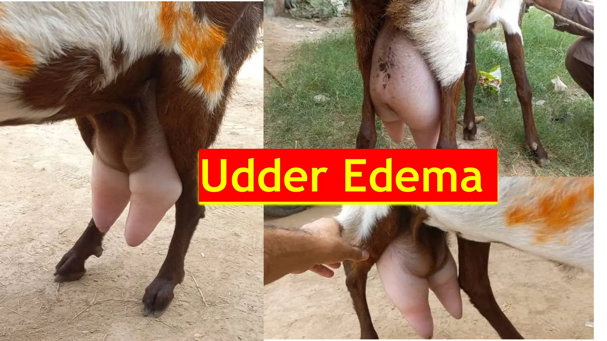
1
Udder edema
• Also called as caked udder.
• It is mostly occurring in Dairy aniamls.
• Odder oedema can be defined as ;
• The accumulation of fluid in the Interstitial tissue spaces.
• It is most serious condition which affects world dairy industry.
• The swollen udder is uncomfortable
• And prone to injury and mastitis.
2
• It makes milking difficult.
• Udder suspensory ligaments become permanently damaged.
• Udder oedema affects mostly;
• Cattle
• Buffaloes
• Sheep
• goats
3
Occurrence ;;
• Udder oedema usually occurs near to parturition.
• Its affects mostly primiparous animals.
• Also affects older cow/does.
• Its affects mostly higher milk producing animals.
• Especially those animals with pendulous udder.
4
Ecnomic impertance ;;
• Udder oedema may cause economic losses in dairy cattle/goats.
• Decrease in milk production b/c of pain.
• It is difficult to milk the oedematous udder properly.
• The oedema can cause permanent damage to udder ligaments.
• subsequent attacks of udder oedema may cause udder skin necrosis,
mostly seen in goats
5
Causes ;;
• The exact cause of udder oedema is unknown.
• Studies shown that udder oedema at parturition results from
decreased blood flow associated with an increased venous blood
pressure in cranial superficial epigastric vein.
6
Inheritance ;;
• The underlying predisposition to udder oedema is hereditary.
• The Alpine goat breed have severe udder oedema.
• A genetic study suggested that there is a significant positive
correlation with milk yield in cow/goat with udder oedema.
• Selection by progeny testing for oedema' could be effective.
7
Mammary blood and lymph flow
• 3-fold increase in mammary blood flow from two weeks before
parturition
• Animals with udder oedema had a 17.1% lower mammary blood flow
than without udder oedema at parturition.
• So mammary blood flow changes are a factor in the development of
udder 'oedema at calving/kidding.
8
Venous blood pressure ;
• Significant increase in the venous blood pressure in the superficial
cranial milk veins associated with the development of udder oedema
.
• Stasis of venous blood or lymph flow during gestation because of
pressure by the gravid uterus been proposed as a cause of udder
oedema.
9
Clinical Signs .;;
• The acute, or physiological form near parturition,
• Chronic or pathological form during lactation.
• This classification is based on the presence of clinical signs of udder.
10
• Clinically, udder oedema has two distinct
stages
• During the first stage
 Gradual congestion of udder.
 The udder becomes greatly
distended,
 Swollen
 filled with colostrum.
11
• During the second stage,
• Digital pressure produces pitting of the oedematous areas, which lasts for
several minutes and is characteristic.
• The udder skin thick and hard on digital palpation.
12
Diagnosis .;
• The oedema is symmetrical,
• involving both udder halves,
• with pitting on pressure,
• absence of heat,
• occurrence near parturition.
13
D.daignosis;
• The condition should be differentiated from
• Hematomas of the udder,
• Ventral abdominal hernia,
• Mastitis
• Considerations must be given to the history of the condition, the
nature of the enlargement, the consistency of tissues involved, and
the type of fluid present.
14
Treatment ;;
• In most cases, udder oedema does not need any treatment.
• The swelling gradually decreases after calving.
• A 20-minute massage, three times daily, together with the alternate
application of hot and cold water.
• Mild exercise
15
• Rubbing the udder with 200 mg diethylstilbestrol in 10 ml corn oil on
the 1st and 3rd days after calving gave favorable results.
• Diuretics have been used extensively.
• Furosemide @500 mg once daily or 250 mg twice daily
• Not to exceed 48 hours post-partum (for udder edema)
16
Prevention and Control
• The condition should be eliminated by selective breeding .
• Udder oedema may be prevented to some extent by good
management at parturition, including moderate exercise
• A mildly laxative in feed.
• Restriction of salts near to parturition/kidding
• Use of udder supports and massage .
17
• Induction of parturition using dexamethasone and estradiol benzoate
injections demonstrated a significant lowering of the incidence of
udder oedema.
• Decreasing the potassium content in feed
18

udder edema in goats

  • 1.
  • 2.
    Udder edema • Alsocalled as caked udder. • It is mostly occurring in Dairy aniamls. • Odder oedema can be defined as ; • The accumulation of fluid in the Interstitial tissue spaces. • It is most serious condition which affects world dairy industry. • The swollen udder is uncomfortable • And prone to injury and mastitis. 2
  • 3.
    • It makesmilking difficult. • Udder suspensory ligaments become permanently damaged. • Udder oedema affects mostly; • Cattle • Buffaloes • Sheep • goats 3
  • 4.
    Occurrence ;; • Udderoedema usually occurs near to parturition. • Its affects mostly primiparous animals. • Also affects older cow/does. • Its affects mostly higher milk producing animals. • Especially those animals with pendulous udder. 4
  • 5.
    Ecnomic impertance ;; •Udder oedema may cause economic losses in dairy cattle/goats. • Decrease in milk production b/c of pain. • It is difficult to milk the oedematous udder properly. • The oedema can cause permanent damage to udder ligaments. • subsequent attacks of udder oedema may cause udder skin necrosis, mostly seen in goats 5
  • 6.
    Causes ;; • Theexact cause of udder oedema is unknown. • Studies shown that udder oedema at parturition results from decreased blood flow associated with an increased venous blood pressure in cranial superficial epigastric vein. 6
  • 7.
    Inheritance ;; • Theunderlying predisposition to udder oedema is hereditary. • The Alpine goat breed have severe udder oedema. • A genetic study suggested that there is a significant positive correlation with milk yield in cow/goat with udder oedema. • Selection by progeny testing for oedema' could be effective. 7
  • 8.
    Mammary blood andlymph flow • 3-fold increase in mammary blood flow from two weeks before parturition • Animals with udder oedema had a 17.1% lower mammary blood flow than without udder oedema at parturition. • So mammary blood flow changes are a factor in the development of udder 'oedema at calving/kidding. 8
  • 9.
    Venous blood pressure; • Significant increase in the venous blood pressure in the superficial cranial milk veins associated with the development of udder oedema . • Stasis of venous blood or lymph flow during gestation because of pressure by the gravid uterus been proposed as a cause of udder oedema. 9
  • 10.
    Clinical Signs .;; •The acute, or physiological form near parturition, • Chronic or pathological form during lactation. • This classification is based on the presence of clinical signs of udder. 10
  • 11.
    • Clinically, udderoedema has two distinct stages • During the first stage  Gradual congestion of udder.  The udder becomes greatly distended,  Swollen  filled with colostrum. 11
  • 12.
    • During thesecond stage, • Digital pressure produces pitting of the oedematous areas, which lasts for several minutes and is characteristic. • The udder skin thick and hard on digital palpation. 12
  • 13.
    Diagnosis .; • Theoedema is symmetrical, • involving both udder halves, • with pitting on pressure, • absence of heat, • occurrence near parturition. 13
  • 14.
    D.daignosis; • The conditionshould be differentiated from • Hematomas of the udder, • Ventral abdominal hernia, • Mastitis • Considerations must be given to the history of the condition, the nature of the enlargement, the consistency of tissues involved, and the type of fluid present. 14
  • 15.
    Treatment ;; • Inmost cases, udder oedema does not need any treatment. • The swelling gradually decreases after calving. • A 20-minute massage, three times daily, together with the alternate application of hot and cold water. • Mild exercise 15
  • 16.
    • Rubbing theudder with 200 mg diethylstilbestrol in 10 ml corn oil on the 1st and 3rd days after calving gave favorable results. • Diuretics have been used extensively. • Furosemide @500 mg once daily or 250 mg twice daily • Not to exceed 48 hours post-partum (for udder edema) 16
  • 17.
    Prevention and Control •The condition should be eliminated by selective breeding . • Udder oedema may be prevented to some extent by good management at parturition, including moderate exercise • A mildly laxative in feed. • Restriction of salts near to parturition/kidding • Use of udder supports and massage . 17
  • 18.
    • Induction ofparturition using dexamethasone and estradiol benzoate injections demonstrated a significant lowering of the incidence of udder oedema. • Decreasing the potassium content in feed 18