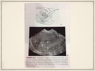
Transvaginal ultrasound (TVS) can be used for early first trimester obstetric ultrasounds, evaluating complications in later pregnancy like placenta previa, assessing the fetal spine position, and evaluating the nuchal region before 14 weeks. It is also used to evaluate suspected ectopic pregnancies, monitor follicle development during fertility treatments, and monitor ovarian response during assisted reproductive technology cycles. The procedure involves first performing a transabdominal ultrasound with a full bladder, then emptying the bladder and inserting the transvaginal probe to obtain images of the cervix, uterus, ovaries, and surrounding structures in both the sagittal and coronal planes. Key anatomical structures like the uterus and ovaries are described in