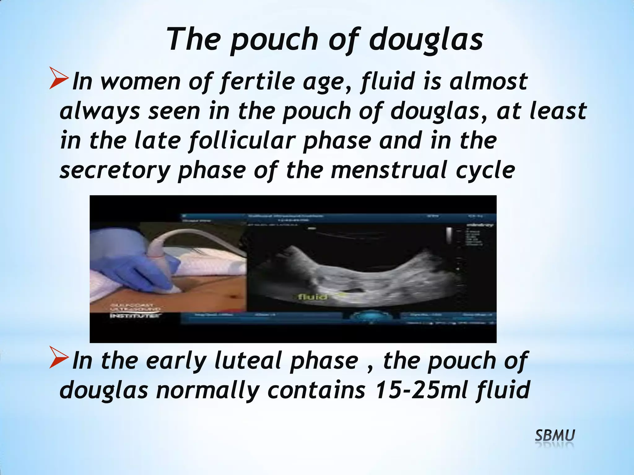
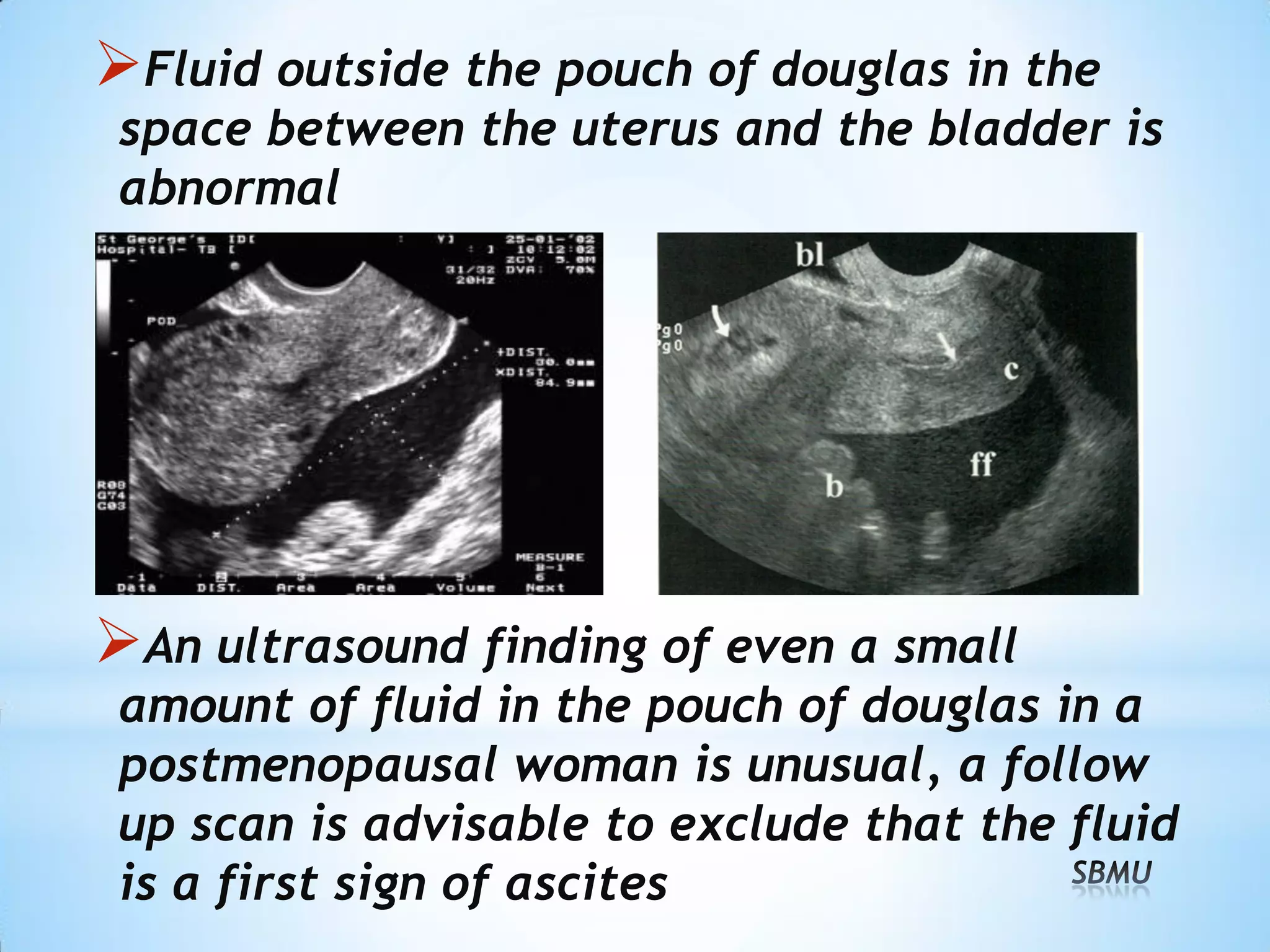
Normal Pelvic Ultrasound
Nazari L,
Assistant professor OB/GYN
Shahid Beheshti University of Medical Sciences
Spring 2016
Introduction
Chief complaint
Symptoms
Check duration of symptoms
Any ultrasound done previously. Check
the records carefully
 LMP
 Any tests done and their reports
 Referring doctors requisition slip
Female Anatomy Review
Uterus: Size, shape, mobility and probe
tenderness
Endometrium: Thickness and morphology.
Any focal abnormality
 Myometrium : Echo pattern and presence of
fibroids and their location
Ovaries: Size and echo pattern. Any
abnormality to be mentioned in terms of size,
echo pattern, walls and focal abnormalities
within it
 Extra-ovarian adnexal
areas: Report whether
any mass is delineated
or not
 Free fluid or fluid in the
pouch of Douglas or adnexa
Transabdominal approach :
•Lower frequency
•Lower resolution image
•Better visualized with full bladder
Transvaginal approach:
•Higher frequency
•higher resolution image
•Better visualized with empty bladder
Indications of TVS
Gynecologic sonography
Early first trimester obstetric
Later OB: Cervix, placenta previa and
evaluation of fetus before 14 weeks’
Suspected EP
Suspected lower abdominal disease in whom
a diagnosis has not been made with TAS
Follicle monitoring
Monitoring for ART
Preparation for TVS
Empty bladder
Explain to the patient
Consent ( Verbal / written)
Be sure about virginity!
Chaperone
Privacy ; Cover the Patient with a sheet
Supine position , bending knees
Anterior limitation : Flex her hips and bring
her knees up toward her chest
Sagittal
Coronal
Uterus
Is a reliable landmark because of its
central location, relatively large size
and the well-known pear-shape
The cervix is less mobile than the
uterine body due to uterosacral
ligaments that position the cervix in
the midline of the pelvis
Uterus
Size: varies with age:
 Neonate: Relatively Large, Body> cervix
 Childhood: Tubular, Body< cervix
 Menarche: Large, Body> Cervix
 Post menarche: Body = 2x Cervix
 Reproductive age: 80x40x40 mm
 Multiparous : +10 mm in each dimension
Version : Angle of the cervix to the vaginal
Flexion: Angle of body of the uterus at the
isthmus
Cysts within the cervix, termed nabothian
cysts, occur frequently
Result from the retention of mucus within
obstructed endocervical glands
Myometrium
•Myometrium is moderately hypoechoic,
especially in relation to the more echogenic
endometrium
•Normal myometrium is homogeneous in
echotexture
Endometrium
Endometrium is the innermost layer of the
uterus that lines the uterine cavity
There is often blood and shed tissue in the
uterine cavity during mensturation
During the late proliferative phase, the
endometrium (calipers) has a multilayered
appearance: echogenic around its periphery
and in the midline, and hypoechoic in between
During the secretory phase, the endometrium
is thick and echogenic
Measurement of the endometrial thickness is
useful in a number of clinical settings
This measurement is best obtained from a
sagittal transvaginal sonographic image,
after sweeping through the uterus to find the
site of maximal thickness
Excluding any fluid that may be present in
the endometrial cavity
Fallopian tubes
Interstitial portion :
seen as a hyperechoic line
extending from the
lateral uterine angle to
the origin of the broad ligament
Isthmus and ampulla : rarely seen without
the use of contrast media
Infundibulum can be seen if it is floating in
peritoneal fluid
Interestitial portion of tube
Fimbria of the tube surrounded by fluid
Ovaries
Iliac vessels are a reliable landmark for
their visualization
Due to mobility of the ovaries and
transducer pressure, position of the ovaries
is often varying for example:
• Cul-de-sac
• In front of the uterus
• Above the uterus
• In the abdominal cavity
Ovaries
Ovaries are imaged as homogeneous,
hypoechogenic ovoid structures with
slightly echogenic central part
Antral follicles 5-12 per ovary
The pouch of douglas
In women of fertile age, fluid is almost
always seen in the pouch of douglas, at least
in the late follicular phase and in the
secretory phase of the menstrual cycle
In the early luteal phase , the pouch of
douglas normally contains 15-25ml fluid
Fluid outside the pouch of douglas in the
space between the uterus and the bladder is
abnormal
An ultrasound finding of even a small
amount of fluid in the pouch of douglas in a
postmenopausal woman is unusual, a follow
up scan is advisable to exclude that the fluid
is a first sign of ascites
Dr.Nazari.Normal_Pelvic_Ultrasound.pdf

Dr.Nazari.Normal_Pelvic_Ultrasound.pdf

  • 1.
    Normal Pelvic Ultrasound NazariL, Assistant professor OB/GYN Shahid Beheshti University of Medical Sciences Spring 2016
  • 2.
    Introduction Chief complaint Symptoms Check durationof symptoms Any ultrasound done previously. Check the records carefully  LMP  Any tests done and their reports  Referring doctors requisition slip
  • 3.
  • 4.
    Uterus: Size, shape,mobility and probe tenderness Endometrium: Thickness and morphology. Any focal abnormality  Myometrium : Echo pattern and presence of fibroids and their location
  • 5.
    Ovaries: Size andecho pattern. Any abnormality to be mentioned in terms of size, echo pattern, walls and focal abnormalities within it  Extra-ovarian adnexal areas: Report whether any mass is delineated or not  Free fluid or fluid in the pouch of Douglas or adnexa
  • 6.
    Transabdominal approach : •Lowerfrequency •Lower resolution image •Better visualized with full bladder
  • 7.
    Transvaginal approach: •Higher frequency •higherresolution image •Better visualized with empty bladder
  • 10.
    Indications of TVS Gynecologicsonography Early first trimester obstetric Later OB: Cervix, placenta previa and evaluation of fetus before 14 weeks’ Suspected EP Suspected lower abdominal disease in whom a diagnosis has not been made with TAS Follicle monitoring Monitoring for ART
  • 11.
    Preparation for TVS Emptybladder Explain to the patient Consent ( Verbal / written) Be sure about virginity! Chaperone Privacy ; Cover the Patient with a sheet Supine position , bending knees Anterior limitation : Flex her hips and bring her knees up toward her chest
  • 12.
  • 13.
  • 14.
    Uterus Is a reliablelandmark because of its central location, relatively large size and the well-known pear-shape The cervix is less mobile than the uterine body due to uterosacral ligaments that position the cervix in the midline of the pelvis
  • 15.
    Uterus Size: varies withage:  Neonate: Relatively Large, Body> cervix  Childhood: Tubular, Body< cervix  Menarche: Large, Body> Cervix  Post menarche: Body = 2x Cervix  Reproductive age: 80x40x40 mm  Multiparous : +10 mm in each dimension
  • 16.
    Version : Angleof the cervix to the vaginal
  • 17.
    Flexion: Angle ofbody of the uterus at the isthmus
  • 18.
    Cysts within thecervix, termed nabothian cysts, occur frequently Result from the retention of mucus within obstructed endocervical glands
  • 19.
    Myometrium •Myometrium is moderatelyhypoechoic, especially in relation to the more echogenic endometrium •Normal myometrium is homogeneous in echotexture
  • 20.
    Endometrium Endometrium is theinnermost layer of the uterus that lines the uterine cavity There is often blood and shed tissue in the uterine cavity during mensturation
  • 21.
    During the lateproliferative phase, the endometrium (calipers) has a multilayered appearance: echogenic around its periphery and in the midline, and hypoechoic in between
  • 22.
    During the secretoryphase, the endometrium is thick and echogenic
  • 23.
    Measurement of theendometrial thickness is useful in a number of clinical settings This measurement is best obtained from a sagittal transvaginal sonographic image, after sweeping through the uterus to find the site of maximal thickness Excluding any fluid that may be present in the endometrial cavity
  • 24.
    Fallopian tubes Interstitial portion: seen as a hyperechoic line extending from the lateral uterine angle to the origin of the broad ligament Isthmus and ampulla : rarely seen without the use of contrast media Infundibulum can be seen if it is floating in peritoneal fluid
  • 25.
  • 26.
    Fimbria of thetube surrounded by fluid
  • 27.
    Ovaries Iliac vessels area reliable landmark for their visualization Due to mobility of the ovaries and transducer pressure, position of the ovaries is often varying for example: • Cul-de-sac • In front of the uterus • Above the uterus • In the abdominal cavity
  • 28.
    Ovaries Ovaries are imagedas homogeneous, hypoechogenic ovoid structures with slightly echogenic central part Antral follicles 5-12 per ovary
  • 29.
    The pouch ofdouglas In women of fertile age, fluid is almost always seen in the pouch of douglas, at least in the late follicular phase and in the secretory phase of the menstrual cycle In the early luteal phase , the pouch of douglas normally contains 15-25ml fluid
  • 30.
    Fluid outside thepouch of douglas in the space between the uterus and the bladder is abnormal An ultrasound finding of even a small amount of fluid in the pouch of douglas in a postmenopausal woman is unusual, a follow up scan is advisable to exclude that the fluid is a first sign of ascites