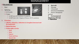
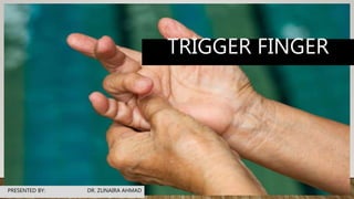
TRIGGER FINGER
PRESENTED BY: DR. ZUNAIRA AHMAD
DEFINITION
• Inflammation and subsequent narrowing of pulley of
affected digit (most commonlyA1) Typically 3rd, 4th digit
• [Annular (A1-A5) ligaments of the tendon sheath
over the flexor tendons]
• Flexor tendon / tendon sheath thickens
• The main characteristic of trigger finger is popping and
/ or catching with movement of the digit especially at
bending.
CLINICAL PRESENTATION
• Initially, patients may present with painless clicking during movement of the digit. This can progress to
painful catching or popping, typically at the MCP or PIP joints.
• Possible additional symptoms are:
• Stiffness and swelling (especially in the morning)
• Intermittent finger locking during active flexion that requires a passive force to extend the finger
• Finger locking or clicking in a bent position then popping straight
• Loss of full flexion / extension
• Palpable painful nodule proximal to A1, and / or finger locked into a flexed position
DIFFERENTIAL DIAGNOSIS
• Dupuytren's contracture
• Flexor tendon / sheath tumor.
• Sesamoid bone anomalies
• Post-traumatic tendon entrapment on the metacarpal head
• History
• Past Medical History
 Diabetic individuals are four times more likely to develop trigger finger
 RA and Gout are also associated with trigger finger
• Observation
 A digit locked in flexion
 Bony proliferative changes in the sub-adjacent PIP joint
• Palpation
• Range of Movement (Loss of motion, particularly in extension)
• Manual Muscle Testing
• Joint Accessory Mobility (PIP, MCP, DIP, and CMC of all affected digits)
• Special Tests (Open and Close hand)
EXAMINATION
Physiotherapy
1. Rest
2. Splinting
 6-10 weeks
 2 types
 At DIP joint (50% resolution of symptoms)
 At MCP joint with 15 degrees of flexion. (92.9% resolution)
3. Exercises
 AROM ( abduction, Adduction to strengthen interossei and
the lumbricals)
 Digital blocking
 Tendon glide exercise
 Duck
 Hook
 Squeeze
 Touch and Trace
 Spread wide
MANAGEMENT
4 Hot/cold
5 Massage
6 Ultrasound
7 Electrical stimulation
8 Passive therapeutic
traction
9 Extracorporeal
Shockwave Therapy
 Operative
Trigger finger/ Stenosing tenosynovitis

Trigger finger/ Stenosing tenosynovitis

  • 1.
  • 2.
    DEFINITION • Inflammation andsubsequent narrowing of pulley of affected digit (most commonlyA1) Typically 3rd, 4th digit • [Annular (A1-A5) ligaments of the tendon sheath over the flexor tendons] • Flexor tendon / tendon sheath thickens • The main characteristic of trigger finger is popping and / or catching with movement of the digit especially at bending.
  • 3.
    CLINICAL PRESENTATION • Initially,patients may present with painless clicking during movement of the digit. This can progress to painful catching or popping, typically at the MCP or PIP joints. • Possible additional symptoms are: • Stiffness and swelling (especially in the morning) • Intermittent finger locking during active flexion that requires a passive force to extend the finger • Finger locking or clicking in a bent position then popping straight • Loss of full flexion / extension • Palpable painful nodule proximal to A1, and / or finger locked into a flexed position
  • 4.
    DIFFERENTIAL DIAGNOSIS • Dupuytren'scontracture • Flexor tendon / sheath tumor. • Sesamoid bone anomalies • Post-traumatic tendon entrapment on the metacarpal head
  • 5.
    • History • PastMedical History  Diabetic individuals are four times more likely to develop trigger finger  RA and Gout are also associated with trigger finger • Observation  A digit locked in flexion  Bony proliferative changes in the sub-adjacent PIP joint • Palpation • Range of Movement (Loss of motion, particularly in extension) • Manual Muscle Testing • Joint Accessory Mobility (PIP, MCP, DIP, and CMC of all affected digits) • Special Tests (Open and Close hand) EXAMINATION
  • 6.
    Physiotherapy 1. Rest 2. Splinting 6-10 weeks  2 types  At DIP joint (50% resolution of symptoms)  At MCP joint with 15 degrees of flexion. (92.9% resolution) 3. Exercises  AROM ( abduction, Adduction to strengthen interossei and the lumbricals)  Digital blocking  Tendon glide exercise  Duck  Hook  Squeeze  Touch and Trace  Spread wide MANAGEMENT 4 Hot/cold 5 Massage 6 Ultrasound 7 Electrical stimulation 8 Passive therapeutic traction 9 Extracorporeal Shockwave Therapy  Operative