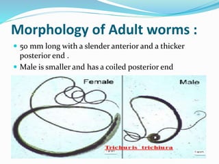
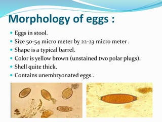
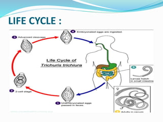
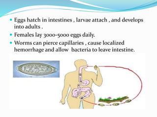
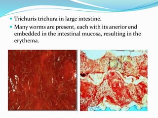
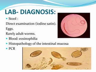
PRESENTED BY:
Man Bdr. Rana
BPH 1st sem 2074
ACAS,Nepal
Morphology of Adult worms :
 50 mm long with a slender anterior and a thicker
posterior end .
 Male is smaller and has a coiled posterior end
Morphology of eggs :
 Eggs in stool.
 Size 50-54 micro meter by 22-23 micro meter .
 Shape is a typical barrel.
 Color is yellow brown (unstained two polar plugs).
 Shell quite thick.
 Contains unembryonated eggs .
LIFE CYCLE :
LIFE CYCLE :
PATHOGENESIS :
 Humans sole host.
 Transmission :
fecal-oral via embryonated ova
• Frequently coexists with ascaris
• Reservoir :
Mainly human , other possible but host specificity not well documented.
• Pathogenic potential:
Low to moderate,dependent on worm numbers and location in large
intestine
• Entirely intraluminal life cycle – eggs are ingested.
 Eggs hatch in intestines , larvae attach , and develops
into adults .
 Females lay 3000-5000 eggs daily.
 Worms can pierce capillaries , cause localized
hemorrhage and allow bacteria to leave intestine.
CLINICAL FEATURES :
 Frequently asymptomatic.
 Clinical sign and symptoms –
adult worms (pathogenic stage)
Dependent on no. of worms none to digestive disturbances , bloody
(frank) mucoid dirrhoea , abdominal pain distention, rectal prolapse ,
and weakness.
• Severe infections :
Tenesmus and rectal prolapse in children
Can be fatal in children
Rarely, elephantiasis in adult.
 Trichuris trichura in large intestine.
 Many worms are present, each with its anerior end
embedded in the intestinal mucosa, resulting in the
erythema.
LAB- DIAGNOSIS:
 Stool :
Direct examination (iodine satin).
Eggs.
Rarely adult worms.
 Blood: eosinophilia
 Histopathology of the intestinal mucosa
 PCR
IMAGING :
 X-Rays Abdomen :
Plain
With contrast/dye
 CT Scan
TREATMENT AND PREVENTION
 Albindazole 400mg once.
 Mebendazole 100mg BD for 3 days.
(600 mg repeated after 2 weeks)
 Pay attention to personal hygiene and eating habits.
Trichuris trichura(whip-worm)

Trichuris trichura(whip-worm)

  • 1.
    PRESENTED BY: Man Bdr.Rana BPH 1st sem 2074 ACAS,Nepal
  • 2.
    Morphology of Adultworms :  50 mm long with a slender anterior and a thicker posterior end .  Male is smaller and has a coiled posterior end
  • 3.
    Morphology of eggs:  Eggs in stool.  Size 50-54 micro meter by 22-23 micro meter .  Shape is a typical barrel.  Color is yellow brown (unstained two polar plugs).  Shell quite thick.  Contains unembryonated eggs .
  • 4.
  • 5.
  • 6.
    PATHOGENESIS :  Humanssole host.  Transmission : fecal-oral via embryonated ova • Frequently coexists with ascaris • Reservoir : Mainly human , other possible but host specificity not well documented. • Pathogenic potential: Low to moderate,dependent on worm numbers and location in large intestine • Entirely intraluminal life cycle – eggs are ingested.
  • 7.
     Eggs hatchin intestines , larvae attach , and develops into adults .  Females lay 3000-5000 eggs daily.  Worms can pierce capillaries , cause localized hemorrhage and allow bacteria to leave intestine.
  • 8.
    CLINICAL FEATURES : Frequently asymptomatic.  Clinical sign and symptoms – adult worms (pathogenic stage) Dependent on no. of worms none to digestive disturbances , bloody (frank) mucoid dirrhoea , abdominal pain distention, rectal prolapse , and weakness. • Severe infections : Tenesmus and rectal prolapse in children Can be fatal in children Rarely, elephantiasis in adult.
  • 9.
     Trichuris trichurain large intestine.  Many worms are present, each with its anerior end embedded in the intestinal mucosa, resulting in the erythema.
  • 10.
    LAB- DIAGNOSIS:  Stool: Direct examination (iodine satin). Eggs. Rarely adult worms.  Blood: eosinophilia  Histopathology of the intestinal mucosa  PCR
  • 11.
    IMAGING :  X-RaysAbdomen : Plain With contrast/dye  CT Scan
  • 12.
    TREATMENT AND PREVENTION Albindazole 400mg once.  Mebendazole 100mg BD for 3 days. (600 mg repeated after 2 weeks)  Pay attention to personal hygiene and eating habits.