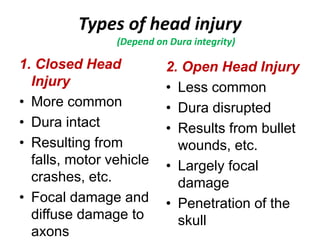
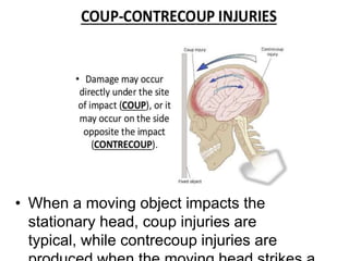
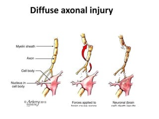
Traumatic brain injuries
Dr. Al-Farah
Neuroscience Module-1
Introduction
• The site has a crucial rule:
–injury of several cubic centimeters of
brain parenchyma may be clinically
silent (e.g. frontal lobe), severely
disabling (e.g. spinal cord), or fatal (e.g.
brain stem)
1. Closed Head
Injury
• More common
• Dura intact
• Resulting from
falls, motor vehicle
crashes, etc.
• Focal damage and
diffuse damage to
axons
2. Open Head Injury
• Less common
• Dura disrupted
• Results from bullet
wounds, etc.
• Largely focal
damage
• Penetration of the
skull
Types of head injury
(Depend on Dura integrity)
Categories of CNS trauma
The physical forces associated with head
injury may result in
1. Skull fractures,
2. Parenchymal injury, and
3. Vascular injury
All three can coexist.
Factors affecting brain trauma
• The magnitude and distribution of
traumatic brain lesions depend on:
the shape of the object causing the
trauma
the force of impact
whether the head is in motion at the
time of injury
1. Skull fractures
-- Linear Skull Fracture
-- Displaced Skull Fracture
-- Basal Skull Fracture (bruising of the
mastoid process and around the eyes, i.e.
"black eyes“, CSF rhinorrhea & otorrhea,
Cranial nerve palsy, Hemotympanum,
Conductive deafness, nystagmus, vomitus.)
-- Diastatic Skull Fracture
• Location of fracture alters the
1. Skull fractures
2. Parenchymal injury
Common location: frontal lobes
along the orbital ridges and the
temporal lobes
Contusion
• When a moving object impacts the
stationary head, coup injuries are
typical, while contrecoup injuries are
2. Parenchymal injury
Plaque jaune or yellow plaque: Remote contusions are
associated with resorption of the injured tissue, resulting in
various degrees of cavitation associated with the presence of a
golden-yellow discoloration due to residual hemosiderin.
3. Diffuse Axonal Injury
• Widespread axonal damage occurring
after a TBI
• Major cause of unconsciousness
and persistent vegetative state after severe
head trauma.
• Cause: The main mechanical force that
causes DAI is rotational acceleration of the
brain, resulting in unrestricted head
movement.
• It constitutes mostly microscopic damage,
Diffuse axonal injury
3. Traumatic Vascular injury
Vascular injury is a frequent component of
CNS trauma.
• Depending on the position of the ruptured
vessel, hemorrhage may be:
1) Epidural hemorrhage
2) Subdural hemorrhage
3) Intracerebral hemorrhage
(Intraparenchymal and intraventricular)
3. Traumatic Vascular injury
Hemorrhages in the
epidural or
subdural space are
typically associated
with trauma
Hemorrhages within
the brain parenchyma
and in the
subarachnoid space, in
contrast, are more
often a manifestation
of underlying
cerebrovascular
disease.
NOTE:
Traumatic hemorrhage
Epidural hemorrhage : hemorrhage
between the durameter and skull following
fracture of the skull
 Most commonly from rupture of middle
meningeal artery.
 The hematoma expands rapidly since
accumulation of blood is arterial and
cause compression of dura and flattening
of underlying gyri.
 Neurosurgical emergency
 Clinically : pt. unconsious followed by
Traumatic hemorrhage
Sub dural hemorrhage
• Hemorrhage within the subdural space.
• Develops most often from rupture of
bridging veins which cross the surface
convexities of the cerebrum.
• Risk : Infants and older individual
Epidural hematoma Subdural hematoma
Traumatic Intracerebral
hemorrhage
Two main types:
I. Intraparencymal hemorrahge- ICH extending
into brain parenchyma
II.Intra-ventricular hemorrhage- ICH extending
into ventricles
Causes:
• Hypertensive vasculopathy (70-80%)
• Ruptured AVM
• Trauma
Intraparencymal hemorrhage-
• Parenchymal vessel rupture from blunt or
penetrating forces.
• May not lose consciousness (unlike Diffuse
axonal injury, contusion)
• Hematoma at primary trauma site (usually
frontal and temporal)
Intra-ventricular hemorrhage-
• Uncommon
• Consequence of severe trauma ass. with DAI
Traumatic Intracerebral
hemorrhage
Traumatic Subarachnoid
Hemorrhage
• Blood collects beneath arachnoid due to tear
of veins in SA space
• Usually associated with other brain injuries
(common with contusions)
• Nearly all cases of traumatic SAH have other
lesions to suggest traumatic cause.
Traumatic brain injury
Sequelae of Brain Trauma
Traumatic brain injuries.ppt

Traumatic brain injuries.ppt

  • 1.
    Traumatic brain injuries Dr.Al-Farah Neuroscience Module-1
  • 2.
    Introduction • The sitehas a crucial rule: –injury of several cubic centimeters of brain parenchyma may be clinically silent (e.g. frontal lobe), severely disabling (e.g. spinal cord), or fatal (e.g. brain stem)
  • 3.
    1. Closed Head Injury •More common • Dura intact • Resulting from falls, motor vehicle crashes, etc. • Focal damage and diffuse damage to axons 2. Open Head Injury • Less common • Dura disrupted • Results from bullet wounds, etc. • Largely focal damage • Penetration of the skull Types of head injury (Depend on Dura integrity)
  • 4.
    Categories of CNStrauma The physical forces associated with head injury may result in 1. Skull fractures, 2. Parenchymal injury, and 3. Vascular injury All three can coexist.
  • 5.
    Factors affecting braintrauma • The magnitude and distribution of traumatic brain lesions depend on: the shape of the object causing the trauma the force of impact whether the head is in motion at the time of injury
  • 6.
    1. Skull fractures --Linear Skull Fracture -- Displaced Skull Fracture -- Basal Skull Fracture (bruising of the mastoid process and around the eyes, i.e. "black eyes“, CSF rhinorrhea & otorrhea, Cranial nerve palsy, Hemotympanum, Conductive deafness, nystagmus, vomitus.) -- Diastatic Skull Fracture • Location of fracture alters the
  • 7.
  • 8.
    2. Parenchymal injury Commonlocation: frontal lobes along the orbital ridges and the temporal lobes
  • 9.
  • 10.
    • When amoving object impacts the stationary head, coup injuries are typical, while contrecoup injuries are
  • 11.
  • 12.
    Plaque jaune oryellow plaque: Remote contusions are associated with resorption of the injured tissue, resulting in various degrees of cavitation associated with the presence of a golden-yellow discoloration due to residual hemosiderin.
  • 13.
    3. Diffuse AxonalInjury • Widespread axonal damage occurring after a TBI • Major cause of unconsciousness and persistent vegetative state after severe head trauma. • Cause: The main mechanical force that causes DAI is rotational acceleration of the brain, resulting in unrestricted head movement. • It constitutes mostly microscopic damage,
  • 14.
  • 15.
    3. Traumatic Vascularinjury Vascular injury is a frequent component of CNS trauma. • Depending on the position of the ruptured vessel, hemorrhage may be: 1) Epidural hemorrhage 2) Subdural hemorrhage 3) Intracerebral hemorrhage (Intraparenchymal and intraventricular)
  • 16.
    3. Traumatic Vascularinjury Hemorrhages in the epidural or subdural space are typically associated with trauma Hemorrhages within the brain parenchyma and in the subarachnoid space, in contrast, are more often a manifestation of underlying cerebrovascular disease. NOTE:
  • 17.
    Traumatic hemorrhage Epidural hemorrhage: hemorrhage between the durameter and skull following fracture of the skull  Most commonly from rupture of middle meningeal artery.  The hematoma expands rapidly since accumulation of blood is arterial and cause compression of dura and flattening of underlying gyri.  Neurosurgical emergency  Clinically : pt. unconsious followed by
  • 18.
    Traumatic hemorrhage Sub duralhemorrhage • Hemorrhage within the subdural space. • Develops most often from rupture of bridging veins which cross the surface convexities of the cerebrum. • Risk : Infants and older individual
  • 20.
  • 22.
    Traumatic Intracerebral hemorrhage Two maintypes: I. Intraparencymal hemorrahge- ICH extending into brain parenchyma II.Intra-ventricular hemorrhage- ICH extending into ventricles Causes: • Hypertensive vasculopathy (70-80%) • Ruptured AVM • Trauma
  • 23.
    Intraparencymal hemorrhage- • Parenchymalvessel rupture from blunt or penetrating forces. • May not lose consciousness (unlike Diffuse axonal injury, contusion) • Hematoma at primary trauma site (usually frontal and temporal) Intra-ventricular hemorrhage- • Uncommon • Consequence of severe trauma ass. with DAI Traumatic Intracerebral hemorrhage
  • 24.
    Traumatic Subarachnoid Hemorrhage • Bloodcollects beneath arachnoid due to tear of veins in SA space • Usually associated with other brain injuries (common with contusions) • Nearly all cases of traumatic SAH have other lesions to suggest traumatic cause.
  • 25.
  • 26.
  • 27.