This document provides an overview of the internal educational program (IEP) of the Vanderbilt University Division of Trauma, Emergency Surgery and Surgical Critical Care. The goal of the IEP is to explore topics related to trauma care from pre-hospital care to injury prevention. The program will outline the full continuum of care provided to trauma patients. It then introduces the trauma team members and multidisciplinary liaisons that will be involved in the educational sessions. The overall goal is to continuously improve trauma patient care and reduce injuries in the local region.
One year mortality rate after hip fracture in the western region of saudi ara...Prof. Hesham N. Mustafa
Background:
The mortality rate of elderly patients who sustain a hip fracture is high compared to the general population. Identifying risk factors can help predict patients at risk of hip fracture to reduce the mortality rate. No studies have shown the mortality rate of patients with hip fractures in the western region of Saudi Arabia. Therefore, this study aimed to identify the risk factors associated with the mortality of patients with hip fractures admitted to the King Abdulaziz Hospital and compare the results with other studies.
Methods:
The mortality rate (within 1 yr or less) in 177 patients over the age of 60 yr who were admitted to the university hospital between July, 2007, and September, 2012, with hip fractures was retrospectively studied. The patients were assessed with regard to gender, age, type of hip fracture, and type of surgical intervention.
Results:
The overall mortality rate 1 yr after hip fracture was 12.43%, and the mean age was 77.77 yr old. The risk factors most associated with mortality were as follows: advanced age (71 to 80 and 81 to 90 yr old), male, peritrochanteric (extracapsular) fracture, and operative fixation with dynamic hip screw.
Conclusions:
The mortality rate of patients with hip fractures within 1 yr has a high-risk potential, especially for male patients over 71 yr of age with peritrochanteric (extracapsular) fractures. Surgical treatment with dynamic hip screw also was shown to be a risk factor between the different treatment options.
Level of Evidence:
Level IV.
Background: Myocarditis is a relatively common inflammatory disease that affects the myocardium. Infectious disease accounts for most of the cases either because of a direct viral infection or post-viral immune-mediated reaction. Cardiovascular magnetic resonance (CMR) has become an established non-invasive diagnosis tool for acute myocarditis. A recent large single centre study with patients with biopsy-proven viral myocarditis undergoing CMR scans found a high rate of mortality. The aim of this study was to assess the rate of clinical events in our population of patients with diagnosed myocarditis by CMR scan.
Methods: Patients who consulted to the emergency department with diagnosis of myocarditis by CMR were retrospectively included in the study from January 2008 to May 2012. A CMR protocol was used in all patients, and were followed up to assess the rate of the composite endpoint of all-cause death, congestive heart failure, sudden cardiac death, hospitalization for cardiac cause, recurrent myocarditis or need of radiofrequency ablation or implantable cardiac defibrillator (ICD). A descriptive statistical analysis was performed.
Results: Thirty-two patients with myocarditis were included in the study. The mean age was 42.6±21.2 years and 81.2% were male. In a mean follow up of 30.4±17.8 months, the rate of the composite endpoint of all-cause death, congestive heart failure, sudden cardiac death, hospitalization for cardiac cause, recurrent myocarditis or need of radiofrequency ablation or ICD was 15.6% (n=5). Two patients had heart failure (one of them underwent heart transplant), one patient needed ICD because of ventricular tachycardia and two other patients were re-hospitalized, for recurrent chest pain and for recurrent myocarditis respectively.
Conclusions: In our series of acute myocarditis diagnosed by CMR we found a low rate of cardiovascular events without mortality. These findings might oppose data from recently published myocarditis trials.
Background: Trauma is a real public health issue, being the third leading cause of death in the world. Blunt Abdominal Trauma (BAT) re presents a significant part of these traumas and constitutes a diagnostic and therapeutic challenge. The aim of this study is to determine the epidemiological and clinical profile of BAT in the Department of General Surgery at Aristide Le Dantec Hospital Patients and methods: This was a retrospective and cross-sectional study from January 2012 to March 2017. Patients who were more than 15 years old with BAT were included. The studied parameters were: age, gender, delay of admission, etiology of the trauma, clinical signs, organ injury, type of treatment and evolution.
Outcomes of Venous Interventions in C5-6 DiseaseVein Global
By: Mark H. Meissner, MD
Visit VeinGlobal at http://www.veinglobal.com/ for more presentations and videos on this topic, or for more information on venous disease news, education and research.
Study of 89 Cases of Peripheral Vascular Disease by CT AngiographyM A Hasnat
The purpose of this study was to observe the morphological pattern by CT angiography
and risk factors for development of peripheral vascular disease in Bangladeshi patient suffering
from peripheral vascular disease using a multidetector scanner in the evaluation of patients with
peripheral vascular disease.
Factors Predicting Neurological Complications Following Percutaneous Coronary Angiography and Interventions in a Large Series of Transfemoral and Transradial Approach.
Ethical Considerations around Urgent Hip Hemiarthroplasty during COVID-19: A ...asclepiuspdfs
Ethical discussion surrounding management of hip fractures in the elderly has always been challenging, but the recent coronavirus disease-19 (COVID-19) pandemic has superimposed additional difficulties that have to be addressed by clinicians. We present a case report of elderly gentleman with a history of moderate to advanced dementia, who was diagnosed with an intertrochanteric femoral fracture after a fall, and was incidentally found to be COVID-19 positive. After a lengthy discussion with next of kin, he had operative management of his hip fracture, followed by a brief convalescence, and eventual discharge to his nursing facility where he passed away.
Is routine thromboprophylaxis warranted in all patients of tibial fracture ma...iosrjce
IOSR Journal of Dental and Medical Sciences is one of the speciality Journal in Dental Science and Medical Science published by International Organization of Scientific Research (IOSR). The Journal publishes papers of the highest scientific merit and widest possible scope work in all areas related to medical and dental science. The Journal welcome review articles, leading medical and clinical research articles, technical notes, case reports and others.
Deep vein thrombosis prophylaxis in a tertiary care center: An observational ...Apollo Hospitals
Deep vein thrombosis (DVT) is a major health problem with substantial mortality and morbidity in medically ill patients. Prevention of DVT by risk factor stratification and subsequent antithrombotic prophylaxis in moderate- to severe-risk category patients is the most rational means of reducing morbidity and mortality.
Deep Venous Thromboembolism in Gynecological Malignanciessemualkaira
Deep venous thrombosis is a severe complication often following
gynecological malignancy. It presents a main reason of post-operative complication, morbidity and mortality in these patients. It is
crucial to know risk factors and to diagnose possible early manifestations of the disease in time [1]. DVT is the second leading
cause of death in patients with gynecologic cancer and the risk of
DVT in women underwent gynecologic surgery ranged from 17%
to 40%, while the rate of pulmonary embolism (PE) was about 1%
to 26% [2].
Surgical Risk Assessment is an Important Factor in any Surgical TreatmentJohnJulie1
Surgical risk is a form of assessing the clinical conditions and health conditions of a person who will undergo surgery, so that the risks of complications are identified throughout the period before, during and after surgery. It is calculated through a physician’s clinical assessment and the requirement for some tests, but to facilitate the assessment, there are also some protocols which have better directing in medical thinking. Any doctor can make this assessment, but most often it is done by a general practitioner, a cardiologist and an anesthesiologist. In this way, it is possible for each person to receive some attention before the surgery, such as seeking more appropriate tests or performing treatments to reduce the risk.
Surgical Risk Assessment is an Important Factor in any Surgical Treatmentsuppubs1pubs1
Surgical risk is a form of assessing the clinical conditions and health conditions of a person who will undergo surgery, so that the risks of complications are identified throughout the period before, during and after surgery. It is calculated through a physician’s clinical assessment and the requirement for some tests, but to facilitate the assessment, there are also some protocols which have better directing in medical thinking. Any doctor can make this assessment, but most often it is done by a general practitioner, a cardiologist and an anesthesiologist. In this way, it is possible for each person to receive some attention before the surgery, such as seeking more appropriate tests or performing treatments to reduce the risk.
TEST BANK For Little and Falace's Dental Management of the Medically Compromi...rightmanforbloodline
TEST BANK For Little and Falace's Dental Management of the Medically Compromised Patient, 10th Edition by Craig Miller, Verified Chapters 1 - 30, Complete Newest Version
Splenomegaly as A Complication Factor in Laparoscopic Splenectomy: Outcomes f...semualkaira
Splenectomy (LS) is believed to be the gold standard in spleen surgery and is considered to be relatively safe with minimal complications, depending on the technology at hand, and the experience of the surgeon.
A brief information about the SCOP protein database used in bioinformatics.
The Structural Classification of Proteins (SCOP) database is a comprehensive and authoritative resource for the structural and evolutionary relationships of proteins. It provides a detailed and curated classification of protein structures, grouping them into families, superfamilies, and folds based on their structural and sequence similarities.
Deep Behavioral Phenotyping in Systems Neuroscience for Functional Atlasing a...Ana Luísa Pinho
Functional Magnetic Resonance Imaging (fMRI) provides means to characterize brain activations in response to behavior. However, cognitive neuroscience has been limited to group-level effects referring to the performance of specific tasks. To obtain the functional profile of elementary cognitive mechanisms, the combination of brain responses to many tasks is required. Yet, to date, both structural atlases and parcellation-based activations do not fully account for cognitive function and still present several limitations. Further, they do not adapt overall to individual characteristics. In this talk, I will give an account of deep-behavioral phenotyping strategies, namely data-driven methods in large task-fMRI datasets, to optimize functional brain-data collection and improve inference of effects-of-interest related to mental processes. Key to this approach is the employment of fast multi-functional paradigms rich on features that can be well parametrized and, consequently, facilitate the creation of psycho-physiological constructs to be modelled with imaging data. Particular emphasis will be given to music stimuli when studying high-order cognitive mechanisms, due to their ecological nature and quality to enable complex behavior compounded by discrete entities. I will also discuss how deep-behavioral phenotyping and individualized models applied to neuroimaging data can better account for the subject-specific organization of domain-general cognitive systems in the human brain. Finally, the accumulation of functional brain signatures brings the possibility to clarify relationships among tasks and create a univocal link between brain systems and mental functions through: (1) the development of ontologies proposing an organization of cognitive processes; and (2) brain-network taxonomies describing functional specialization. To this end, tools to improve commensurability in cognitive science are necessary, such as public repositories, ontology-based platforms and automated meta-analysis tools. I will thus discuss some brain-atlasing resources currently under development, and their applicability in cognitive as well as clinical neuroscience.
Earliest Galaxies in the JADES Origins Field: Luminosity Function and Cosmic ...Sérgio Sacani
We characterize the earliest galaxy population in the JADES Origins Field (JOF), the deepest
imaging field observed with JWST. We make use of the ancillary Hubble optical images (5 filters
spanning 0.4−0.9µm) and novel JWST images with 14 filters spanning 0.8−5µm, including 7 mediumband filters, and reaching total exposure times of up to 46 hours per filter. We combine all our data
at > 2.3µm to construct an ultradeep image, reaching as deep as ≈ 31.4 AB mag in the stack and
30.3-31.0 AB mag (5σ, r = 0.1” circular aperture) in individual filters. We measure photometric
redshifts and use robust selection criteria to identify a sample of eight galaxy candidates at redshifts
z = 11.5 − 15. These objects show compact half-light radii of R1/2 ∼ 50 − 200pc, stellar masses of
M⋆ ∼ 107−108M⊙, and star-formation rates of SFR ∼ 0.1−1 M⊙ yr−1
. Our search finds no candidates
at 15 < z < 20, placing upper limits at these redshifts. We develop a forward modeling approach to
infer the properties of the evolving luminosity function without binning in redshift or luminosity that
marginalizes over the photometric redshift uncertainty of our candidate galaxies and incorporates the
impact of non-detections. We find a z = 12 luminosity function in good agreement with prior results,
and that the luminosity function normalization and UV luminosity density decline by a factor of ∼ 2.5
from z = 12 to z = 14. We discuss the possible implications of our results in the context of theoretical
models for evolution of the dark matter halo mass function.
(May 29th, 2024) Advancements in Intravital Microscopy- Insights for Preclini...Scintica Instrumentation
Intravital microscopy (IVM) is a powerful tool utilized to study cellular behavior over time and space in vivo. Much of our understanding of cell biology has been accomplished using various in vitro and ex vivo methods; however, these studies do not necessarily reflect the natural dynamics of biological processes. Unlike traditional cell culture or fixed tissue imaging, IVM allows for the ultra-fast high-resolution imaging of cellular processes over time and space and were studied in its natural environment. Real-time visualization of biological processes in the context of an intact organism helps maintain physiological relevance and provide insights into the progression of disease, response to treatments or developmental processes.
In this webinar we give an overview of advanced applications of the IVM system in preclinical research. IVIM technology is a provider of all-in-one intravital microscopy systems and solutions optimized for in vivo imaging of live animal models at sub-micron resolution. The system’s unique features and user-friendly software enables researchers to probe fast dynamic biological processes such as immune cell tracking, cell-cell interaction as well as vascularization and tumor metastasis with exceptional detail. This webinar will also give an overview of IVM being utilized in drug development, offering a view into the intricate interaction between drugs/nanoparticles and tissues in vivo and allows for the evaluation of therapeutic intervention in a variety of tissues and organs. This interdisciplinary collaboration continues to drive the advancements of novel therapeutic strategies.
Richard's aventures in two entangled wonderlandsRichard Gill
Since the loophole-free Bell experiments of 2020 and the Nobel prizes in physics of 2022, critics of Bell's work have retreated to the fortress of super-determinism. Now, super-determinism is a derogatory word - it just means "determinism". Palmer, Hance and Hossenfelder argue that quantum mechanics and determinism are not incompatible, using a sophisticated mathematical construction based on a subtle thinning of allowed states and measurements in quantum mechanics, such that what is left appears to make Bell's argument fail, without altering the empirical predictions of quantum mechanics. I think however that it is a smoke screen, and the slogan "lost in math" comes to my mind. I will discuss some other recent disproofs of Bell's theorem using the language of causality based on causal graphs. Causal thinking is also central to law and justice. I will mention surprising connections to my work on serial killer nurse cases, in particular the Dutch case of Lucia de Berk and the current UK case of Lucy Letby.
Seminar of U.V. Spectroscopy by SAMIR PANDASAMIR PANDA
Spectroscopy is a branch of science dealing the study of interaction of electromagnetic radiation with matter.
Ultraviolet-visible spectroscopy refers to absorption spectroscopy or reflect spectroscopy in the UV-VIS spectral region.
Ultraviolet-visible spectroscopy is an analytical method that can measure the amount of light received by the analyte.
THE IMPORTANCE OF MARTIAN ATMOSPHERE SAMPLE RETURN.Sérgio Sacani
The return of a sample of near-surface atmosphere from Mars would facilitate answers to several first-order science questions surrounding the formation and evolution of the planet. One of the important aspects of terrestrial planet formation in general is the role that primary atmospheres played in influencing the chemistry and structure of the planets and their antecedents. Studies of the martian atmosphere can be used to investigate the role of a primary atmosphere in its history. Atmosphere samples would also inform our understanding of the near-surface chemistry of the planet, and ultimately the prospects for life. High-precision isotopic analyses of constituent gases are needed to address these questions, requiring that the analyses are made on returned samples rather than in situ.
This pdf is about the Schizophrenia.
For more details visit on YouTube; @SELF-EXPLANATORY;
https://www.youtube.com/channel/UCAiarMZDNhe1A3Rnpr_WkzA/videos
Thanks...!
Observation of Io’s Resurfacing via Plume Deposition Using Ground-based Adapt...Sérgio Sacani
Since volcanic activity was first discovered on Io from Voyager images in 1979, changes
on Io’s surface have been monitored from both spacecraft and ground-based telescopes.
Here, we present the highest spatial resolution images of Io ever obtained from a groundbased telescope. These images, acquired by the SHARK-VIS instrument on the Large
Binocular Telescope, show evidence of a major resurfacing event on Io’s trailing hemisphere. When compared to the most recent spacecraft images, the SHARK-VIS images
show that a plume deposit from a powerful eruption at Pillan Patera has covered part
of the long-lived Pele plume deposit. Although this type of resurfacing event may be common on Io, few have been detected due to the rarity of spacecraft visits and the previously low spatial resolution available from Earth-based telescopes. The SHARK-VIS instrument ushers in a new era of high resolution imaging of Io’s surface using adaptive
optics at visible wavelengths.
Lateral Ventricles.pdf very easy good diagrams comprehensive
Traumagram spring 2016
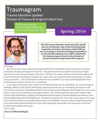
1. Greetings,
Welcome to the internal educational program (IEP) of the Vanderbilt University Division of Trauma,
Emergency Surgery and Surgical Critical Care. Our goal is to provide an opportunity to pursue topics
germane to trauma from all aspects of the team. My hope is to explore all areas of interest throughout the
course of the year including pre-hospital care, acute care issues, post-discharge requirements, as well as
injury prevention. We will attempt to outline the care provided to our trauma patient population from
point of injury until the patients care is completed.
As you know, the trauma team consists of the Chief of the Division, Dr. Rick Miller, our Trauma Program
Manager, Melissa Smith, RN, the Performance Improvement Director, Dr. Tim Nunez, the Outreach and
Prevention coordinator, Cathy Wilson, RN, the Trauma Resuscitation Manager, Kevin High, RN, as well as the
entire trauma faculty and Acute Care Surgery Fellows. Our multidisciplinary liaison team includes Tyler
Barrett (EM), Robert Boyce (Ortho), Reid Thompson (Neurosurgery), Shannon Kilkelly (Anesthesia), Peter
Bream (Radiology) and the LifeFlight team. Our goal is to improve the care of the trauma patient in a caring
and consistent manner and to help minimize injury in the Middle Tennessee region through outreach and
prevention efforts determined by the needs of the community. Please take a few minutes to review this
material and complete the survey.
Yours Truly,
Oscar Guillamondegui
The ACS trauma education requirement (for faculty
who are not liaisons) may be met by documenting
acquisition of 16 hours of trauma-related CME per
year on average or by demonstrating participation in
an internal educational process (IEP) conducted by
the trauma program based on the principles of
practice-based learning and the PIPs program.
A Message from the
Trauma Medical Director,
Oscar Guillamondegui, MD
Spring 2016
2. 2
1. The predominant immunoglobin associated with life threatening hemolytic transfusion reaction
is:
a. IgA
b. IgG
c. IgM
d. IVIG
2. True or False: IVIG can induce hemolytic transfusion reactions or TRALI.
3. According to the Vanderbilt Blood Product Administration policy,
a. The unused blood and IV set including IV solution must be sent to the lab
b. The transfusion should not be restarted under any circumstance
c. The first step is to obtain the patient’s vital signs
d. The post-transfusion patient blood sample should be sent in a blue top.
4. The most common transfusion reaction is:
a. Hemolytic transfusion reaction leading to hematuria, respiratory distress or death
b. Transfusion related acute lung injury
c. Mild temperature elevation and flushing
d. Red urine with urticarial
5. If you suspect a blood transfusion reaction what should be what should be turned into the blood
bank:
a. Remainder of the blood product, including the IV solution and IV set (needle removed);
b. Scan or copy of the TAR;
c. Completed Report of Suspected Transfusion Reaction form; and
d. Post-transfusion patient blood product sample, purple top (EDTA), properly labeled for Blood
Bank.
e. All the above
Answer Key for Winter 2016 Trauma IEP Newsletter
(answers are in bold and Italics below)
3. 3
Rich Lesperance, MD
Case Study: A 49 year-old female was brought to Vanderbilt by EMS after a high-speed motor-vehicle collision.
She has a past medical history of morbid obesity (BMI 40), asthma and Type 2 Diabetes. Her injuries included
multiple long-bone, spine and pelvic fractures, and a ruptured diaphragm. She was admitted to the trauma ICU
and made multiple trips to the operating room over the next few days for repair of her orthopedic, spine and
visceral injuries. Subcutaneous low-molecular weight heparin (LMWH) was initiated as Deep Vein Thrombosis
(DVT) prophylaxis.
The 19th century pathologist, Rudolph Virchow, was the first to scientifically describe venous
thrombosis. The “triad” of venous stasis, hypercoagulability and endothelial injury was named in his honor
(but not actually described by him1) and is still used today to understand the pathophysiology of venous
thrombosis2.
Deep vein thrombosis remains a serious, widespread problem amongst trauma patients. Estimates of
its frequency vary from 12% to 65% in hospitalized trauma patients not receiving prophylaxis3, this
variance may be due to variable screening regimens for DVT (see below). This included an associated
pulmonary embolism rate of 17%4. The risk drops precipitously to approximately 10-12% overall with the
introduction of chemoprophylaxis and mechanical devices to stave off the effects of venous
stasis/immobility.
There are many risk factors for DVT identified in trauma patients. Severe head injury, pelvic injury,
long bone fractures, increased age, greater than one operative procedure and greater than 3 days on a
ventilator have all been associated with an increased risk of a DVT.5,6 It should be noted that many of these
could be considered surrogates of “immobility”. Comorbidities such as diabetes, obesity, renal failure and
malignancies have all been shown to increase the risk of a DVT formation.7
The incidence of DVT increases as screening measures increase. Universal screening of all trauma
patients has been proposed to help identify and start treatment, but this is not a straightforward
proposition. Many of the studies suggesting a benefit to screening were performed prior to widespread use
of pharmacologic DVT prophylaxis. More recent studies showed that universal screening is not cost-
effective, requiring between $20,000 and $65,000 per positive diagnosis.8,9 There is a wide range of opinion
among trauma surgeons on whether universal screening is beneficial, with only about half of surgeons
surveyed stating they routinely screen all trauma patients.10 The most current edition of practice guidelines
from the American College of Chest Physicians recommends against universal screening of trauma
patients.11
Classic physical exam findings for a DVT include swelling, pain or erythema of the affected limb. The
“Homans’ Sign” (calf discomfort on forced dorsiflexion of the foot) has also been taught to generations of
medical providers as a physical exam finding suggestive of a DVT. Unfortunately, DVTs are quite difficult to
diagnose based on physical findings alone -even the surgeon that the Homans’ sign was named after refuted
4. 4
it later in his career.12 Multiple studies on the accuracy of physical exam findings revealed Homans’ sign was
positive less than 50% of the time in all patients (including medical) later confirmed to have a DVT12. In
trauma patients the accuracy is even lower: one of the largest studies to use ultrasound to define the
incidence found that only 2% of the DVTs had physical exam findings suggestive of the diagnosis.4 Because
they are notoriously difficult to diagnose, clinical suspicion and non-invasive imaging are of critical
importance in diagnosing a DVT.
The most common imaging study to screen for and diagnose a DVT is duplex ultrasonography.
Duplex ultrasonography, is highly (>95%) sensitive and specific for DVT in the proximal leg veins, when
compared to contrast venography studies13. Another non-invasive diagnostic modality is impedance
plethysmography, which uses electrical sensors to measure the change in blood volume in the leg when a
venous occlusion tourniquet is released. Impedance plethysmography can also be highly sensitive and
specific14, but requires special equipment and training. Contrast venography has for many years been the
gold standard for diagnosis, but is an invasive angiogram requiring venous cannulation and administration
of contrast material. It is rarely performed. Other modalities of venography including CT or MRI are also
able to determine the presence of a DVT with a high degree of accuracy. However, these studies are
expensive, and require movement of the patient to the scanner and administration of contrast material,
which can be problematic in the trauma population.
The patient underwent several operative procedures during her first few days in the Trauma ICU, and several
doses of LMWH were held as a result. On her fifth hospital day an occlusive thrombus in her common femoral
vein was identified on ultrasound, and a continuous infusion of unfractionated heparin (UH) was started for
therapeutic anticoagulation.
Missed doses of chemoprophylaxis, or delayed initiation, have been found to increase the risk for
DVT 6,15. Common reasons include: not ordering DVT prophylaxis promptly, or holding doses for surgical
procedures due to hemorrhage fears. The Vanderbilt Trauma and Orthopedic services have recently
formalized an understanding that the risk of DVT outweighs the risk of perioperative bleeding in most
trauma patients, and prophylaxis will not be held prior to most abdominal and orthopedic surgeries.
LMWH or low-dose unfractionated heparin (LDUH) are the typical agents used for pharmacologic
DVT prophylaxis. In addition to drugs, mechanical adjuncts to prevent stasis in the lower extremities (ie,
SCDs, compression stockings) have been used as well. Pharmacologic DVT prophylaxis is considered more
effective than mechanical, but the effects are additive so patients should benefit from both.16 LMWH is
thought to be superior to LDUH for DVT prophylaxis17, and enoxaparin is the preferred agent at Vanderbilt.
Twice daily enoxaparin dosing is thought to be the optimal starting dose in the trauma population.
Exceptions include renal failure or high risk bleeding situations such as spinal epidural catheter placement
and intracranial pressure monitor placement, in which case LDUH is used.
The Vanderbilt Department of Trauma and Surgical Critical Care Practice Management Guideline for
DVT prophylaxis can be accessed (by clicking on “Protocols”) via any clinical workstation by clicking the
MDSCC icon, or accessed anywhere at the following URL: https://medschool.vanderbilt.edu/trauma-and-
scc/trauma-and-surgical-critical-care-practice-management-guidelines
5. 5
VUMC Clinical Management Guideline
Group
(see protocol for
risk details)
Risk Interventions
Low /
Moderate
Risk
2-4% DVT
1-2% PE
LMWH + SCDs
for all patients without
contraindications
Renal Failure: lower LMWH dose or
use LDUH.
Indwelling CNS catheter: use LDUH
Obesity: increase LMWH or LDUH
dose
(see protocol for details)
High Risk
4-6% DVT
1-2% PE
Consider surveillance duplex
Very High
Risk
10-20% DVT
5-10% PE
(0.2-5%
fatal)
Consider surveillance duplex
and/or IVC filter
6. 6
References:
Bagot CN, Arya R. Virchow and his triad: a question of attribution. Br J Haematol. Oct 2008;143(2):180-190.
2. Wolberg AS, Aleman MM, Leiderman K, Machlus KR. Procoagulant activity in hemostasis and
thrombosis: Virchow's triad revisited. Anesth Analg. Feb 2012;114(2):275-285.
3. Lozano LM, Perel P, Ker K, Cirocchi R, Farinella E, Morales CH. Thromboprophylaxis for trauma
patients. Cochrane Database Syst Rev. 2010;2010(1).
4. Geerts WH, Code KI, Jay RM, Chen E, Szalai JP. A prospective study of venous thromboembolism after
major trauma. N Engl J Med. Dec 15 1994;331(24):1601-1606.
5. Haut ER, Chang DC, Pierce CA, et al. Predictors of posttraumatic deep vein thrombosis (DVT): hospital
practice versus patient factors-an analysis of the National Trauma Data Bank (NTDB). J Trauma. Apr
2009;66(4):994-999; discussion 999-1001.
6. Michetti CP, Franco E, Coleman J, Bradford A, Trickey AW. Deep vein thrombosis screening and risk
factors in a high-risk trauma population. J Surg Res. Dec 2015;199(2):545-551.
7. Paffrath T, Wafaisade A, Lefering R, et al. Venous thromboembolism after severe trauma: incidence,
risk factors and outcome. Injury. Jan 2010;41(1):97-101.
8. Brasel KJ, Borgstrom DC, Weigelt JA. Cost-effective prevention of pulmonary embolus in high-risk
trauma patients. J Trauma. Mar 1997;42(3):456-460; discussion 460-452.
9. Satiani B, Falcone R, Shook L, Price J. Screening for major deep vein thrombosis in seriously injured
patients: a prospective study. Ann Vasc Surg. Nov 1997;11(6):626-629.
10. Haut ER, Schneider EB, Patel A, et al. Duplex ultrasound screening for deep vein thrombosis in
asymptomatic trauma patients: a survey of individual trauma surgeon opinions and current trauma
center practices. J Trauma. Jan 2011;70(1):27-33; discussion 33-24.
11. Gould MK, Garcia DA, Wren SM, et al. Prevention of VTE in nonorthopedic surgical patients:
Antithrombotic Therapy and Prevention of Thrombosis, 9th ed: American College of Chest Physicians
Evidence-Based Clinical Practice Guidelines. Chest. Feb 2012;141(2 Suppl):e227S-277S.
12. McGee SR. Evidence-based physical diagnosis. 3rd ed. Philadelphia: Elsevier/Saunders; 2012.
13. Gaitini D. Current approaches and controversial issues in the diagnosis of deep vein thrombosis via
duplex Doppler ultrasound. J Clin Ultrasound. Jul-Aug 2006;34(6):289-297.
14. Locker T, Goodacre S, Sampson F, Webster A, Sutton AJ. Meta-analysis of plethysmography and
rheography in the diagnosis of deep vein thrombosis. Emerg Med J. Aug 2006;23(8):630-635.
15. Louis SG, Sato M, Geraci T, et al. Correlation of missed doses of enoxaparin with increased incidence
of deep vein thrombosis in trauma and general surgery patients. JAMA Surg. Apr 2014;149(4):365-
370.
16. Barrera LM, Perel P, Ker K, Cirocchi R, Farinella E, Morales Uribe CH. Thromboprophylaxis for trauma
patients. Cochrane Database Syst Rev. 2013;3:CD008303.
17. Geerts WH, Jay RM, Code KI, et al. A comparison of low-dose heparin with low-molecular-weight
heparin as prophylaxis against venous thromboembolism after major trauma. N Engl J Med. Sep 5
1996;335(10):701-707.
7. 7
The Society of Trauma Nurses is a professional nonprofit organization whose mission is to ensure optimal
trauma care to all people through initiatives focused on trauma nurses related to prevention, education and
collaboration.
At this year’s Society of Trauma Nurses’ conference held in Anaheim, California a few nurses from the Division
of Trauma presented topics on palliative care and research on the Rural Trauma Team Development Course.
Melissa Smith – Trauma Program Mgr
melissa.d.smith@vanderbilt.edu
Oscar Guillamondegui – Trauma Medical
Director
Oscar.guillamondegui@vanderbilt.edu
Tim Nunez – Trauma PI Director
Timothy.c.nunez@vanderbilt.edu
Cathy Wilson – Trauma Outreach & Injury
Prevention Coordinator
Catherine.s.wilson@vanderbilt.edu
Rich Lesperance– ACS Fellow/IEP editor
Richard.lesperance@vanderbilt.edu
Spring 2016 Contributors Trauma
IEP Newsletter
Melissa Smith & Cathy Wilson won the
Research Poster Award for their work
on the Rural Trauma Team
Development course along with Dr.
Brad Dennis.
Teresa Hobt-Bingham presented 2 sessions entitled,
“Improving Palliative Care Consultation in a Trauma ICU
and Step-Down Unit. This is work that is currently going
on in the Trauma ICU.
8. "Safe Steps for Seniors"
The American Trauma Society, in collaboration with
the Society of Trauma Nurses, is once again pleased to present
National Trauma Awareness Month. This May, National
Trauma Awareness Month celebrates its 28th anniversary
with the campaign slogan, “Safe Steps for Seniors” and focuses
on senior safety and falls. Falls are the leading cause of fatal
and non-fatal injuries for older Americans. Falls result in more
than 2.5 million injuries treated in emergency departments
annually, including over 734,000 hospitalizations and more
than 21,700 deaths.
Falls threaten seniors’ safety and independence and
generate enormous economic and personal costs. However,
falling is not an inevitable result of aging. Through practical
lifestyle adjustments, evidence-based falls prevention
programs, and clinical-community partnerships, the number
of falls among seniors can be substantially reduced.
One-third of Americans aged 65+
falls each year.
One out of five falls causes a serious
injury such as broken bones or a
head injury.
Each year, 2.5 million older people
are treated in emergency
departments for fall injuries. That
equates to 1 older adult every 13
seconds.
Over 734,000 patients a year are
hospitalized because of a fall injury,
more than 21,700 suffer fatalities.
Each year at least 250,000 older
people are hospitalized for hip
fractures.
More than 95% of hip fractures are
caused by falling, usually by falling
sideways.
Falls are the most common cause of
traumatic brain injuries (TBI).
Adjusted for inflation, the direct
medical costs for fall injuries are
$34 billion annually. Hospital costs
account for two-thirds of the total.
The financial toll for older adult
falls is expected to increase as the
population ages and may reach
$67.7 billion by 2020.
9. 9
Trauma Admissions are Increasing!!
Melissa Smith in the Division of Trauma has created a diagram to show the increase in Trauma Admissions
that have been steadily going up over the past 5+ years. As Nashville is one of the largest growing cities, we
believe the same will be happening to us!
According to CNN Money Nashville is one of the fastest growing cities: In 2015, 30,000 new residents moved here,
population is now at 1.8 million: http://money.cnn.com/gallery/real_estate/2014/03/27/fastest-growing-cities/7.html
The Tennessean and others have named Nashville the new “IT CITY” bringing in 82 residents a day
http://www.tennessean.com/story/insider/extras/2016/03/26/nashville-has-exciting-growth-uncertainty-city/81109814/
10. 10
Division of Trauma and Surgical Critical Care
For any questions in regards to the IEP or Trauma cases
please contact:
Melissa Smith: 322.6745
or
Oscar Guillamondegui: 936.0180